. 2019 May 20;14(5):e0217183.
doi: 10.1371/journal.pone.0217183. eCollection 2019.

Evaluation of the biocompatibility and stability of allogeneic tissue-engineered cartilage in humanized mice

Affiliations

Evaluation of the biocompatibility and stability of allogeneic tissue-engineered cartilage in humanized mice

Emeline Perrier-Groult et al. PLoS One. .

Abstract

Articular cartilage (AC) has poor capacities of regeneration and lesions often lead to osteoarthritis. Current AC reconstruction implies autologous chondrocyte implantation which requires tissue sampling and grafting. An alternative approach would be to use scaffolds containing off-the-shelf allogeneic human articular chondrocytes (HACs). To investigate tolerance of allogeneic HACs by the human immune system, we developed a humanized mouse model implanted with allogeneic cartilage constructs generated in vitro. A prerequisite of the study was to identify a scaffold that would not provoke inflammatory reaction in host. Therefore, we first compared the response of hu-mice to two biomaterials used in regenerative medicine, collagen sponge and agarose hydrogel. Four weeks after implantation in hu-mice, acellular collagen sponges, but not acellular agarose hydrogels, showed positive staining for CD3 (T lymphocytes) and CD68 (macrophages), suggesting that collagen scaffold elicits weak inflammatory reaction. These data led us to deepen our evaluation of the biocompatibility of allogeneic tissue-engineered cartilage by using agarose as scaffold. Agarose hydrogels were combined with allogeneic HACs to reconstruct cartilage in vitro. Particular attention was paid to HLA-A2 compatibility between HACs to be grafted and immune human cells of hu-mice: HLA-A2+ or HLA-A2- HACs agarose hydrogels were cultured in the presence of a chondrogenic cocktail and implanted in HLA-A2+ hu-mice. After four weeks implantation and regardless of the HLA-A2 phenotype, chondrocytes were well-differentiated and produced cartilage matrix in agarose. In addition, no sign of T-cell or macrophage infiltration was seen in the cartilaginous constructs and no significant increase in subpopulations of T lymphocytes and monocytes was detected in peripheral blood and spleen. We show for the first time that humanized mouse represents a useful model to investigate human immune responsiveness to tissue-engineered cartilage and our data together indicate that allogeneic cartilage constructs can be suitable for cartilage engineering.


Conflict of interest statement

The authors have declared that no competing interests exist.

Figures

Fig 1
Fig 1. Experimental procedure of the study.
(A) Newborn NSG mice were sub-lethally irradiated and intra-hepatically injected with human CD34+ hematopoietic stem cells (hCD34+ HSC) isolated from cord blood samples of HLA-A2+ donors. In parallel, human articular chondrocytes were extracted from knee joint cartilage of HLA-A2+ or HLA-A2- donors, amplified and combined with scaffold. After 3 weeks of in vitro culture in the presence of BIT (200 ng/mL BMP-2, 5 μg/mL insulin, 100 nM T3), HLA-A2+ or HLA-A2- cartilaginous constructs were subcutaneously implanted in the back of the HLA-A2+ humanized mice. 4 weeks after implantation, mice were sacrificed, cartilaginous constructs were harvested for western-blotting and immunohistochemistry analysis and spleens and blood samples were collected for flow cytometry analysis. The photograph shows macroscopic aspect of the implants just after harvesting from the mouse: the cartilaginous construct (black hatched circle) remains adherent to the skin and is vascularized (black arrow: blood capillary). (B) Human cell subpopulations in humanized mice. Flow cytometry analysis was performed 10 weeks after hCD34+ HSC inoculation. Cells were first gated to exclude the doublets. B (CD19+), T (CD3+) cells and macrophages (CD163+) were detected among human CD45+ cells. CD4 and CD8 cells were gated among T cells.
Fig 2
Fig 2. Infiltration of immune cells in acellular collagen sponge.
Immunostaining of human CD3+ T cells and CD68+ macrophages in (A) acellular collagen sponge and (B) in acellular agarose gels, 4 weeks after implantation in hu-mice. Right panels show high magnifications of the zones framed by solid lines in the left pictures. Scale bars are 100 μm.
Fig 3
Fig 3. Stability of HLA-A2- and HLA-A2+ chondrocyte-agarose constructs implanted in HLA-A2+ hu-mice.
Haematoxylin and Eosin (H&S) staining of HLA-A2- (A, B) and HLA-A2+ (C, D) human articular chondrocytes embedded in agarose hydrogels. These constructs were cultured in presence of BIT for 3 weeks in vitro then implanted in HLA-A2- hu-mice for 4 weeks. (B) and (D) show high magnifications of the zones framed by solid lines at the periphery of the implants in (A) and (C), respectively. These zones are fibrous tissues rich in fibroblasts. Black arrows indicate the presence of micro-vessels and yellow arrows the presence of giant cells composed of fused macrophages. (A, C) scale bars are 500 μm and (B, D) scale bars are 20 μm.
Fig 4
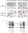
Fig 4. Cartilage-characteristic matrix is produced in vitro in HLA-A2- and HLA-A2+ chondrocyte-agarose constructs then accumulates with time in HLA-A2+ hu-mice.
(A, B) Type II collagen immunostaining of HLA-A2- and HLA-A2+ chondrocyte-agarose constructs after 3 weeks of in vitro culture (before implantation) and after 3 weeks of in vitro culture followed by 4 weeks of in vivo implantation (at the end of the implantation period) in HLA-A2- hu-mice. Note that cells exhibit more intense intra- and peri-cellular staining after, rather than before, implantation. (C) Western blot analysis of type II collagen synthesis before and 4 weeks after implantation. Note that type II collagen was more abundant in the cartilage disks after the implantation period, in accord with the immunohistochemical data. This abundant production also explain why bands are smeared. Scale bars are 20 μm.
Fig 5
Fig 5. Immune cells do not infiltrate agarose gels containing HLA-A2- or HLA-A2+ chondrocytes.
Immunostaining of human CD3+ T cells and CD68+ macrophages in the core (red squares) and at the periphery (blue squares) of HLA-A2- or HLA-A2+ chondrocyte-agarose constructs that were implanted for 4 weeks in HLA-A2+ hu-mice. Note sparse staining of CD3+ and CD68+ cells in the fibrous membranes surrounding the implants. Black scale bars are 500 μm. White scale bars are 100 μm.
Fig 6
Fig 6. Allogeneic cartilage constructed in agarose gel do not trigger peripheral inflammatory/immune response.
Flow cytometry analysis of characteristic human cell surface markers in peripheral blood and spleen of HLA-A2+ mice that were implanted for 4 weeks with HLA-A2+ (A2+) or HLA-A2- (A2-) chondrocyte-agarose constructs. (A) Percentage of cells expressing CD45. (B) Percentage of cells expressing CD3 (total T lymphocyte population) among CD45 positive cells. (C) Comparison of subpopulations of T lymphocytes, CD4+ and CD8+. (D) Determination of the proportion of cells expressing the CD163 marker (monocytes / macrophages) among human cells (hCD45 positive cells). Data are presented as mean±SD. For each graph, the dashed blue line represents the average percentage of cells expressing the cell surface marker when hu-mice were implanted with acellular agarose hydrogels.

References

    1. Görtz S and Bugbee WD. Allografts in articular cartilage repair. J Bone Joint Surg Am. 2006;88: 1374–1384. - PubMed
    1. Langer F and Gross AE. Immunogenicity of allograft articular cartilage. J Bone Joint Surg Am. 1974;56: 297–304. - PubMed
    1. Malejczyk J, Osiecka A, Hyc A, Moskalewski S. Effect of immunosuppression on rejection of cartilage formed by transplanted allogeneic rib chondrocytes in mice. Clin Orthop Relat Res. 1991;269: 266–273. - PubMed
    1. Rahfoth B, Weisser J, Sternkopf F, Aigner T, von der Mark K, Bräuer R. Transplantation of allograft chondrocytes embedded in agarose gel into cartilage defects of rabbits. Osteoarthritis Cartilage. 1998;6: 50–65. 10.1053/joca.1997.0092 - DOI - PubMed
    1. Kawabe N and Yoshinao M. The repair of full-thickness articular cartilage defects. Immune responses to reparative tissue formed by allogeneic growth plate chondrocyte implants. Clin Orthop Relat Res. 1991;268: 279–293. - PubMed

Publication types