Comparative Study
. 2019 Aug 1:376:70-81.
doi: 10.1016/j.taap.2019.05.013. Epub 2019 May 18.

A comparison of particulate hexavalent chromium cytotoxicity and genotoxicity in human and leatherback sea turtle lung cells from a one environmental health perspective

Affiliations
Comparative Study

A comparison of particulate hexavalent chromium cytotoxicity and genotoxicity in human and leatherback sea turtle lung cells from a one environmental health perspective

Rachel M Speer et al. Toxicol Appl Pharmacol. .

Abstract

Evaluating health risks of environmental contaminants can be better achieved by considering toxic impacts across species. Hexavalent chromium [Cr(VI)] is a marine pollutant and global environmental contaminant. While Cr(VI) has been identified as a human lung carcinogen, health effects in marine species are poorly understood. Little is known about how Cr(VI) might impact humans and marine species differently. This study used a One Environmental Health Approach to compare the cytotoxicity and genotoxicity of particulate Cr(VI) in human and leatherback sea turtle (Dermochelys coriacea) lung fibroblasts. Leatherbacks may experience prolonged exposures to environmental contaminants and provide insight to how environmental exposures affect health across species. Since humans and leatherbacks may experience prolonged exposure to Cr(VI), and prolonged Cr(VI) exposure leads to carcinogenesis in humans, in this study we considered both acute and prolonged exposures. We found particulate Cr(VI) induced cytotoxicity in leatherback cells comparable to human cell data supporting current research that shows Cr(VI) impacts health across species. To better understand mechanisms of Cr(VI) toxicity we assessed the genotoxic effects of particulate Cr(VI) in human and leatherback cells. Particulate Cr(VI) induced similar genotoxicity in both cell lines, however, human cells arrested at lower concentrations than leatherback cells. We also measured intracellular Cr ion concentrations and found after prolonged exposure human cells accumulated more Cr than leatherback cells. These data indicate Cr(VI) is a health concern for humans and leatherbacks. The data also suggest humans and leatherbacks respond to chemical exposure differently, possibly leading to the discovery of species-specific protective mechanisms.

Keywords: Chromate; Genotoxicity; Hexavalent chromium; Leatherback sea turtle; Marine pollution; One health.


Conflict of interest statement

Conflict of Interest Statement

Dr. John Wise, Sr. reports grants from the National Institute of Environmental Health Sciences (NIEHS), the Maine Space Grant Consortium (JPW), The Ocean Foundation (JPW), the Henry Foundation (JPW), and the Curtis and Edith Munson Foundation (JPW). There are no other conflicts to declare.

Figures
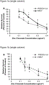
Figure 1:
Figure 1:. Particulate Cr(VI) is similarly cytotoxic in human and leatherback lung cells.
This figure shows relative survival decreases with increasing concentrations of particulate Cr(VI) after (A) 24 h and (B) 120 h exposure in human lung and leatherback cells. Regression analysis was used to determine r2 and LC50 values for 24 h (C) and 120 h (D) exposure in human lung and leatherback cells. Data represent an average of at least three independent experiments ± SEM. *Indicates data point is statistically different (p<0.05) between human lung and leatherback cells; ǂ indicate data point is statistically different (p<0.05) from control.
Figure 2:
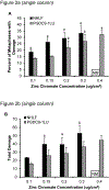
Figure 2:. Particulate Cr(VI) induces similar levels of genotoxicity in human and leatherback lung cells.
This figure shows 24 h exposure particulate Cr(VI) induces increasing levels of (A) percent of metaphases with damage and (B) total damage in human lung and leatherback cells. No metaphases were observed at the highest concentration tested of particulate Cr(VI) (0.4 ug/cm2) in human lung cells. Control values for percent of metaphases with damage after 24 h exposure were 10.3 ± 0.3 and 6.7 ± 1.9 in human and leatherback lung cells, respectively. Control values for total damage after 24 h exposure were 12.3 ± 0.9 and 8.3 ± 2.7 in human and leatherback lung cells, respectively. Similarly, after 120 h exposure the (C) percent of metaphases with damage and (D) total damage also increased in human and leatherback lung cells. No metaphases (NM) were observed in human lung cells at 0.3 ug/cm2 or 0.4 ug/cm2 particulate Cr(VI) and no metaphases were observed in leatherback lung cells at 0.4 ug/cm2 particulate Cr(VI). Control values for percent of metaphases with damage after 120 h exposure were 4 ± 0.6 and 8 ± 0.6 in human and leatherback lung cells, respectively. Control values for total damage after 120 h exposure were 4.3 ± 0.9 and 8.5 ± 1 in human lung and leatherback cell, respectively. Data represent an average of at least three independent experiments ± SEM. *Indicates data point is statistically different (p<0.05) between human lung and leatherback cells; ǂ indicate data point is statistically different (p<0.05) from control. One hundred metaphases were assessed per concentrations in each independent experiment at both 24 h and 100 h exposure. Due to a lack of metaphases at higher treatment concentrations only 88 and 72 metaphases were assessed for two of the human lung experiments after 24 h exposure to 0.3 ug/cm2 zinc chromate. Similarly, after 120 h exposure to 0.3 ug/cm2 zinc chromate only 76 metaphases were assessed in one experiment in leatherback cells.
Figure 3:
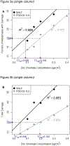
Figure 3:. Regression analysis of the genotoxicity of particulate Cr(VI) in leatherback and human lung cells.
This figure shows the regression analysis used to determine r2 and TC20 values for the (A) percent of metaphases with damage and (B) total damage in 100 metaphases for human and leatherback lung cells after 24 h exposure. Similarly, regression analysis of the 120 h exposure data show the r2 and TC20 values for (C) percent of metaphases with damage and (D) total damage in 100 metaphases for human and leatherback lung cells.
Figure 4:
Figure 4:. Intracellular chromium ion concentrations increase with increasing particulate Cr(VI) exposure in human lung and leatherback cells.
This figure shows with increasing particulate Cr(VI) exposure intracellular chromium ion concentrations increase in a concentration-dependent manner after 24 h and 120 h exposure in human lung and leatherback cells. Data represent an average of at least three independent experiments ± SEM. *Indicates data point is statistically different (p<0.05) between human lung and leatherback cells; ǂ indicate data point is statistically different (p<0.05) from control.
Figure 5:
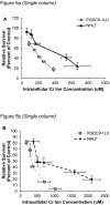
Figure 5:. Particulate Cr(VI) is more cytotoxic to leatherback cells than human lung cells based on intracellular chromium ion concentrations.
This figure shows when comparing intracellular concentrations of Cr ions, particulate Cr(VI) is more cytotoxic in leatherback lung cells than human lung cells after (A) 24 h and (B) 120 h exposure. Data represent an average of at least three independent experiments ± SEM. Regression analysis determined r2 values for human lung and leatherback cells after (C) 24 h exposure and (D) 120 h exposure to be significant predictors of relative survival. *Indicates data point is statistically different (p<0.05) between human lung and leatherback cells; ǂ indicate data point is statistically different (p<0.05) from control.
Figure 6:
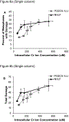
Figure 6:. More genotoxicity occurs at lower intracellular Cr ion concentrations in leatherback cells than human lung cells after prolonged exposure.
This figure shows after 24 h exposure to particulate Cr(VI) the (A) percent of metaphases with damage and (B) total damage is higher in human lung cells than leatherback cells at similar intracellular Cr ion levels compared. However, after 120 h exposure to particulate Cr(VI) the (C) percent of metaphases with damage and (D) total damage is greater in leatherback lung cells than human lung cells at similar intracellular Cr ion concentrations. Data represent an average of three independent experiments ± SEM. *Indicates data point is statistically different (p<0.05) between human and leatherback lung cells; ǂ indicate data point is statistically different (p<0.05) from control.
Figure 7:
Figure 7:. Regression analysis of the genotoxicity of particulate Cr(VI) in human and leatherback lung fibroblasts based on intracellular Cr ion concentrations.
This figure shows the regression analysis used to determine r2 and TC20 values for the (A) percent of metaphases with damage and (B) total damage in 100 metaphases for human and leatherback lung cells after 24 h exposure based on intracellular Cr ion concentrations. Similarly, regression analysis of the 120 h exposure data show the r2 and TC20 values for (C) percent of metaphases with damage and (D) total damage in 100 metaphases for human lung and leatherback cells based on intracellular Cr ion concentration.

Similar articles

Cited by

References

    1. Aguirre AA, Tabor GM, 2004. Introduction: Marine vertebrates as sentinels of marine ecosystem health. EcoHealth. 1: 236–238. DOI: 10.1007/s10393-004-0091-9. - DOI
    1. Al-Hamood MH, Elbetieha A, Bataineh H, 1998. Sexual maturation and fertility of male and female mice exposed prenatally and postnatally to trivalent and hexavalent chromium compounds. Reprod. Fertil. Dev 10, 179–183., doi:10.1071/r97001. - DOI - PubMed
    1. Archibald DW, James MC, 2016. Evaluating inter-annual relative abundance of leatherback sea turtles in Atlantic Canada. Marine Ecology, 547: 233–246.
    1. Bataineh H, Al-Hamood MH, Elbetieha A, Bani Hani I, 1997. Effect of long-term ingestion of chromium compounds on aggression, sex behavior and fertility in adult male rat. Drug Chem. Toxicol 20, 133–149., doi:10.3109/01480549709003875. - DOI - PubMed
    1. Bickler PE, and Buck LT 2007. Hypoxia tolerance in reptiles, amphibians, and fishes: life with variable oxygen availability. Annu. Rev. Physiol 69, 145–170. - PubMed

Publication types

MeSH terms