. 2019 Aug:80:763-776.
doi: 10.1016/j.bbi.2019.05.025. Epub 2019 May 17.

Handling stress impairs learning through a mechanism involving caspase-1 activation and adenosine signaling

Affiliations

Handling stress impairs learning through a mechanism involving caspase-1 activation and adenosine signaling

Albert E Towers et al. Brain Behav Immun. 2019 Aug.

Abstract

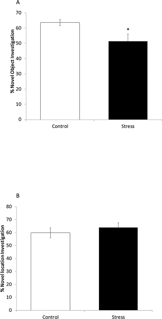
Acute stressors can induce fear and physiologic responses that prepare the body to protect from danger. A key component of this response is immune system readiness. In particular, inflammasome activation appears critical to linking stress to the immune system. Here, we show that a novel combination of handling procedures used regularly in mouse research impairs novel object recognition (NOR) and activates caspase-1 in the amygdala. In male mice, this handling-stress paradigm combined weighing, scruffing and sham abdominal injection once per hr. While one round of weigh/scruff/needle-stick had no impact on NOR, two rounds compromised NOR without impacting location memory or anxiety-like behaviors. Caspase-1 knockout (KO), IL-1 receptor 1 (IL-1R1) KO and IL-1 receptor antagonist (IL-RA)-administered mice were resistant to handling stress-induced loss of NOR. In addition, examination of the brain showed that handling stress increased caspase-1 activity 85% in the amygdala without impacting hippocampal caspase-1 activity. To delineate danger signals relevant to handling stress, caffeine-administered and adenosine 2A receptor (A2AR) KO mice were tested and found resistant to impaired learning and caspase-1 activation. Finally, mice treated with the β-adrenergic receptor antagonist, propranolol, were resistant to handling stress-induced loss of NOR and caspase-1 activation. Taken together, these results indicate that handling stress-induced impairment of object learning is reliant on a pathway requiring A2AR-dependent activation of caspase-1 in the amygdala that appears contingent on β-adrenergic receptor functionality.

Keywords: Adenosine; Amygdala; Caffeine; Caspase-1; Learning; Stress; β-Adrenergic receptor.


Figures

Figure 1.
Figure 1.. Handling stress paradigm.
At the start of the dark cycle (time 0 hr), mice were subjected to weighing, scruffing, and IP needle stick, as indicated. Six hour stress (6 hr stress) was defined as two rounds of weigh/scruff/needle stick and is illustrated. Three hour stress (3 hr stress) was defined as only one round of weigh/scruff/needle stick.
Figure 2.
Figure 2.. Repeated stress handling impairs recognition memory but not spatial memory or anxiety-like behavior.
(A) Mice were exposed to control conditions (control) or 6 hr handling stress (stress) then tested for NOR. Results are expressed as mean ± SEM % novel object investigation; n=7 (*p<0.05). (B) Mice were treated as in A and tested for NLR was performed. Results are expressed as mean ± SEM % Novel location investigation; n=8. (C) Mice were treated as in A & B mice and tested for EZM performance. Results are expressed as mean ± SEM % time in open arms; n=12. (D) Mice were treated as in A & B mice and blood glucose was measured. Results are expressed as mean ± SEM Blood Glucose; n=9 (*p<0.05). (E) Mice were treated as in A & B mice and home-cage locomotor activity was measured. Results are expressed as mean ± SEM cm moved during each time period; n=6 (*p<0.05). A-E were analyzed using a One-way ANOVA.
Figure 3.
Figure 3.. Repeated stress handling activates caspase-1 in the brain.
(A) Mice were exposed to control conditions (control) or 6 hr handling stress (stress). Activated caspase-1 was examined by IHC. Results are expressed as mean ± SEM vs. control group; n=10–12 (*p<0.05). Analyzed using a One-way ANOVA for each region. (B) is a representative image from A (40x).
Figure 4.
Figure 4.. Mice deficient in caspase-1 or the IL-1R1 lack stress handling-induced memory impairment.
(A) C57 Wild-type (WT) and Caspase-1 KO (Casp-1 KO) mice were exposed to control conditions (control) or stress handling (stress) for 6hr. NOR was performed 3 hr following stress exposure. NOR was performed 3 hr following stress exposure. Results are expressed as mean ± SEM % novel object investigation; n=8. (B) As in A, WT and IL1R1 KO (IL1R1) mice were subjected to either control conditions or stress handling for 6 hr and NOR was performed 3 hr following stress exposure. Results are expressed as mean ± SEM % novel object recognition; n=7–9. (C) Mice were exposed stress or control conditions as in A & B immediately following administration of the IL1R1 inhibitor Kineret. NOR was performed 3 hr following stress exposure. Results are expressed as mean ± SEM % novel object recognition. For both A-C values without a common superscript are significantly different (p<0.05), and were analyzed using a Two-way ANOVA.
Figure 5.
Figure 5.. Adenosine A2AR antagonism prevents stress handling-induced memory impairment and the activation of caspase-1 in the amygdala.
Mice were exposed to control conditions (control) or stress stimuli (stress) for 6 hr. (A) prior to stress exposure mice were administered the pan-adenosine receptor antagonist caffeine. NOR was performed 3 hr following stress exposure. Results are expressed as mean ± SEM % novel object investigation; n=7–8. (B) Mice were treated as in A and caspase-1 activity was measured by IHC. Results are expressed as mean ± SEM % control vs. saline treated controls; n=8–10. (C) Representative images (40x) from the amygdala from B. (D)Mice were stressed as in A and administered the A2AR specific antagonist DMPX prior to stress exposure or a 2% DMSO vehicle control. NOR was performed 3 hr after stress exposure. Results are expressed as mean ± SEM % novel object investigation; n= 7–8. (E) Mice were treated as in D and caspase-1 activity was measured via IHC. Results are expressed as % control vs. DMSO treated control mice; n=7–8. (F) Representative amygdala images (40x) from E. (G) BALB/c control (WT) and A2AR KO mice were stressed as in A. NOR was performed 3 hr following stress exposure. Results are expressed as mean ± SEM % novel object investigation; n=8. (H) WT and A2AR KO mice were treated as in G and caspase-1 activity was measured via IHC. Results are expressed as mean ± SEM % control vs. BALB/c controls; n=10–11. (I) Representative amygdala images (40x) from H. (J) Mice were administered IP saline or IP adenosine. NOR was performed 30 min following adenosine administration. Results are expressed as mean ± SEM % novel object investigation; n= 8 (*p<0.05). (K) Mice were treated as in J and caspase-1 activity was measured by IHC. Results are expressed as % control vs. saline; n= 6–8 (*p<0.05). (L) Representative amygdala images (40x) from K. (M) Mice were treated as in J but were also ICV injected with biotin-YVAD-cmk immediately following adenosine injection. NOR was performed 30 min following YVAD injection. Results are expressed as mean ± SEM % novel object investigation; n= 4–5). For A-M values without a common superscript are significantly different (p<0.05). A-I & M were analyzed using a Two-way ANOVA procedure, J-K were analyzed using a One-way ANOVA.
Figure 6.
Figure 6.. Catecholamine but not glucocorticoid antagonism blocks stress handling-induced memory impairment and caspase-1 activation.
Mice exposed to control conditions (control) or stress stimuli (stress) for 6 hr. (A) Prior to stress exposure mice were administered the β-adrenergic receptor antagonist propranolol or vehicle control. NOR was performed 3 hr following stress exposure. Results are expressed as means ± SEM % novel object investigation; n=8. (B) Mice were treated identically to A and active caspase-1 was measured via IHC. Results are expressed as mean ± SEM % Control vs. saline treated control mice; n=7–9. (C) Representative images (40x) from the amygdala of B. (D) Mice were treated identically to A, but prior to stress exposure mice were administered the glucocorticoid receptor antagonist mifepristone or a 5% DMSO vehicle control. Results are expressed as means ± SEM % novel object investigation; n=8. For A-D values without a common superscript are significantly different (p<0.05), and all were analyzed using a Two-way ANOVA procedure.

Similar articles

Cited by

References

    1. Abdul-Muneer PM, Long M, Conte AA, Santhakumar V, Pfister BJ, 2017. High Ca2+ Influx During Traumatic Brain Injury Leads to Caspase-1-Dependent Neuroinflammation and Cell Death. Mol. Neurobiol. 54, 3964–3975. 10.1007/s12035-016-9949-4 - DOI - PMC - PubMed
    1. AHA hospital statistics, 2017. Health Forum LLC.
    1. Ambrogi Lorenzini C, Bucherelli C, Giachetti A, Tassoni G, 1987. Spontaneous and conditioned behavior of Wistar and Long Evans rats. Arch. Ital. Biol 125, 155–70. - PubMed
    1. An D, Chen W, Yu D-Q, Wang S-W, Yu W-Z, Xu H, Wang D-M, Zhao D, Sun Y-P, Wu J-C, Tang Y-Y, Yin S-M, 2017. Effects of social isolation, re-socialization and age on cognitive and aggressive behaviors of Kunming mice and BALB/c mice. Anim. Sci. J 88, 798–806. 10.1111/asj.12688 - DOI - PubMed
    1. Arnsten AF, Dudley AG, 2005. Methylphenidate improves prefrontal cortical cognitive function through alpha2 adrenoceptor and dopamine D1 receptor actions: Relevance to therapeutic effects in Attention Deficit Hyperactivity Disorder. Behav. Brain Funct 1, 2 10.1186/1744-9081-1-2 - DOI - PMC - PubMed

Publication types

MeSH terms