Review
. 2020 Jan;1865(1):158461.
doi: 10.1016/j.bbalip.2019.05.005. Epub 2019 May 18.

Lipid synthesis and transport are coupled to regulate membrane lipid dynamics in the endoplasmic reticulum

Affiliations
Review

Lipid synthesis and transport are coupled to regulate membrane lipid dynamics in the endoplasmic reticulum

Tamas Balla et al. Biochim Biophys Acta Mol Cell Biol Lipids. 2020 Jan.

Abstract

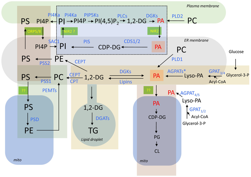
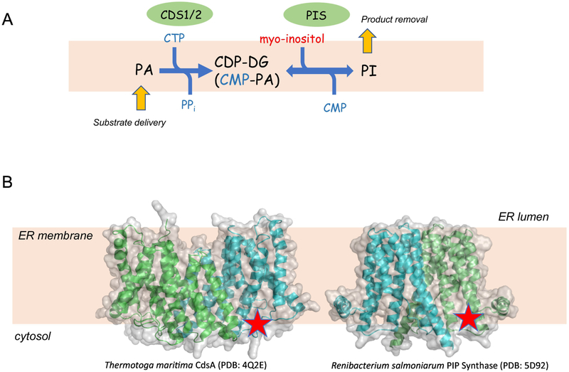
Structural lipids are mostly synthesized in the endoplasmic reticulum (ER), from which they are actively transported to the membranes of other organelles. Lipids can leave the ER through vesicular trafficking or non-vesicular lipid transfer and, curiously, both processes can be regulated either by the transported lipid cargos themselves or by different secondary lipid species. For most structural lipids, transport out of the ER membrane is a key regulatory component controlling their synthesis. Distribution of the lipids between the two leaflets of the ER bilayer or between the ER and other membranes is also critical for maintaining the unique membrane properties of each cellular organelle. How cells integrate these processes within the ER depends on fine spatial segregation of the molecular components and intricate metabolic channeling, both of which we are only beginning to understand. This review will summarize some of these complex processes and attempt to identify the organizing principles that start to emerge. This article is part of a Special Issue entitled Endoplasmic reticulum platforms for lipid dynamics edited by Shamshad Cockcroft and Christopher Stefan.

Keywords: Endoplasmic reticulum; Lipid transfer protein; Membrane contact sites; Non-vesicular lipid transfer; Phosphatidylcholine; Phosphatidylinositol; Phosphatidylserine.


Conflict of interest statement

The Authors declare no conflict of interest related to this work.

Figures

Figure 1:
Figure 1:
Enzymatic pathways of lipid synthesis and their membrane localization in a generic mammalian cell. The enzymes are labeled blue and the lipids are black except PA (red) which represents a major hub for several metabolic pathways. Major organelles are designated by the rounded shapes. Metabolic units that have been linked are designated by rectangles of different colors. The dotted lines denote inositol lipids that serve as signaling entities. Lipid transfer proteins working in contact sites are labeled yellow on green background. Note that lipids in the ER can be part of multiple metabolic units and how these are kept separated is one of the important questions yet to be understood. Abbreviations of enzymes: PI4K, PI 4-kinase; PIP5K, PI4P 5-kinase; PLC, phospholipase C; DGK, diacylglycerol kinase; PLD, phospholipase D; Sac1, Sac1 phosphatase; PIS, phosphatidylinositol synthase; CDS, CDP-DG synthase; PSS, phosphatidylserine synthase; CEPT, choline/ethanolamine phosphotransferase; CPT, CDP-choline:1,2-diacylglycerolphosphocholine transferase; PSD, phosphatidylserine decarboxylase; PEMT, Phosphatidylethanolamine N-methyltransferase; DGAT, diacylglycerol acyl transferase; AGPATs, 1-Acylglycerol-3-Phosphate O-Acyltransferase (* denotes that several forms of these enzymes exist in the ER. AGPAT4 and −5 are found in mitochondria); GPAT, Glycerol-3-Phosphate Acyltransferase. Abbreviation of lipids: PI, phosphatidylinositol; PI4P, phosphatidylinositol 4-phosphate; PI(4,5)P2, phosphatidylinositol 4,5-bisphosphate; DG, diacylglycerol; PA, phosphatidic acid; PS, phosphatidylserine; PC, phosphatidylcholine; PE, phosphatidylethanolamine; TG, triacylglycerol; PG, phosphatidylglycerol; CL, cardiolipin; CDP-DG, cytidine-diphosphate-diacylglycerol.
Figure 2:
Figure 2:
The enzymatic reactions of PI synthesis. (A) The two enzymes that work in tandem are CDS1/2 that convert PA to CDP-DG and, PIS, which converts CDP-DG to PI. Note that the PIS-catalyzed step is reversible and hence, the PI lipid product has to be removed in order to keep the reaction flowing to the right. Yellow arrows show that the direction of the reaction is determined by substrate delivery and product removal. (B) The X-ray structures of bacterial homologues (or ancestors) of the two enzymes are shown with the active site (red star) facing the cytoplasmic aspect of the ER membrane. The structures of the Thermotoga maritima CdsA (PDB: 4Q2E) (the closest bacterial homologue of the CDS enzymes) and the Renibacterium salmoniarum PIP Synthase (PDB: 5D92) (the closest bacterial homologue of the PIS enzyme) are shown using Pymol for rendering.

Similar articles

Cited by

References

    1. Kent C, Eukaryotic phospholipid biosynthesis, Annu Rev Biochem, 64 (1995) 315–343. - PubMed
    1. Yang Y, Lee M, Fairn GD, Phospholipid subcellular localization and dynamics, J Biol Chem, 293 (2018) 6230–6240. - PMC - PubMed
    1. Gonzalez-Baro MR, Coleman RA, Mitochondrial acyltransferases and glycerophospholipid metabolism, Biochim Biophys Acta, 1862 (2017) 49–55. - PubMed
    1. Wang H, Airola MV, Reue K, How lipid droplets "TAG" along: Glycerolipid synthetic enzymes and lipid storage, Biochim Biophys Acta, 1862 (2017) 1131–1145. - PMC - PubMed
    1. Wilfling F, Wang H, Haas JT, Krahmer N, Gould TJ, Uchida A, Cheng JX, Graham M, Christiano R, Frohlich F, Liu X, Buhman KK, Coleman RA, Bewersdorf J, Farese RV Jr., Walther TC, Triacylglycerol synthesis enzymes mediate lipid droplet growth by relocalizing from the ER to lipid droplets, Dev Cell, 24 (2013) 384–399. - PMC - PubMed

Publication types

LinkOut - more resources