

Microstructural neuroimaging of white matter tracts in persistent post-concussion syndrome: A prospective controlled cohort study
- PMID: 31108457
- PMCID: PMC6526293
- DOI: 10.1016/j.nicl.2019.101842
Microstructural neuroimaging of white matter tracts in persistent post-concussion syndrome: A prospective controlled cohort study
Abstract
Introduction: Children with mild traumatic brain injury (mTBI) typically recover quickly, however approximately 15% experience persistent post-concussive symptoms (PPCS) past 3 months. The microstructural pathology associated with underlying persistent symptoms is poorly understood but is suggested to involve axonal injury to white matter tracts. Diffusion tensor imaging (DTI) can be used to visualize and characterize damage to white matter microstructure of the brain.
Objective: We aimed to investigate white matter microstructure in children with persistent concussive symptoms as compared to typically developing controls, alongside evaluating differences in white matter changes over time and how this relates to symptom recovery.
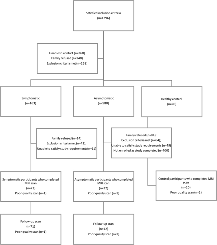
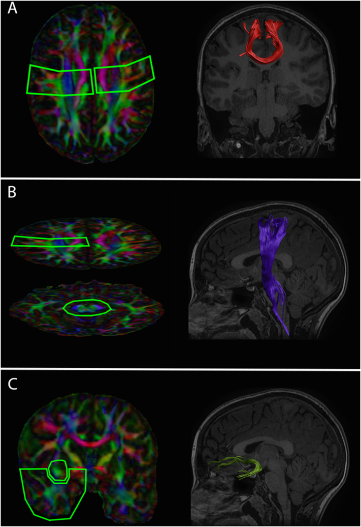
Methods: The current study is a prospective, longitudinal, controlled cohort study of children with mTBI. 104 children aged 8 to 18 years with a mTBI (72 symptomatic; 32 asymptomatic) were recruited from the Alberta Children's Hospital and compared to 20 healthy controls. Microstructural evidence of white matter injury was evaluated using DTI one month post injury and repeated 4 to 6 weeks later. Primary outcomes included fractional anisotropy and mean diffusivity of the corticospinal tracts, uncinate fasciculi, and motor fibers of the corpus callosum. Post-concussive symptoms were also measured using the Post-Concussion Symptom Inventory (PCSI) taken at both time points.
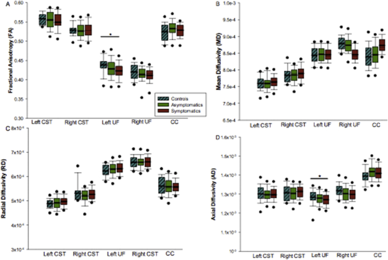
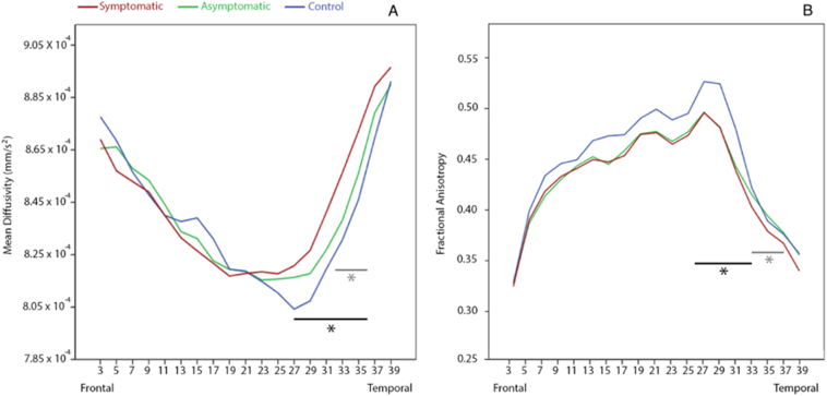
Results: Fractional anisotropy of the left uncinate fasciculi was lower in symptomatic children compared to controls (F(2,119) = 3.582, p = 0.031). No other significant differences were observed.
Conclusions: Our findings provide evidence of microstructural injury following mTBI in children with ongoing post-concussive symptoms one month post injury. The changes were persistent 4-6 weeks later. Further longitudinal studies of white matter microstructure in PPCS will be helpful to clarify whether these white matter alterations resolve over time.
Keywords: Diffusion tensor imaging; Mild traumatic brain injury; Pediatrics; Persistent post-concussive symptoms; Uncinate fasciculus.
Copyright © 2019. Published by Elsevier Inc.
Figures
Similar articles
-
Evidence of Ongoing Cerebral Microstructural Reorganization in Children With Persisting Symptoms Following Mild Traumatic Brain Injury: A NODDI DTI Analysis.J Neurotrauma. 2024 Jan;41(1-2):41-58. doi: 10.1089/neu.2023.0196. Epub 2023 Nov 29. J Neurotrauma. 2024. PMID: 37885245
-
Longitudinal white matter microstructural changes in pediatric mild traumatic brain injury: An A-CAP study.Hum Brain Mapp. 2022 Aug 15;43(12):3809-3823. doi: 10.1002/hbm.25885. Epub 2022 Apr 25. Hum Brain Mapp. 2022. PMID: 35467058 Free PMC article.
-
Post-acute white matter microstructure predicts post-acute and chronic post-concussive symptom severity following mild traumatic brain injury in children.Neuroimage Clin. 2020;25:102106. doi: 10.1016/j.nicl.2019.102106. Epub 2019 Dec 13. Neuroimage Clin. 2020. PMID: 31896466 Free PMC article.
-
Evidence for Altered White Matter Organization After Mild Traumatic Brain Injury: A Scoping Review on the Use of Diffusion Magnetic Resonance Imaging and Blood-Based Biomarkers to Investigate Acute Pathology and Relationship to Persistent Post-Concussion Symptoms.J Neurotrauma. 2025 Apr;42(7-8):640-667. doi: 10.1089/neu.2024.0039. Epub 2024 Aug 21. J Neurotrauma. 2025. PMID: 39096132
-
Brain dysfunction underlying prolonged post-concussive syndrome: A systematic review.J Affect Disord. 2020 Feb 1;262:71-76. doi: 10.1016/j.jad.2019.10.058. Epub 2019 Nov 4. J Affect Disord. 2020. PMID: 31710931 Free PMC article.
Cited by
-
White matter microstructure of the neural emotion regulation circuitry in mild traumatic brain injury.Eur J Neurosci. 2021 May;53(10):3463-3475. doi: 10.1111/ejn.15199. Epub 2021 Apr 2. Eur J Neurosci. 2021. PMID: 33759227 Free PMC article.
-
Assessment of white matter microstructure integrity in subacute postconcussive vestibular dysfunction using NODDI.Imaging Neurosci (Camb). 2024;2:imag-2-00147. doi: 10.1162/imag_a_00147. Epub 2024 Apr 10. Imaging Neurosci (Camb). 2024. PMID: 40746965 Free PMC article.
-
A Scoping Review of Magnetic Resonance Modalities Used in Detection of Persistent Postconcussion Symptoms in Pediatric Populations.J Child Neurol. 2023 Feb;38(1-2):85-102. doi: 10.1177/08830738221120741. Epub 2022 Nov 15. J Child Neurol. 2023. PMID: 36380680 Free PMC article.
-
Examining brain white matter after pediatric mild traumatic brain injury using neurite orientation dispersion and density imaging: An A-CAP study.Neuroimage Clin. 2021;32:102887. doi: 10.1016/j.nicl.2021.102887. Epub 2021 Nov 19. Neuroimage Clin. 2021. PMID: 34911193 Free PMC article.
-
White Matter Abnormalities Associated With Prolonged Recovery in Adolescents Following Concussion.Front Neurol. 2021 Jun 24;12:681467. doi: 10.3389/fneur.2021.681467. eCollection 2021. Front Neurol. 2021. PMID: 34248824 Free PMC article.
References
-
- Adams H., Mitchell D.E., Graham D.I., Doyle D. Diffuse brain damage of immediate impact type. Its relationship to'primary brain-stem damage'in head injury. Brain. 1977;100(3):489–502. - PubMed
-
- Adams J.H., Doyle D., Ford I., Gennarelli T.A., Graham D.I., McLellan D.R. Diffuse axonal injury in head injury: definition, diagnosis and grading. Histopathol. 1989;15(1):49–59. - PubMed
-
- Armstrong R.C., Mierzwa A.J., Marion C.M., Sullivan G.M. White matter involvement after TBI: clues to axon and myelin repair capacity. Exp. Neurol. 2016;275:328–333. - PubMed
-
- Barlow K.M., Crawford S., Stevenson A., Sandhu S.S., Belanger F., Dewey D. Epidemiology of postconcussion syndrome in pediatric mild traumatic brain injury. Pediatr. 2010;126(2):e374. (peds-2009) - PubMed
-
- Barlow K.M., Brooks B.L., MacMaster F.P., Kirton A., Seeger T., Esser M.…Kirk V. A double-blind, placebo-controlled intervention trial of 3 and 10 mg sublingual melatonin for post-concussion syndrome in youths (PLAYGAME): study protocol for a randomized controlled trial. Trials. 2014;15(1):271. - PMC - PubMed
Publication types
MeSH terms
Grants and funding
LinkOut - more resources
Full Text Sources
Miscellaneous
