Review
. 2019 Jun;45(4):354-372.
doi: 10.1055/s-0039-1688497. Epub 2019 May 20.

Use of Viscoelastography in Malignancy-Associated Coagulopathy and Thrombosis: A Review

Affiliations
Review

Use of Viscoelastography in Malignancy-Associated Coagulopathy and Thrombosis: A Review

Mark Walsh et al. Semin Thromb Hemost. 2019 Jun.

Abstract

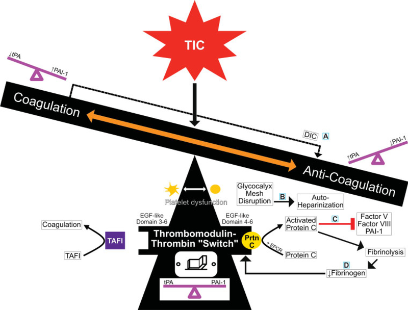
The relationship between malignancy and coagulopathy is one that is well documented yet incompletely understood. Clinicians have attempted to quantify the hypercoagulable state produced in various malignancies using common coagulation tests such as prothrombin time, activated partial thromboplastin time, and platelet count; however, due to these tests' focus on individual aspects of coagulation during one specific time point, they have failed to provide clinicians the complete picture of malignancy-associated coagulopathy (MAC). Viscoelastic tests (VETs), such as thromboelastography (TEG) and rotational thromboelastometry (ROTEM), are whole blood analyses that have the advantage of providing information related to the cumulative effects of plasma clotting factors, platelets, leukocytes, and red cells during all stages of the coagulation and fibrinolytic processes. VETs have gained popularity in the care of trauma patients to objectively measure trauma-induced coagulopathy (TIC), but the utility of VETs remains yet unrealized in many other medical specialties. The authors discuss the similarities and differences between TIC and MAC, and propose a mechanism for the hypercoagulable state of MAC that revolves around the thrombomodulin-thrombin complex as it switches between activating the protein C anticoagulation pathway or the thrombin activatable fibrinolysis inhibitor coagulation pathway. Additionally, they review the current literature on the use of TEG and ROTEM in patients with various malignancies. Although limited research is currently available, early results demonstrate the utility of both TEG and ROTEM in the prediction of hypercoagulable states and thromboembolic complications in oncologic patients.


Conflict of interest statement

Dr. Walsh reports grants from Haemonetics, outside the submitted work. Dr. H. Moore received research support from Instrument Laboratories and Haemonetics. He also shares intellectual property with Haemonetics and Thrombo Therapeutics Incorporated. He is also on the board of Thrombo Therapeutics Incorporated. Dr. E. Moore reports research grant from Haemonetics, outside the submitted work. In addition, he has a patent tPA TEG issued. Dr. Achneck reports having served as an employee of Haemonetics in the position of Director of Medical Affairs and Clinical Development during writing of the manuscript. The other authors have no conflict of interest to disclose.

Figures

Fig. 1
Fig. 1
Proposed molecules and cells involved in the hypercoagulable/hypofibrinolytic phenotype present in malignancy-associated coagulopathy (MAC). ADP, adenosine diphosphate; NETs, neutrophil extracellular traps; PAI-1, plasminogen activator inhibitor-1; TNF, tissue necrosis factor.
Fig. 2
Fig. 2
Proposed mechanisms for trauma-induced coagulopathy (TIC). This teeter-totter spectrum shows the balance and molecular interplay between hypocoagulable/hyperfibrinolytic and hypercoagulable/hypofibrinolytic phenotypes in TIC. There are four current hypotheses to explain TIC which are each centered around the balance of key molecules as follows: (A)DIC–fibrinolysis, (B) glycocalyx, (C) activated protein C, and (D) fibrinogen. These hypotheses are not mutually exclusive, but instead explain the mechanisms at play in TIC within minutes to hours following trauma. At the center, the TM–thrombin complex is the fulcrum of coagulation regulation. Structural and/or posttranslational covalent modifications of the TM–thrombin complex, along with certain cofactors and receptors, allow the TM–thrombin complex to switch between activating the protein C anticoagulation pathway or the TAFI coagulation pathway. (A) The DIC–fibrinolysis hypothesis centers around the consumption of clotting factors and platelets following hypoperfusion and endothelial injury; this consumption leads to a hypocoagulable/hyperfibrinolytic phenotype where fibrinolytic activity exceeds clot formation resulting in hemorrhage. (B) The glycocalyx hypothesis centers around endothelium injury resulting in glycocalyx shedding and systemic autoheparinization. (C) According to the protein C activation hypothesis, endothelium injury causes the activation of protein C by the TM–thrombin complex at the EPCR; protein C activation favors anticoagulation by inactivating PAI-1 and increasing levels of tPA. (D) The fibrinogen-centric hypothesis refers to the inverse relationship between fibrinogen levels and the activation of protein C. Accordingly, when fibrinolysis predominates, fibrinogen levels are low and protein C is activated on the TM–thrombin complex. DIC, disseminated intravascular coagulation; EGF, epidermal growth factor; EPCR, endothelial protein C receptor; PAI-1, plasminogen activator inhibitor 1; Prtn C, protein C; TAFI, thrombin activatable fibrinolysis inhibitor; TM, thrombomodulin; tPA, tissue plasminogen activator.
Fig. 3
Fig. 3
A double teeter-totter (or scale) spectrum showing the balance and molecular interplay between hypocoagulable/hyperfibrinolytic and hypercoagulable/hypofibrinolytic phenotypes in malignancy-associated coagulopathy (MAC) as compared with trauma-induced coagulopathy (TIC). This figure depicts the proposed mechanisms, endothelial interactions, and TEG/ROTEM tracings of both MAC and TIC; both MAC and TIC are centered around the thrombomodulin–thrombin complex as it switches between activating the protein C anticoagulation pathway and the TAFI coagulation pathway. While the majority of MAC patients favor coagulation and many TIC patients favor anticoagulation, they both exist within a fluid spectrum where the “switch” can be flipped back and forth from a procoagulant to an anticoagulant state when the conditions are favorable (i.e., protein C vs. TAFI activation and PAI-1 vs. tPA activity). The relative importance of PAI-1 and tPA ratios creates a milieu which favors thrombosis in patients with longstanding cancer and in trauma patients with persistent fibrinolytic shutdown following resuscitation. During the acute stage of severe trauma, the hyperfibrinolytic/anticoagulant phenotype gives way in minutes to hours to a fibrinolytic shutdown/procoagulant phenotype with successful resuscitation; this is similar to the hyperfibrinolytic phenotype of patients with early untreated acute promyelocytic leukemia. The four TIC hypotheses (right image) were included for the purposes of comparison with MAC. As explained in ►Fig. 2 and in-text, these hypotheses center around (A) DIC–fibrinolysis, (B) glycocalyx, (C) activated protein C, and (D) fibrinogen. ADP, adenosine diphosphate; DIC, disseminated intravascular coagulation; EGF, epidermal growth factor; EPCR, endothelial protein C receptor; FDPs, fibrin degradation products; G-CSF, granulocyte colony-stimulating factor; ICAM, intercellular adhesion molecule 1; IL-1, interleukin-1; NETs, neutrophil extracellular traps; PAI-1, plasminogen activator inhibitor-1; Prtn C, protein C; ROTEM, rotational thromboelastometry; TAFI, thrombin activatable fibrinolysis inhibitor; TEG, thromboelastography; TM, thrombomodulin; TNF, tumor necrosis factor; tPA, tissue plasminogen activator; VCAM, vascular cell adhesion molecule 1; VEGF, vascular endothelial growth factor; VTE, venous thromboembolism.
Fig. 3
Fig. 3
A double teeter-totter (or scale) spectrum showing the balance and molecular interplay between hypocoagulable/hyperfibrinolytic and hypercoagulable/hypofibrinolytic phenotypes in malignancy-associated coagulopathy (MAC) as compared with trauma-induced coagulopathy (TIC). This figure depicts the proposed mechanisms, endothelial interactions, and TEG/ROTEM tracings of both MAC and TIC; both MAC and TIC are centered around the thrombomodulin–thrombin complex as it switches between activating the protein C anticoagulation pathway and the TAFI coagulation pathway. While the majority of MAC patients favor coagulation and many TIC patients favor anticoagulation, they both exist within a fluid spectrum where the “switch” can be flipped back and forth from a procoagulant to an anticoagulant state when the conditions are favorable (i.e., protein C vs. TAFI activation and PAI-1 vs. tPA activity). The relative importance of PAI-1 and tPA ratios creates a milieu which favors thrombosis in patients with longstanding cancer and in trauma patients with persistent fibrinolytic shutdown following resuscitation. During the acute stage of severe trauma, the hyperfibrinolytic/anticoagulant phenotype gives way in minutes to hours to a fibrinolytic shutdown/procoagulant phenotype with successful resuscitation; this is similar to the hyperfibrinolytic phenotype of patients with early untreated acute promyelocytic leukemia. The four TIC hypotheses (right image) were included for the purposes of comparison with MAC. As explained in ►Fig. 2 and in-text, these hypotheses center around (A) DIC–fibrinolysis, (B) glycocalyx, (C) activated protein C, and (D) fibrinogen. ADP, adenosine diphosphate; DIC, disseminated intravascular coagulation; EGF, epidermal growth factor; EPCR, endothelial protein C receptor; FDPs, fibrin degradation products; G-CSF, granulocyte colony-stimulating factor; ICAM, intercellular adhesion molecule 1; IL-1, interleukin-1; NETs, neutrophil extracellular traps; PAI-1, plasminogen activator inhibitor-1; Prtn C, protein C; ROTEM, rotational thromboelastometry; TAFI, thrombin activatable fibrinolysis inhibitor; TEG, thromboelastography; TM, thrombomodulin; TNF, tumor necrosis factor; tPA, tissue plasminogen activator; VCAM, vascular cell adhesion molecule 1; VEGF, vascular endothelial growth factor; VTE, venous thromboembolism.
Fig. 4
Fig. 4
(A) Depiction of a TEG. TEG and ROTEM use independently labeled parameters that are equivalent. Reaction time (R) is the clotting time to cause a 2-mmdisplacement during initial clot formation. Kinetics (K) is the clot formation time. α-Angle is the rate of clot formation. Maximum amplitude (MA) is the maximum clot strength. Lysis at 30 minutes (LY30) is the percent decrease in amplitude 30minutes after achieving MA. As illustrated on the right-hand side, citrated blood is placed in a warmed cup into which a pin descends. As the cup rotates and the clot forms, the connection between the rotating cup and the pin induces torque on the pin, which is registered by a transducer as a characteristic shovel-shaped curve. Abbreviations: TEG, thromboelastography; ROTEM, rotational thromboelastometry; R, reaction time; K, kinetics; α-angle, rate of clot formation; MA, maximal amplitude; LY30, lysis at 30 minutes. (B)Depiction of a ROTEM tracing. TEG and ROTEM use independently labeled parameters that are equivalent. Clotting time (CT) is the reaction time to cause a 2-mm displacement during initial clot formation. Clot formation time (CFT) measures clot kinetics. α-Angle is the rate of clot formation. Maximum clot firmness (MCF) measures maximum clot strength. Lysis at 30 minutes (LI30) is the percent decrease in amplitude 30 minutes after MCF. As illustrated on the right-hand side, citrated blood is placed in a warmed cup with a pin. Unlike the TEG, the ROTEM pin revolves within a fixed cup. The resultant force of the clot on the pin is transduced and then traced as a characteristic shovel-shaped curve. TEG, thromboelastography; ROTEM, rotational thromboelastometry.
Fig. 5
Fig. 5
Schematic of simplified TEG tracing. The TEG shovel-shaped tracing in black, with a normal handle length and moderately wide shovel blade with slight narrowing of the tip of the blade, represents physiologic hemostasis with normal R, α-angle, MA, and LY30. The superimposed TEG shovel tracing in red depicts a tracing with a prolonged R, narrow α-angle, a narrow MA, and pointed and increased LY30; these parameters are indicative of a hypocoagulopathic/hyperfibrinolytic phenotype. The superimposed TEG shovel tracing in blue with a very short handle and wide blade with very little tapering of the tip of the blade depicts a tracing with a decreased R and a wide α-angle, MA, and very low LY30. The blue shovel tracing is indicative of a hypercoagulable/fibrinolytic shutdown phenotype. α-Angle, rate of clot formation; K, kinetics; LY30, lysis at 30 minutes; MA, maximal amplitude; R, reaction time; TEG, thromboelastography.

Similar articles

Cited by

References

    1. Virchow RLK. Cellular Pathology: As Based upon Physiological and Pathological Histology. Twenty Lectures Delivered in the Pathological Institute of Berlin during the Months of February, March and April, 1858. New York, NY: RM De Witt; 1860 - PubMed
    1. Bagot CN, Arya R. Virchow and his triad: a question of attribution. Br J Haematol 2008;143(02):180–190 - PubMed
    1. Kashuk JL, Moore EE, Sabel A, et al. Rapid thrombelastography (r-TEG) identifies hypercoagulability and predicts thromboembolic events in surgical patients. Surgery 2009;146(04):764–772, discussion 772–774 - PubMed
    1. Trousseau A Clinique médicale de l’Hôtel-Dieu de Paris. Vol 2 Paris: Baillière; 1861
    1. Sørensen HT, Mellemkjaer L, Olsen JH, Baron JA. Prognosis of cancers associated with venous thromboembolism. N Engl J Med 2000;343(25):1846–1850 - PubMed

MeSH terms