Review
. 2019 May 17;10(5):377.
doi: 10.3390/genes10050377.

Retinal miRNA Functions in Health and Disease

Affiliations
Review

Retinal miRNA Functions in Health and Disease

Marta Zuzic et al. Genes (Basel). .

Abstract

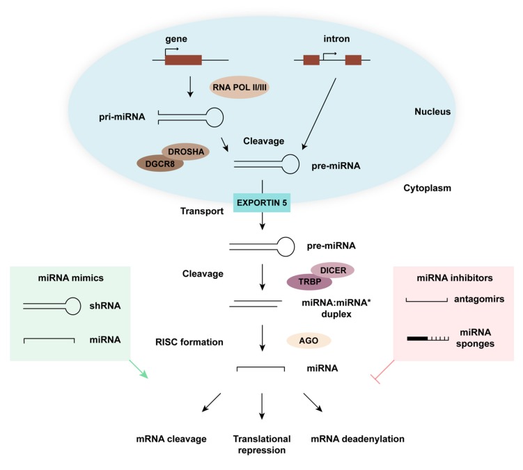
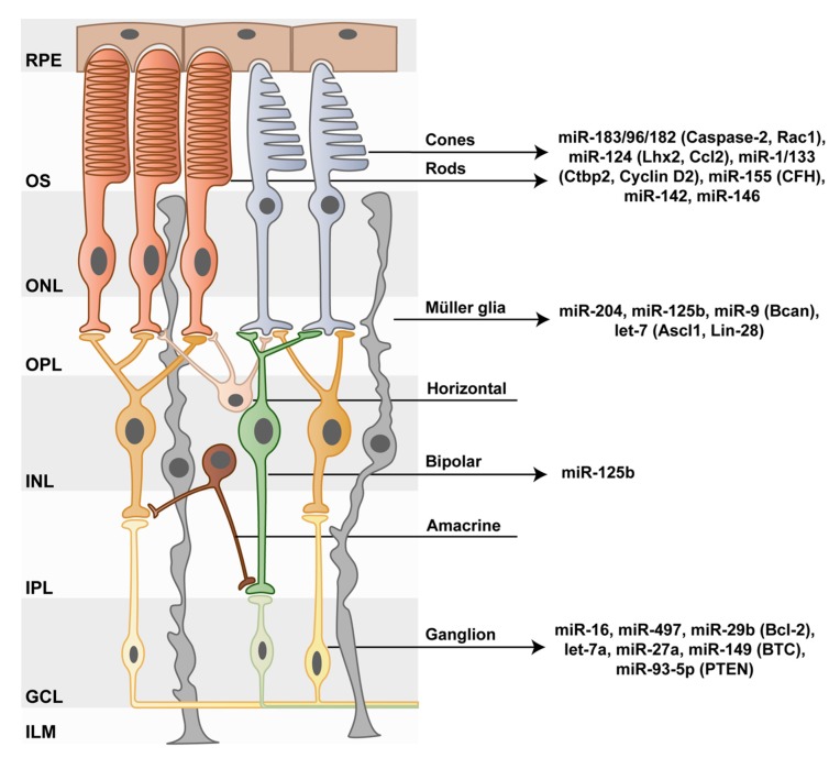
The health and function of our visual system relies on accurate gene expression. While many genetic mutations are associated with visual impairment and blindness, we are just beginning to understand the complex interplay between gene regulation and retinal pathologies. MicroRNAs (miRNAs), a class of non-coding RNAs, are important regulators of gene expression that exert their function through post-transcriptional silencing of complementary mRNA targets. According to recent transcriptomic analyses, certain miRNA species are expressed in all retinal cell types, while others are cell type-specific. As miRNAs play important roles in homeostasis, cellular function, and survival of differentiated retinal cell types, their dysregulation is associated with retinal degenerative diseases. Thus, advancing our understanding of the genetic networks modulated by miRNAs is central to harnessing their potential as therapeutic agents to overcome visual impairment. In this review, we summarize the role of distinct miRNAs in specific retinal cell types, the current knowledge on their implication in inherited retinal disorders, and their potential as therapeutic agents.

Keywords: Müller glia; bipolar cells; cones; microRNA; photoreceptors; retina; retinal degeneration; retinal inherited disorders; retinitis pigmentosa; rods.


Conflict of interest statement

The authors declare no conflict of interest, except for V.B., who is an inventor of a patent application that is related to some aspects of this manuscript filed by Friedrich Miescher Institute for Biomedical Research, Basel, Switzerland.

Figures

Figure 1
Figure 1
MicroRNA (miRNA) pathway. miRNA biogenesis starts in the nucleus where partially self-complementary RNA polymerase II or III transcripts from a miRNA gene or from an intronic region assemble into a hairpin-like structure known as primary (pri)-miRNA. Pri-miRNAs are cleaved by the DiGeorge Critical Region 8 (Dgcr8)/Drosha complex and transported to the cytoplasm via Exportin 5. In the cytoplasm, pre-miRNAs are cleaved by the Dicer/HIV-1 TAR RNA binding protein (TRBP) nuclease complex, thereby giving rise to a miRNA duplex. This duplex is then loaded onto the Argonaute (AGO) protein, a component of the RNA-induced silencing complex (RISC), where one of the two strands is discarded while the other serves to search complementary transcripts. Targets bound by miRNAs exhibit reduced translational efficiency, mainly as a consequence of mRNA cleavage or deadenylation. The miRNA pathway can be modulated by introducing miRNA mimics (green) or miRNA inhibitors (red). shRNA is small hairpin RNA.
Figure 2
Figure 2
MiRNAs acting as modulators of retinal cell behavior. In the vertebrate eye, the retinal pigment epithelium (RPE) separates the retina from the subretinal space. Within the outermost layer of the retina, rod and cone photoreceptors sense light with their outer segments (OS). Photoreceptor bodies reside within the outer nuclear layer (ONL), and their axons protrude into the outer plexiform layer (OPL), where they synapse with excitatory bipolar cells and inhibitory horizontal cells. The bodies of these cells, as well as of amacrine cells, which create inhibitory synapses with the axons of bipolar cells, reside in turn within the inner nuclear layer (INL). The electrochemical signal produced by photoreceptors during phototransduction is transmitted through bipolar cells to ganglion cells via synaptic connections in the inner plexiform layer (IPL). In a final step, ganglion cells send this information to higher brain areas through their axons, which bundle up to form the optic nerve. An additional cell type within the retina is Müller glia. These cells play key roles in the support of neuronal functions and in mediating the reaction to a number of physiological signals, including immune responses. Müller glia feet form the outer limiting membrane (OLM), which separates photoreceptor OS from their somata. Within the retina, miRNAs play central roles in health and disease. Recognized miRNA species associated to the functionality of specific retinal cell types are shown with validated targets between parentheses. GCL, ganglion cell layer; ILM, inner limiting membrane.

References

    1. Ha M., Kim V.N. Regulation of microRNA biogenesis. Nat. Rev. Mol. Cell Biol. 2014;15:509–524. doi: 10.1038/nrm3838. - DOI - PubMed
    1. Han J., Lee Y., Yeom K.H., Kim Y.K., Jin H., Kim V.N. The Drosha-DGCR8 complex in primary microRNA processing. Genes Dev. 2004;18:3016–3027. doi: 10.1101/gad.1262504. - DOI - PMC - PubMed
    1. Westholm J.O., Lai E.C. Mirtrons: microRNA biogenesis via splicing. Biochimie. 2011;93:1897–1904. doi: 10.1016/j.biochi.2011.06.017. - DOI - PMC - PubMed
    1. Chiang H.R., Schoenfeld L.W., Ruby J.G., Auyeung V.C., Spies N., Baek D., Johnston W.K., Russ C., Luo S., Babiarz J.E., et al. Mammalian microRNAs: Experimental evaluation of novel and previously annotated genes. Genes Dev. 2010;24:992–1009. doi: 10.1101/gad.1884710. - DOI - PMC - PubMed
    1. Bartel D.P. MicroRNAs: Target recognition and regulatory functions. Cell. 2009;136:215–233. doi: 10.1016/j.cell.2009.01.002. - DOI - PMC - PubMed

Publication types

MeSH terms