. 2019 May 17;11(5):691.
doi: 10.3390/cancers11050691.

RAC1B Suppresses TGF-β1-Dependent Cell Migration in Pancreatic Carcinoma Cells through Inhibition of the TGF-β Type I Receptor ALK5

Affiliations

RAC1B Suppresses TGF-β1-Dependent Cell Migration in Pancreatic Carcinoma Cells through Inhibition of the TGF-β Type I Receptor ALK5

Hendrik Ungefroren et al. Cancers (Basel). .

Abstract

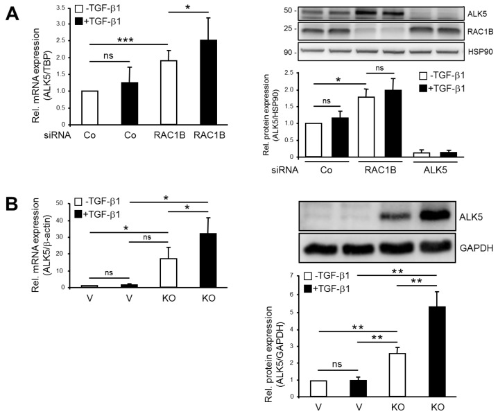
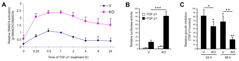
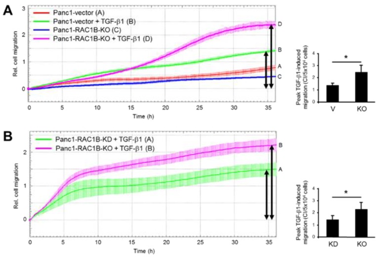
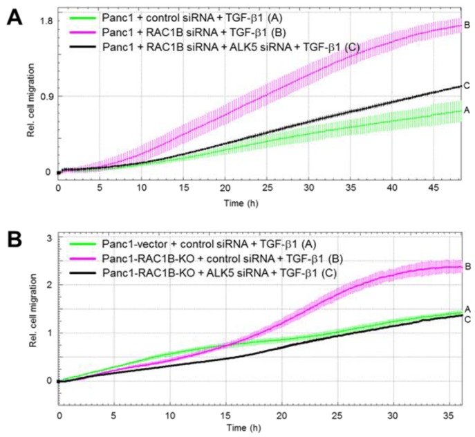
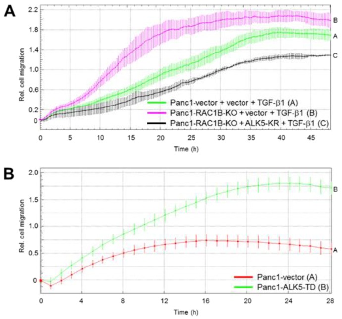
The small GTPase Ras-related C3 botulinum toxin substrate 1B (RAC1B) has been shown previously by RNA interference-mediated knockdown (KD) to function as a powerful inhibitor of transforming growth factor (TGF)-β1-induced cell migration and epithelial-mesenchymal transition in epithelial cells, but the underlying mechanism has remained enigmatic. Using pancreatic carcinoma cells, we show that both KD and Clustered Regularly Interspaced Short Palindromic Repeats (CRISPR)/Cas9-mediated knockout (KO) of RAC1B increased the expression of the TGF-β type I receptor ALK5 (activin receptor-like kinase 5), but this effect was more pronounced in CRISPR-KO cells. Of note, in KO, but not KD cells, ALK5 upregulation was associated with resensitization of TGFBR1 to induction by TGF-β1 stimulation. RAC1B KO also increased TGF-β1-induced C-terminal SMAD3 phosphorylation, SMAD3 transcriptional activity, growth inhibition, and cell migration. The KD of ALK5 expression by RNA interference or inactivation of the ALK5 kinase activity by dominant-negative interference or ATP-competitive inhibition rescued the cells from the RAC1B KD/KO-mediated increase in TGF-β1-induced cell migration, whereas the ectopic expression of kinase-active ALK5 mimicked this RAC1B KD/KO effect. We conclude that RAC1B downregulates the abundance of ALK5 and SMAD3 signaling, thereby attenuating TGF-β/SMAD3-driven cellular responses, such as growth inhibition and cell motility.

Keywords: ALK5; CRISPR/Cas9; RAC1B; RNA interference; TGF-β; cell migration; pancreatic carcinoma.


Conflict of interest statement

The authors declare no conflict of interest.

Figures

Figure 1
Figure 1
Effect of RAC1B knockdown (KD) and knockout (KO) on activin receptor-like kinase 5 (ALK5) expression in Panc1 cells. (A) Panc1-RAC1B-KD cells were generated by transfecting Panc1 cells twice with 50 nM of either control (Co) siRNA or RAC1B siRNA. Forty-eight hours after the second transfection, the cells were treated with transforming growth factor (TGF)-β1 for 24 h and subsequently processed for qPCR of ALK5 and TATA-box-binding protein (TBP) (left-hand panel) or immunoblot analysis of ALK5, RAC1B, and heat shock protein (HSP)90 (right-hand panel). The graphs show quantification of ALK5 data normalized to those for TBP (ΔΔCt values from qPCR) or HSP90 (densitometric values from immunoblots) and represent the mean ±SD from three independent experiments. (B) Panc1-RAC1B-KO and vector control cells were treated with TGF-β1 for 24 h and processed for either qPCR of ALK5 and β-actin (left-hand panel) or immunoblot analysis of ALK5 and Glyceraldehyde 3-phosphate dehydrogenase (GAPDH) (right-hand panel). Expression of RAC1B in these cells is shown in Figure S1. The graphs indicate the normalized data (mean ±SD, n = 3). The asterisks indicate significance (student’s t-test). V, vector control; ns, not significant.
Figure 2
Figure 2
Effect of RAC1B knockout (KO) on transforming growth factor (TGF)-β1-induced phosphorylation of SMAD3C, SMAD3-dependent transcriptional activity, and growth inhibition in Panc1 cells. (A) Panc1-RAC1B-KO (KO) and vector control cells (V) were treated with TGF-β1 for various times (as indicated) and subjected to phospho-immunoblotting for SMAD3C and total SMAD3. Densitometric data for p-SMAD3C were normalized to those for total SMAD3 and represent the mean ± SD from three independent experiments. (B) Panc1-RAC1B-KO and vector control cells (V) were cotransfected with p(CAGA)12-luc and pRL-TK-luc, and after a 24 h treatment with TGF-β1, they were subjected to dual luciferase measurement. Data are the mean ±SD of six replicates per condition and are from one representative assay out of four assays performed in total with very similar results. (C) Panc1-RAC1B-KO and vector control cells were seeded at 50,000 cells per 12-well in normal growth medium and treated, or not, on the following day with TGF-β1 for 24 h or 48 h. Following detachment by trypsinization, viable cells were counted. Data are the mean ±SD of four wells processed in parallel and are displayed as % inhibition by TGF-β1 treatment relative to untreated control cells set arbitrarily to 100%. Data are representative of three experiments. The asterisks indicate significance (student’s t-test).
Figure 3
Figure 3
Transforming growth factor (TGF)-β1-induced migration in Panc1-RAC1B-knockout (KO) cells and knockdown (KD) cells. (A) Kinetics of TGF-β1-induced migration of Panc1-RAC1B-KO cells and corresponding vector control cells as measured by real-time cell migration assay. Shown is a representative assay, and data are the mean ± SD of three parallel wells. Differences between Panc1-RAC1B-KO + TGF-β1 (magenta curve, tracing D) and Panc1-vector + TGF-β1 (green curve, tracing B) are significant at 17:45 and all later time points. The bar graph shows quantification of peak TGF-β1-induced migratory activities (indicated by black arrows) of Panc1-vector vs. Panc1-RAC1B-KO cells by depicting the maximal cell index (CI) values (mean ± SD, n = 4). (B) Comparative analysis of TGF-β1-induced migration of Panc1-RAC1B-KD and KO cells by real-time cell migration assay. Panc1-RAC1B-KD were generated by transfecting cells twice with 50 nM of either control siRNA or RAC1B siRNA. Forty-eight hours after the second round of transfection, the cells were subjected together with Panc1-RAC1B-KO cells to real-time cell migration assay in the presence of 5 ng/mL TGF-β1. Data shown are the mean ± SD of three parallel wells and are significantly different at 05:15 and remain so at all later time points. The bar graph shows quantification of peak TGF-β1-induced migratory activities of Panc1-RAC1B-KD and KO cells by depicting their maximal CI values. Data represent the mean ± SD from four (KO) and eight (KD) assays. The asterisk indicates significance (student’s t-test).
Figure 4
Figure 4
Effect of activin receptor-like kinase 5 (ALK5) knockdown (KD) on transforming growth factor (TGF)-β1-induced migration (chemokinesis) in Panc1-RAC1B-KD and knockout (KO) cells. (A) Panc1 cells were transfected twice with either 50 nM of control siRNA, 25 nM RAC1B siRNA+ 25 nM control siRNA, or 25 nM RAC1B siRNA + 25 nM ALK5 siRNA. Forty-eight hours after the second transfection, the cells were processed for migration assay on the xCELLigence platform. Immediately before the start of the assay, one-half of the cells received 5 ng/mL TGF-β1. (B) As in (A) except that Panc1-RAC1B-KO cells received 50 nM of either siCo or siALK5. Data in (A) and (B) are from one representative experiment and are the mean ±SD from 3–4 wells per condition. Differences between Panc1 + RAC1B siRNA + ALK5 siRNA + TGF-β1 (black curve, tracing C) and Panc1 + RAC1B siRNA + TGF-β1 (magenta curve, tracing B) are significant at 16:30 and all later time points, while in (B) those between Panc1-RAC1B-KO + ALK5 siRNA + TGF-β1 (black curve, tracing C) and Panc1-RAC1B-KO + control siRNA + TGF-β1 (magenta curve, tracing B) are significant at 07:00 and all later time points. Successful inhibition of RAC1B and ALK5 was verified by immunoblotting (data not shown). For functional validation of the ALK5 siRNA, see Figure 1A.
Figure 5
Figure 5
Effect of kinase-dead and kinase-active activin receptor-like kinase 5 (ALK5) on transforming growth factor (TGF)-β1-induced migration in Panc1 cells. (A) Panc1-RAC1B-knockout (KO) cells were transiently transfected with either empty vector or ALK5-K232R (ALK5-KR) and 48 h later subjected to real-time cell migration assay. Data are from one representative experiment out of three experiments performed in total and represent the mean ±SD from 3–4 wells per condition. Differences between Panc1-RAC1B-KO + ALK5-KR + TGF-β1 (black curve, tracing D) and Panc1-RAC1B-KO + vector + TGF-β1 (magenta curve, tracing B) are significant at 07:00 and all later time points. (B) Effect of kinase-active ALK5 on Panc1 cell migration. Panc1 cells stably expressing either empty vector or ALK5-T204D (ALK5-TD) were subjected to real-time cell migration assay. Data are from one representative experiment (three performed in total) and represent the mean ±SD from three parallel wells. Differences between Panc1-ALK5-TD (green curve, tracing B) and Panc1-vector (red curve, tracing A) are significant at 03:15 and all later time points.

Similar articles

Cited by

References

    1. Melzer C., Hass R., Lehnert H., Ungefroren H. RAC1B: A Rho GTPase with versatile functions in malignant transformation and tumor progression. Cells. 2019;8:21. doi: 10.3390/cells8010021. - DOI - PMC - PubMed
    1. Ungefroren H., Sebens S., Giehl K., Helm O., Groth S., Fandrich F., Rocken C., Sipos B., Lehnert H., Gieseler F. Rac1b negatively regulates TGF-beta1-induced cell motility in pancreatic ductal epithelial cells by suppressing Smad signalling. Oncotarget. 2014;5:277–290. doi: 10.18632/oncotarget.1696. - DOI - PMC - PubMed
    1. Witte D., Otterbein H., Forster M., Giehl K., Zeiser R., Lehnert H., Ungefroren H. Negative regulation of TGF-beta1-induced MKK6-p38 and MEK-ERK signalling and epithelial-mesenchymal transition by Rac1b. Sci. Rep. 2017;7:17313. doi: 10.1038/s41598-017-15170-6. - DOI - PMC - PubMed
    1. Melzer C., von der Ohe J., Hass R., Ungefroren H. TGF-beta-dependent growth arrest and cell migration in benign and malignant breast epithelial cells are antagonistically controlled by Rac1 and Rac1b. Int. J. Mol. Sci. 2017;18:1574. doi: 10.3390/ijms18071574. - DOI - PMC - PubMed
    1. Melzer C., Hass R., von der Ohe J., Lehnert H., Ungefroren H. The role of TGF-beta and its crosstalk with RAC1/RAC1b signaling in breast and pancreas carcinoma. Cell Commun. Signal. 2017;15:19. doi: 10.1186/s12964-017-0175-0. - DOI - PMC - PubMed