Review
. 2019 May 18;11(5):456.
doi: 10.3390/v11050456.

Genetic Variability and Evolution of Hepatitis E Virus

Affiliations
Review

Genetic Variability and Evolution of Hepatitis E Virus

Putu Prathiwi Primadharsini et al. Viruses. .

Abstract

Hepatitis E virus (HEV) is a single-stranded positive-sense RNA virus. HEV can cause both acute and chronic hepatitis, with the latter usually occurring in immunocompromised patients. Modes of transmission range from the classic fecal-oral route or zoonotic route, to relatively recently recognized but increasingly common routes, such as via the transfusion of blood products or organ transplantation. Extrahepatic manifestations, such as neurological, kidney and hematological abnormalities, have been documented in some limited cases, typically in patients with immune suppression. HEV has demonstrated extensive genomic diversity and a variety of HEV strains have been identified worldwide from human populations as well as growing numbers of animal species. The genetic variability and constant evolution of HEV contribute to its physiopathogenesis and adaptation to new hosts. This review describes the recent classification of the Hepeviridae family, global genotype distribution, clinical significance of HEV genotype and genomic variability and evolution of HEV.

Keywords: evolution; genetic variability; genotypes; hepatitis E virus.


Conflict of interest statement

The authors declare no conflict of interest.

Figures
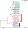
Figure 1
Figure 1
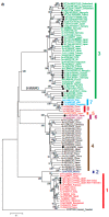
Phylogenetic tree for members of the Hepeviridae family. The maximum-likelihood tree was created using MEGA 7 [24] based on amino acid sequences of the entire ORF2 region from members of the Hepeviridae family. Each reference sequence is shown with the genotype (if available) followed by the accession number, species (animal/human) and the country where it was isolated. Unassigned HEV strains are highlighted with closed circles. The bootstrap values (>70%) for the nodes are indicated as percentage data obtained from 1000 resampling analyses. The scale bar indicates the number of nucleotide substitutions per site.
Figure 2
Figure 2
Phylogenetic trees for member of the Orthohepevirus genus. Maximum-likelihood trees were created using MEGA 7 [24] based on entire genomic sequences from members of the Orthohepevirus A species (a), Orthohepevirus B species (b) and Orthohepevirus C species (c). Each reference sequence is shown with genotype/subtype (if available) followed by accession number and the country where it was isolated. (a) Subtypes 3la, 3lb, 3kc and 1gd have been proposed by Wang et al. [68], De Sabato et al. [69], Miura et al. [67] and Nishizawa et al. [65], respectively, and those provisionally proposed by HEVNet (https://www.rivm.nl/en/hevnet) are indicated in brackets. (b) Provisional genotypes 1–4 for avian HEV strains are abbreviated as Gt1, Gt2, Gt3 and Gt4, respectively. (c) Genotypes C3 and C4 proposed by Wang et al. [78] are indicated in brackets. Reference HEV sequences from moose (KF951328) (a), little egret (KX589065) (b) and human (HEV-1) (M73218) (c) were used as an outgroup. Unassigned HEV strains are highlighted with closed circles. The bootstrap values (>70%) for the nodes are indicated as percentage data obtained from 1000 resampling analyses. The scale bar indicates the number of nucleotide substitutions per site.
Figure 2
Figure 2
Phylogenetic trees for member of the Orthohepevirus genus. Maximum-likelihood trees were created using MEGA 7 [24] based on entire genomic sequences from members of the Orthohepevirus A species (a), Orthohepevirus B species (b) and Orthohepevirus C species (c). Each reference sequence is shown with genotype/subtype (if available) followed by accession number and the country where it was isolated. (a) Subtypes 3la, 3lb, 3kc and 1gd have been proposed by Wang et al. [68], De Sabato et al. [69], Miura et al. [67] and Nishizawa et al. [65], respectively, and those provisionally proposed by HEVNet (https://www.rivm.nl/en/hevnet) are indicated in brackets. (b) Provisional genotypes 1–4 for avian HEV strains are abbreviated as Gt1, Gt2, Gt3 and Gt4, respectively. (c) Genotypes C3 and C4 proposed by Wang et al. [78] are indicated in brackets. Reference HEV sequences from moose (KF951328) (a), little egret (KX589065) (b) and human (HEV-1) (M73218) (c) were used as an outgroup. Unassigned HEV strains are highlighted with closed circles. The bootstrap values (>70%) for the nodes are indicated as percentage data obtained from 1000 resampling analyses. The scale bar indicates the number of nucleotide substitutions per site.
Figure 3
Figure 3
Positions of mutations in the eight recombinant cDNA clones of the JE03-1760F strain. Upper panel: the genomic structure of HEV. Abbreviations: MeT, methyltransferase; Y, Y domain; PCP, papain-like cysteine protease; HVR, hypervariable region; X, X domain; Hel, helicase; and RdRp, RNA-dependent RNA polymerase. Lower panel: the open circles denote synonymous mutations without structural changes in HEV RNA, the shaded circles denote synonymous mutations with structural changes in HEV RNA and the closed circles denote non-synonymous mutations. Modified from Nagashima et al. [187].

References

    1. Lhomme S., Marion O., Abravanel F., Chapuy-Regaud S., Kamar N., Izopet J. Hepatitis E pathogenesis. Viruses. 2016;8:212. doi: 10.3390/v8080212. - DOI - PMC - PubMed
    1. Kamar N., Izopet J., Pavio N., Aggarwal R., Labrique A., Wedemeyer H., Dalton H.R. Hepatitis E virus infection. Nat. Rev. Dis. Primers. 2017;3:17086. doi: 10.1038/nrdp.2017.86. - DOI - PubMed
    1. Nimgaonkar I., Ding Q., Schwartz R.E., Ploss A. Hepatitis E virus: Advances and challenges. Nat. Rev. Gastroenterol. Hepatol. 2018;15:96–110. doi: 10.1038/nrgastro.2017.150. - DOI - PMC - PubMed
    1. Hoofnagle J.H., Nelson K.E., Purcell R.H. Hepatitis E. N. Engl. J. Med. 2012;367:1237–1244. doi: 10.1056/NEJMra1204512. - DOI - PubMed
    1. Kamar N., Marion O., Abravanel F., Izopet J., Dalton H.R. Extrahepatic manifestations of hepatitis E virus. Liver Int. 2016;36:467–472. doi: 10.1111/liv.13037. - DOI - PubMed

Publication types

LinkOut - more resources