

Ganoderma lucidum Extract Reduces the Motility of Breast Cancer Cells Mediated by the RAC⁻Lamellipodin Axis
- PMID: 31109134
- PMCID: PMC6567024
- DOI: 10.3390/nu11051116
Ganoderma lucidum Extract Reduces the Motility of Breast Cancer Cells Mediated by the RAC⁻Lamellipodin Axis
Abstract
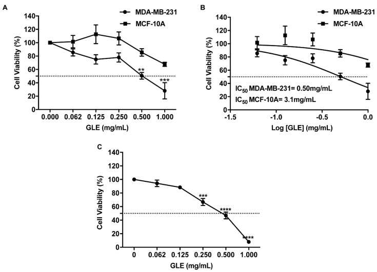
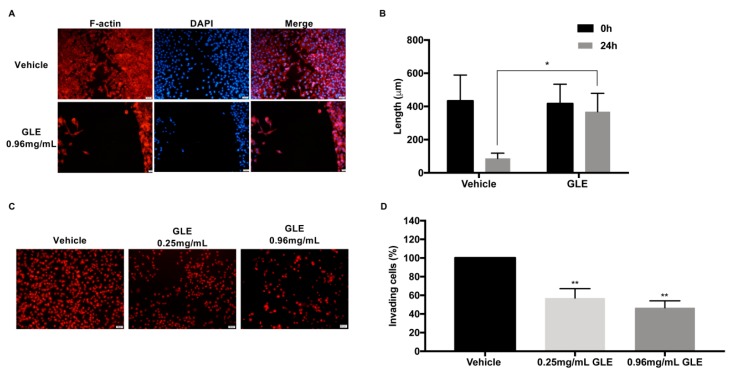
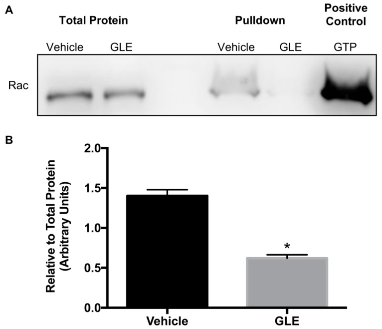
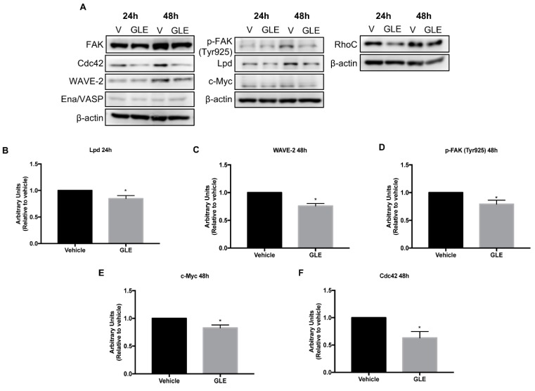
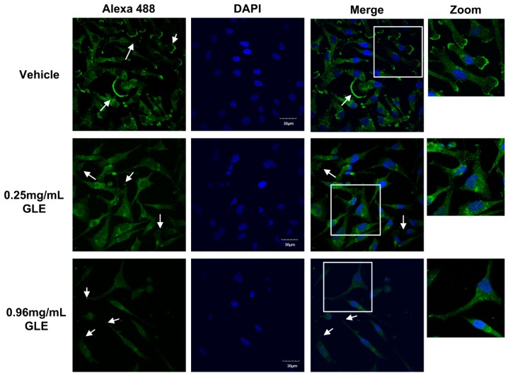
Breast cancer (BC) is the second leading cause of cancer death among women worldwide. The main cause of BC morbidity and mortality is the invasiveness capacity of cancer cells that may lead to metastasis. Here, we aimed to investigate the therapeutic efficacy of Ganoderma lucidum extract (GLE)-a medicinal mushroom with anticancer properties-on BC motility via the Rac/Lamellipodin pathway. GLE treatment effects were tested on MDA-MB-231 breast cancer cells. The effects were tested on cell viability, migration and invasion. Pulldowns, immunoblotting, and immunofluorescence were used to measure Rac activity and the expression of proteins involved in cell migration and in lamellipodia formation, respectively. As a result, GLE suppressed BC cell viability, migration, and invasion capacity. GLE impaired Rac activity, as well as downregulated Lamellipodin, ENA/VASP, p-FAK (Tyr925), Cdc42, and c-Myc expression. Lamellipodia formation was significantly reduced by GLE. In conclusion, we demonstrate that GLE reduces Rac activity and downregulates signaling molecules involved in lamellipodia formation. These novel findings serve as basis for further studies to elucidate the potential of GLE as a therapeutic agent regulating the Rac/Lamellipodin pathway in BC metastasis.
Keywords: Ganoderma lucidum; Rac; breast cancer; cancer cell migration; lamellipodin.
Conflict of interest statement
The authors declare no conflicts of interest. The funders had no role in the design of the study; in the collection, analyses, or interpretation of data; in the writing of the manuscript, or in the decision to publish the results.
Figures
Similar articles
-
The mushroom Ganoderma lucidum suppresses breast-to-lung cancer metastasis through the inhibition of pro-invasive genes.Int J Oncol. 2014 Jun;44(6):2009-15. doi: 10.3892/ijo.2014.2375. Epub 2014 Apr 9. Int J Oncol. 2014. PMID: 24718855 Free PMC article.
-
A supercritical-CO2 extract of Ganoderma lucidum spores inhibits cholangiocarcinoma cell migration by reversing the epithelial-mesenchymal transition.Phytomedicine. 2016 May 15;23(5):491-7. doi: 10.1016/j.phymed.2016.02.019. Epub 2016 Mar 3. Phytomedicine. 2016. PMID: 27064008
-
Combined effect of green tea and Ganoderma lucidum on invasive behavior of breast cancer cells.Int J Oncol. 2007 Apr;30(4):963-9. Int J Oncol. 2007. PMID: 17332936
-
Ganoderma lucidum (Reishi) in cancer treatment.Integr Cancer Ther. 2003 Dec;2(4):358-64. doi: 10.1177/1534735403259066. Integr Cancer Ther. 2003. PMID: 14713328 Review.
-
A Review of Twenty Years of Research on the Regulation of Signaling Pathways by Natural Products in Breast Cancer.Molecules. 2022 May 25;27(11):3412. doi: 10.3390/molecules27113412. Molecules. 2022. PMID: 35684353 Free PMC article. Review.
Cited by
-
Functional and clinical characteristics of focal adhesion kinases in cancer progression.Front Cell Dev Biol. 2022 Nov 2;10:1040311. doi: 10.3389/fcell.2022.1040311. eCollection 2022. Front Cell Dev Biol. 2022. PMID: 36407100 Free PMC article. Review.
-
Accumulation of amyloid beta (Aβ) and amyloid precursor protein (APP) in tumors formed by a mouse xenograft model of inflammatory breast cancer.FEBS Open Bio. 2022 Jan;12(1):95-105. doi: 10.1002/2211-5463.13308. Epub 2021 Oct 26. FEBS Open Bio. 2022. PMID: 34592066 Free PMC article.
-
Polysaccharides from sporoderm-removed spores of Ganoderma lucidum induce apoptosis in human gastric cancer cells via disruption of autophagic flux.Oncol Lett. 2021 May;21(5):425. doi: 10.3892/ol.2021.12686. Epub 2021 Mar 29. Oncol Lett. 2021. PMID: 33850566 Free PMC article.
-
High-Pressure Supercritical CO2 Extracts of Ganoderma lucidum Fruiting Body and Their Anti-hepatoma Effect Associated With the Ras/Raf/MEK/ERK Signaling Pathway.Front Pharmacol. 2020 Dec 14;11:602702. doi: 10.3389/fphar.2020.602702. eCollection 2020. Front Pharmacol. 2020. PMID: 33381043 Free PMC article.
-
Early Preclinical Studies of Ergosterol Peroxide and Biological Evaluation of Its Derivatives.ACS Omega. 2024 Aug 19;9(35):37117-37127. doi: 10.1021/acsomega.4c04350. eCollection 2024 Sep 3. ACS Omega. 2024. PMID: 39246459 Free PMC article.
References
MeSH terms
Substances
Grants and funding
- SC3GM111171/National Institute of General Medical Sciences
- P20 GM103475/GM/NIGMS NIH HHS/United States
- G12 MD007583/MD/NIMHD NIH HHS/United States
- SC3 GM111171/GM/NIGMS NIH HHS/United States
- P031S130068/U.S. Department of Education
- Hurricane Relief Funds/Puerto Rico Science, Technology and Research Trust
- G12MD007583/National Institute on Minority Health and Health Disparities
- 2017-043/Puerto Rico Science, Technology and Research Trust
- P20GM103475/National Institute of General Medical Sciences
- P031M105050/U.S. Department of Education
- G12 RR003035/RR/NCRR NIH HHS/United States
LinkOut - more resources
Full Text Sources
Medical
Research Materials
Miscellaneous
