Observational Study
. 2019 May;12(5):e008130.
doi: 10.1161/CIRCIMAGING.118.008130.

Left Ventricular Flow Analysis

Affiliations
Observational Study

Left Ventricular Flow Analysis

Victoria M Stoll et al. Circ Cardiovasc Imaging. 2019 May.

Abstract

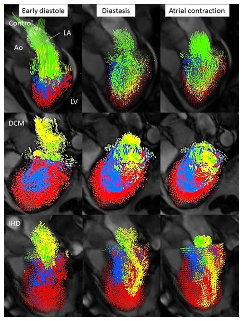
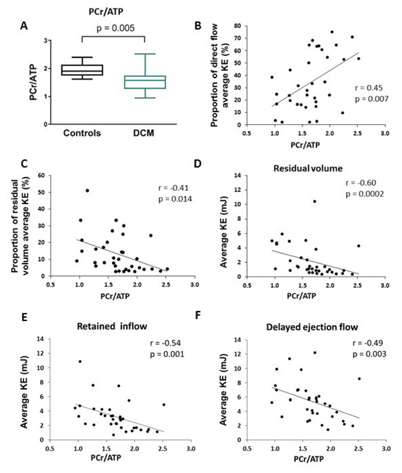
Background: Cardiac remodeling, after a myocardial insult, often causes progression to heart failure. The relationship between alterations in left ventricular blood flow, including kinetic energy (KE), and remodeling is uncertain. We hypothesized that increasing derangements in left ventricular blood flow would relate to (1) conventional cardiac remodeling markers, (2) increased levels of biochemical remodeling markers, (3) altered cardiac energetics, and (4) worsening patient symptoms and functional capacity. Methods Thirty-four dilated cardiomyopathy patients, 30 ischemic cardiomyopathy patients, and 36 controls underwent magnetic resonance including 4-dimensional flow, BNP (brain-type natriuretic peptide) measurement, functional capacity assessment (6-minute walk test), and symptom quantification. A subgroup of dilated cardiomyopathy and control subjects underwent cardiac energetic assessment. Left ventricular flow was separated into 4 components: direct flow, retained inflow, delayed ejection flow, and residual volume. Average KE throughout the cardiac cycle was calculated.

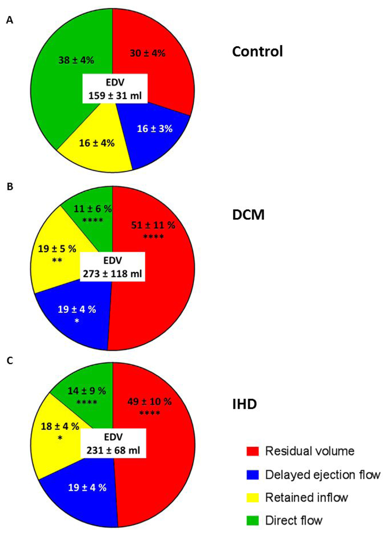
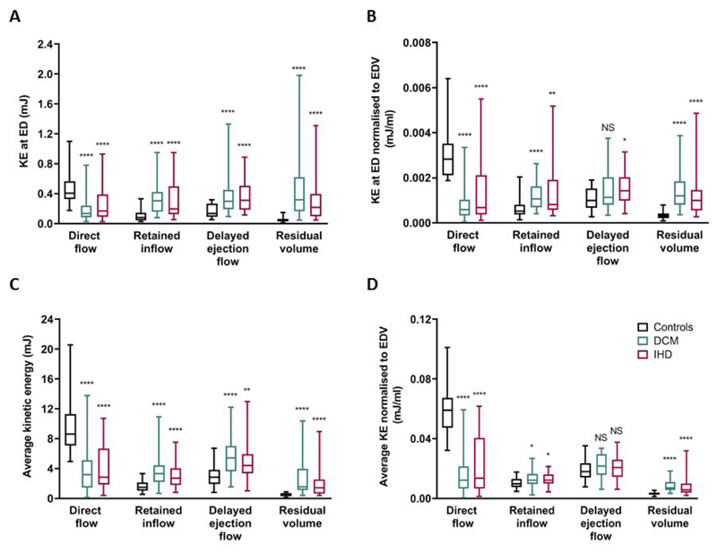
Results: Patients had reduced direct flow proportion and direct-flow average KE compared with controls ( P<0.0001). The residual volume proportion and residual volume average KE were increased in patients ( P<0.0001). Importantly, in a multiple linear regression model to predict the patient's 6-minute walk test, the independent predictors were age (β=-0.3015; P=0.019) and direct-flow average KE (β=0.280, P=0.035; R2 model, 0.466, P=0.002). In contrast, neither ejection fraction nor left ventricular volumes were independently predictive.

Conclusions: This study demonstrates an independent predictive relationship between the direct-flow average KE and a prognostic measure of functional capacity. Intracardiac 4-dimensional flow parameters are novel biomarkers in heart failure and may provide additive value in monitoring new therapies and predicting prognosis.

Keywords: biomarkers; heart failure; magnetic resonance imaging; prognosis; walk test.


Figures

Figure 1
Figure 1. Representative diastolic LV visualisations in a control (direct flow 35%, residual volume 29%), DCM (direct flow 10%, residual volume 55%) and IHD patient with an antero-apical infarct (direct flow 8%, residual volume 56%).
Despite similar proportions of residual volume between the IHD and DCM patient the distribution differs; with a global distribution in the DCM patient and a more localised distribution in the IHD patient, corresponding to the area of infarction. Direct flow, green; retained inflow, yellow; delayed ejection flow, blue and residual volume, red. Ao, aorta; DCM, dilated cardiomyopathy; IHD, ischaemic cardiomyopathy; LA, left atrium; LV left ventricle.
Figure 2
Figure 2. Flow components by percentage of the end-diastolic volume for (A) Control, (B) DCM and (C) IHD.
Data are mean ± standard deviation. ****P<0.0001, **P<0.01, *P<0.05 compared to corresponding component in controls. All comparisons between DCM and IHD patients were non-significant. DCM, dilated cardiomyopathy; EDV, end diastolic volume; IHD ischaemic cardiomyopathy.
Figure 3
Figure 3. Kinetic energy profiles. (A)Kinetic energy at end diastole; (B)Kinetic energy at end diastole normalised to EDV; (C)Average kinetic energy throughout the cardiac cycle and (D)Average kinetic energy normalised to EDV.
Bars show minimum and maximum values.. ****P<0.0001, ***P<0.001 compared to corresponding component value in controls. All comparisons between DCM and IHD patients were non-significant. DCM, dilated cardiomyopathy; KE ED, kinetic energy end diastole; IHD ischaemic cardiomyopathy.
Figure 4
Figure 4. Differences in flow component percentage, kinetic energy at end diastole and average kinetic energy according to left ventricular ejection fraction (LVEF).
LVEF>55%, n=4; EF 45-54% n=11; EF 36-44% n=21; EF≤35%, n=28. Panels A, D, G and J bars show mean value and error bars indicate standard deviation. Other panels bars show minimum and maximum values. *P<0.05 compared to controls, §P<0.05 LVEF≤35% compared to 45-54%, ∂P<0.05 LVEF≤35% compared to ≥55%, #P<0.05 LVEF 36-44% compared to 45-54%. EDV, end diastolic volume; EF, ejection fraction; KE ED, kinetic energy end diastole.
Figure 5
Figure 5. Myocardial energetics results and correlations in DCM patients. (A)PCr/ATP ratio in controls compared to DCM. Correlations between PCr/ATP ratio and (B)proportion of direct flow average KE; (C)proportion of residual volume average KE; (D)residual volume average KE; (E)retained inflow average KE and (F)delayed ejection flow average KE.
PCr/ATP, phosphocreatine to adenosine triphosphate concentration ratio; KE kinetic energy.

Comment in

References

    1. Nichols M, Townsend N, Scarborough P, Rayner M. Cardiovascular disease in Europe 2014: epidemiological update. Eur Heart J. 2014;35:2950–2959. - PubMed
    1. Burchfield JS, Xie M, Hill JA. Pathological Ventricular Remodeling: Mechanisms: Part 1 of 2. Circulation. 2013;128:388–400. - PMC - PubMed
    1. Hill JA, Olson EN. Mechanisms of disease: Cardiac plasticity. New Engl J Med. 2008;358:1370–1380. - PubMed
    1. Yang GZ, Merrifield R, Masood S, Kilner PJ. Flow and myocardial interaction: an imaging perspective. Philos T R Soc B. 2007;362:1329–1341. - PMC - PubMed
    1. Eriksson J, Bolger AF, Ebbers T, Carlhall CJ. Four-dimensional blood flow-specific markers of LV dysfunction in dilated cardiomyopathy. Eur Heart J-Card Img. 2013;14:417–424. - PMC - PubMed

Publication types

MeSH terms