. 2019 May 20;16(1):107.
doi: 10.1186/s12974-019-1504-6.

Proteoglycans involved in bidirectional communication between mast cells and hippocampal neurons

Affiliations

Proteoglycans involved in bidirectional communication between mast cells and hippocampal neurons

Juan Antonio Flores et al. J Neuroinflammation. .

Abstract

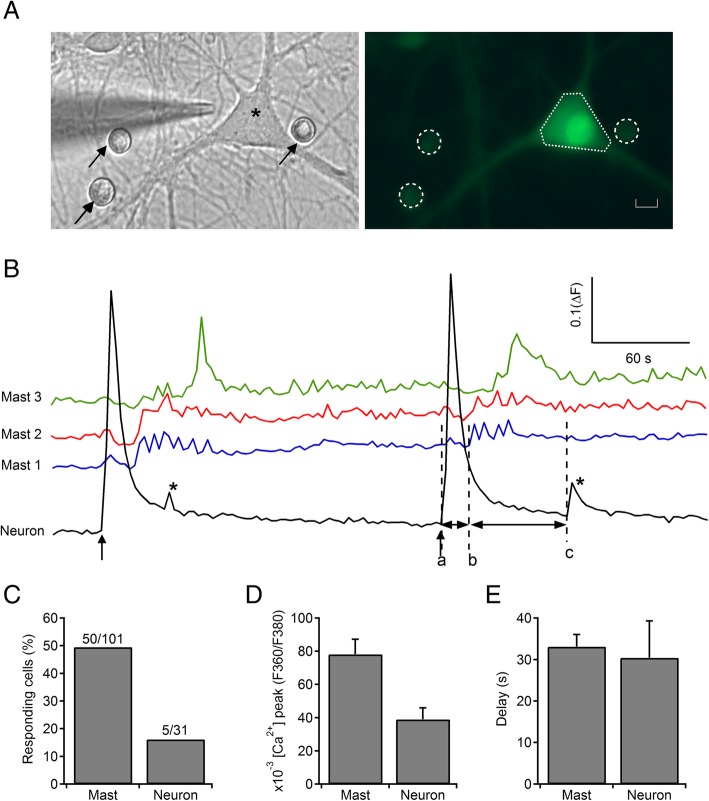
Background: Mast cells (MCs) in the brain can respond to environmental cues and relay signals to neurons that may directly influence neuronal electrical activity, calcium signaling, and neurotransmission. MCs also express receptors for neurotransmitters and consequently can be activated by them. Here, we developed a coculture model of peritoneal MCs, incubated together with dissociated hippocampal neurons for the study of cellular mechanisms involved in the mast cell-neuron interactions.

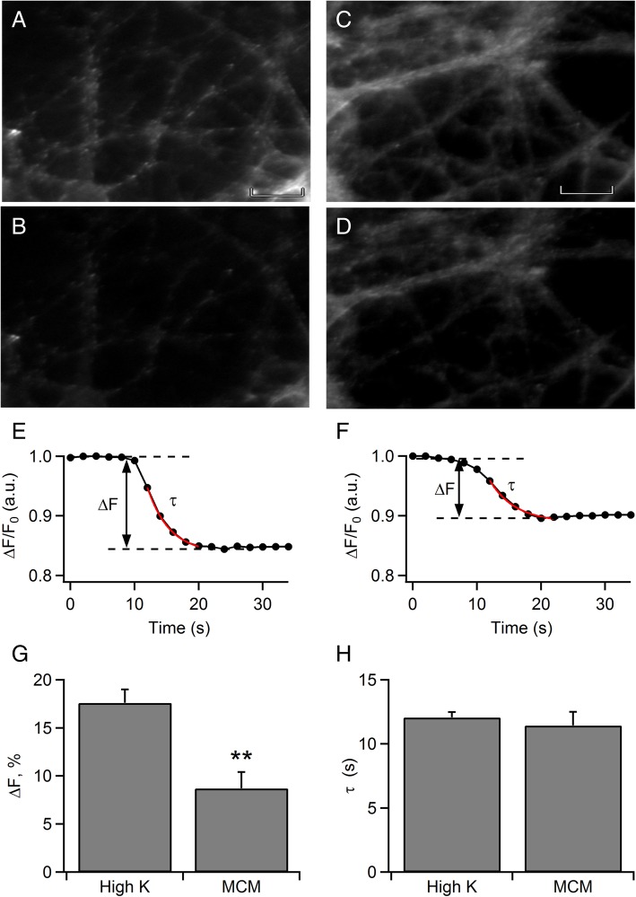
Methods: Calcium imaging was used to simultaneously record changes in intracellular calcium [Ca2+]i in neurons and MCs. To provide insight into the contribution of MCs on neurotransmitter release in rat hippocampal neurons, we used analysis of FM dye release, evoked by a cocktail of mediators from MCs stimulated by heat.

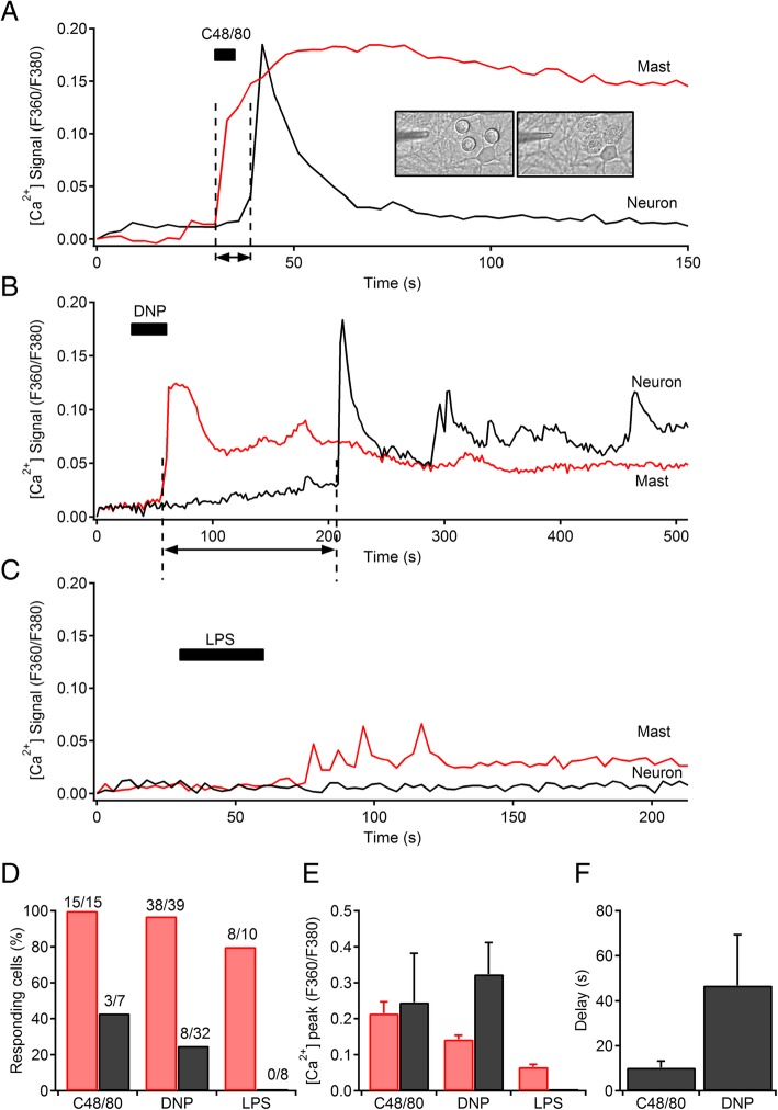
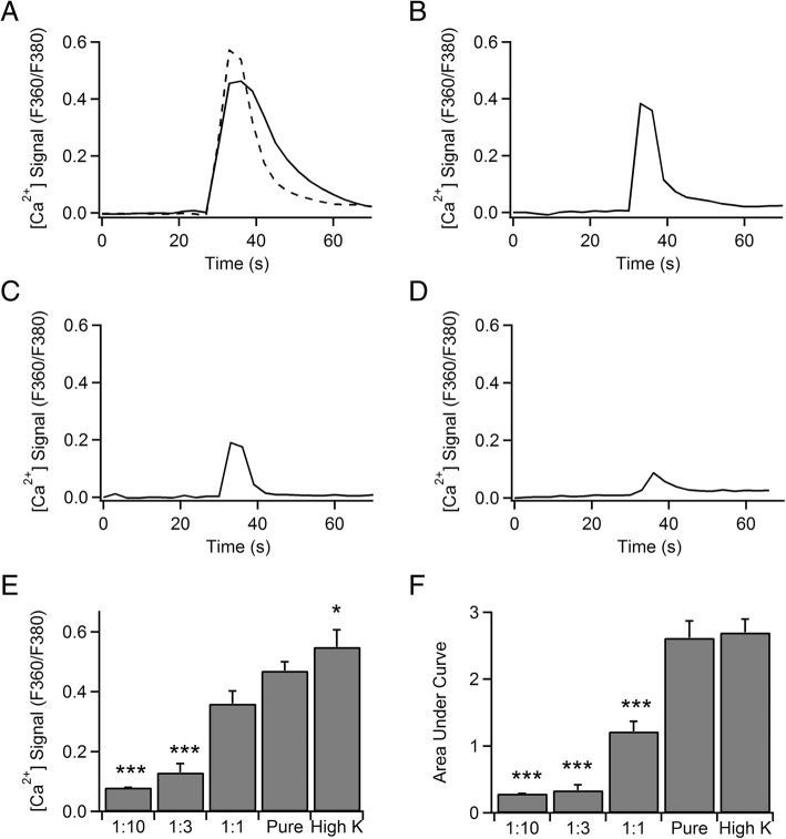
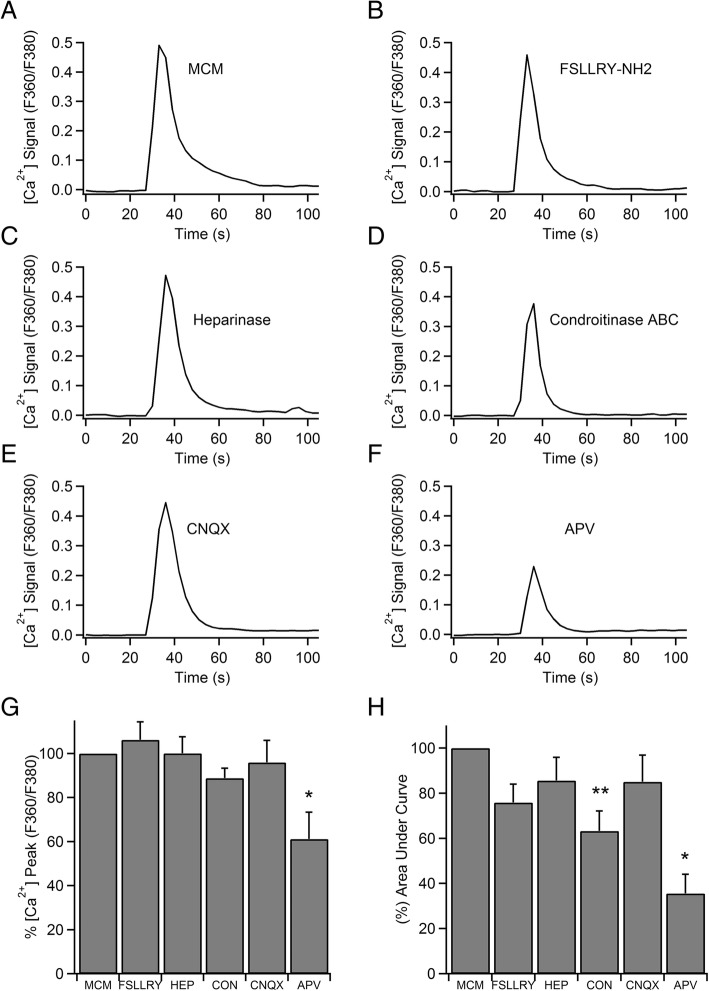
Results: Bidirectional communication is set up between MCs and hippocampal neurons. Neuronal depolarization caused intracellular calcium [Ca2+]i oscillations in MCs that produced a quick response in neurons. Furthermore, activation of MCs with antigen or the secretagogue compound 48/80 also resulted in a neuronal [Ca2+]i response. Moreover, local application onto neurons of the MC mediator cocktail elicited Ca2+ transients and a synaptic release associated with FM dye destaining. Neuronal response was partially blocked by D-APV, a N-methyl-D-aspartate receptor (NMDAR) antagonist, and was inhibited when the cocktail was pre-digested with chondroitinase ABC, which induces enzymatic removal of proteoglycans of chondroitin sulfate (CS).

Conclusions: MC-hippocampal neuron interaction affects neuronal [Ca2+]i and exocytosis signaling through a NMDAR-dependent mechanism.

Keywords: Ca2+ imaging; NMDA receptors; exocytosis; hippocampal neurotransmission; neuro-immune; proteoglycans.


Conflict of interest statement

The authors declare that they have no competing interests.

Figures

Fig. 1
Fig. 1
Calcium imaging of MC and Neuronal activity. a Phase-contrast image on the left panel of a hippocampal neuron (asterisk) at 2 weeks in culture following 24 h coculture with mast cells (black arrows). A glass micropipette of 1 μm ϕ was positioned very close to the neuronal soma before pressure ejection (2.5 psi) pulses of 5 s. The right panel shows the same cells loaded with the Ca2+ sensitive dye Fura-2 AM (MCs selected by dashed circles and neuron by the dotted polygon). Scale bar is 5 μm. b Traces of [Ca2+]i transients in three mast cells (blue, red, and green traces) and a hippocampal neuronal cell body (black trace) triggered by the initial neuronal depolarization by microperfusion (70 mM [K+] Locke solution). Note the [Ca2+]i oscillation pattern elicited in MCs after initial neuronal activation and the short [Ca2+]i transient (*) evoked in neuron later. Both responses were reproduced in a second stimulation. c Percentage of responding cells. d Mean [Ca2+]i peak obtained in MC- and neuron-evoked responses. e Latencies measured from the beginning of neuronal [Ca2+]i transient induced by depolarization (a) to the origin of MC oscillations (b) and from the latter to the second rise in neuronal trace (c) (*)
Fig. 2
Fig. 2
MC to neuronal [Ca2+]i signaling. a Traces of [Ca2+]i transients in a mast cell (red trace) and a hippocampal neuronal cell body (black trace) in response to mast cell activation by C48/80 microperfusion (100 μg/ml for 30 s). Inset shows pictures of MCs morphology and appearance before and after stimulation. b Traces of [Ca2+]i transients in a mast cell (red trace) and a hippocampal neuron (black trace) in response to mast cell activation by FcεRI receptor crosslinking by DNP microperfusion (1 μg/ml for 30 s). c Traces of [Ca2+]i transients in a MC (red trace) and a hippocampal neuronal cell body (black trace) in response to MC activation by LPS (1 μg/ml for 1 min). d Percentage of responding cells to C48/80, DNP, and LPS. e Mean [Ca2+]i peak obtained in MCs (red bars) and hippocampal neurons (black bars) evoked by C48/80, DNP, and LPS. f Latencies measured by the time interval between the rise of MC [Ca2+]i transient and the onset of neuronal [Ca2+]i response
Fig. 3
Fig. 3
Mast cell mediator cocktail (MCM) directly elicits [Ca2+]i transients in hippocampal neurons. a [Ca2+]i signals evoked by single pressure ejection pulses of 5 s of pure extract of MCM (solid trace) and 70 mM [K+] Locke solution (dashed trace). The resulting calcium responses obtained by MCM was compared with calcium responses elicited by depolarization. b Representative [Ca2+]i signal evoked by a single pressure ejection pulse of 5 s of MCM at 1:1 dilution. In order to estimate the minimal number of MCs necessary to obtain enough amount of mediators to induce a calcium transient in neurons, different dilutions from pure MCM were made. c Representative [Ca2+]i signal evoked by a single pressure ejection pulse of 5 s of MCM at 1:3 dilution. d [Ca2+]i signal evoked by a single pressure ejection pulse of 5 s of MCM a 1:10 dilution. e Mean [Ca2+]i peak obtained in neuronal responses evoked by 70 mM [K+] and MCM at 1:0 (pure), 1:1, 1:3, and 1:10 dilutions. f Mean area under curve obtained from [Ca2+]i transients in neurons evoked by 70 mM [K+] and MCM at 1:0 (pure), 1:1, 1:3, and 1:10 dilutions. The latter was the lowest effective dilution capable of stimulating neurons. *p < 0.05, ***p < 0.001
Fig. 4
Fig. 4
Results of FM imaging system in cultured hippocampal neurons. a, c Synaptic boutons of hippocampal neurons stained by FM1-43 dye in a loading process which consisted in 2 min of stimulation with 70 mM [K+ ] and subsequent washing. The presence of numerous puncta of fluorescence that appear to be lined up along the neurites result from localized internalization of the dye and are not washed out. b Synaptic boutons destained after a stimulation pulse of 5 s with 70 mM [K+]-enriched solution or d with MCM. Note the marked decrease in fluorescence intensity of most of the puncta visible in a and c. Scale bar is 5 μm and applies to all frames e Normalized destaining curves of FM dye loaded synapses upon stimulation with high K+ and f MCM. FM dye release curves of all single synapses were fitted mono-exponentially. g Amplitude (ΔF) and h τ were measured from monoexponential fitting curves obtained by depolarization and MCM, and plotted. Data represent average values of 6 (70 mM [K+ ]) and 14 (MCM) neurons. **p < 0.01
Fig. 5
Fig. 5
PGs of CS and NMDA-receptors contribute to outline the [Ca2+]i signal elicited by mast cells mediator cocktail. a Representative time course of the elevation of [Ca2+]i upon application of a brief pulse of MCM to a neuronal soma. b The response was not changed after incubation with FSLLRY-NH2 and c Heparinase, but d was decreased when MCM was pre-digested with enzyme chondroitinase ABC. e, f Examples of [Ca2+]i signals elicited after CNQX 20 μM and D-APV 50 μM treatment. g Averaged results on the magnitude of the peak of [Ca2+]i and h area under curve are presented as percentage of means ± SEM. Chondroitinase inhibited partially the [Ca2+]i signal as well as the NMDA receptor selective antagonist, D-APV, that blocked [Ca2+]i response by 65%; however, the AMPA and kainate receptors blocker, CNQX, did not affect it. *p < 0.05; **p < 0.01

Similar articles

Cited by

References

    1. Steinman L. Elaborate interactions between the immune and nervous systems. Nat Immunol. 2004;5:575–581. doi: 10.1038/ni1078. - DOI - PubMed
    1. Skaper SD, Facci L, Giusti P. Mast cells, glia and neuroinflammation: partners in crime? Immunology. 2014;141:314–327. doi: 10.1111/imm.12170. - DOI - PMC - PubMed
    1. Nimmerjahn A, Kirchhoff F, Helmchen F. Resting microglial cells are highly dynamic surveillants of brain parenchyma in vivo. Science. 2005;308:1314–1318. doi: 10.1126/science.1110647. - DOI - PubMed
    1. Hanisch U-K, Kettenmann H. Microglia: active sensor and versatile effector cells in the normal and pathologic brain. Nat Neurosci. 2007;10:1387–1394. doi: 10.1038/nn1997. - DOI - PubMed
    1. Silver R, Silverman AJ, Vitković L, Lederhendler II. Mast cells in the brain: evidence and functional significance. Trends Neurosci. 1996;19:25–31. doi: 10.1016/0166-2236(96)81863-7. - DOI - PubMed

LinkOut - more resources