Review
. 2019 May 20;14(1):46.
doi: 10.1186/s13000-019-0816-2.

Tumor-to-tumor metastasis: a rare case of breast carcinoma metastasizing to a pheochromocytoma, and a literature review

Affiliations
Review

Tumor-to-tumor metastasis: a rare case of breast carcinoma metastasizing to a pheochromocytoma, and a literature review

Weiwei Tan et al. Diagn Pathol. .

Abstract

Background: Tumor-to-tumor metastasis is a well-recognized but uncommon entity. Breast carcinoma is one of the most common metastatic donors. Breast carcinoma metastasizes commonly to adrenal glands. However, the co-existence of a metastatic lesion with an existing adrenal tumor is a rare finding.

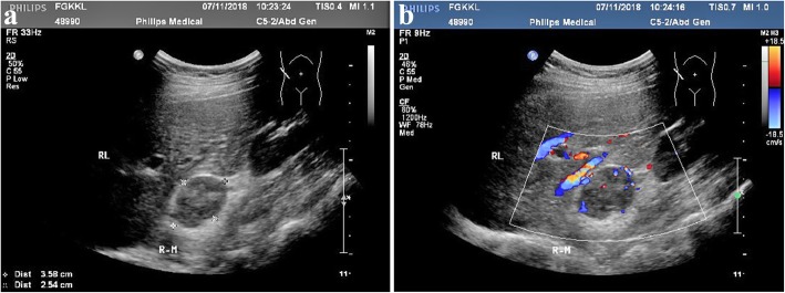
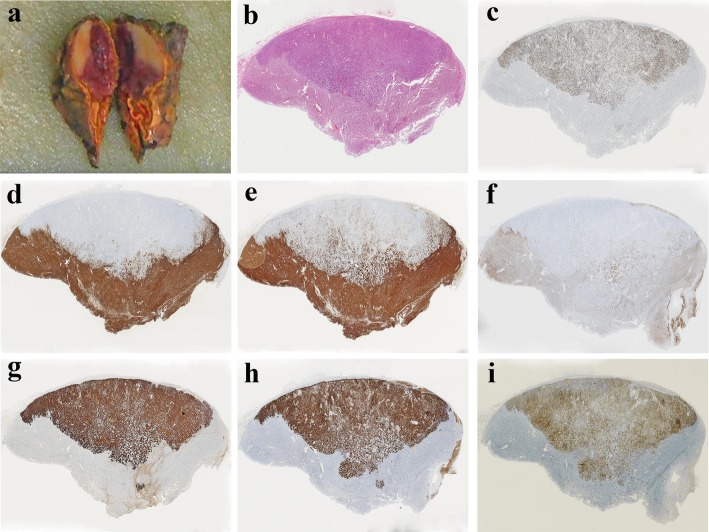
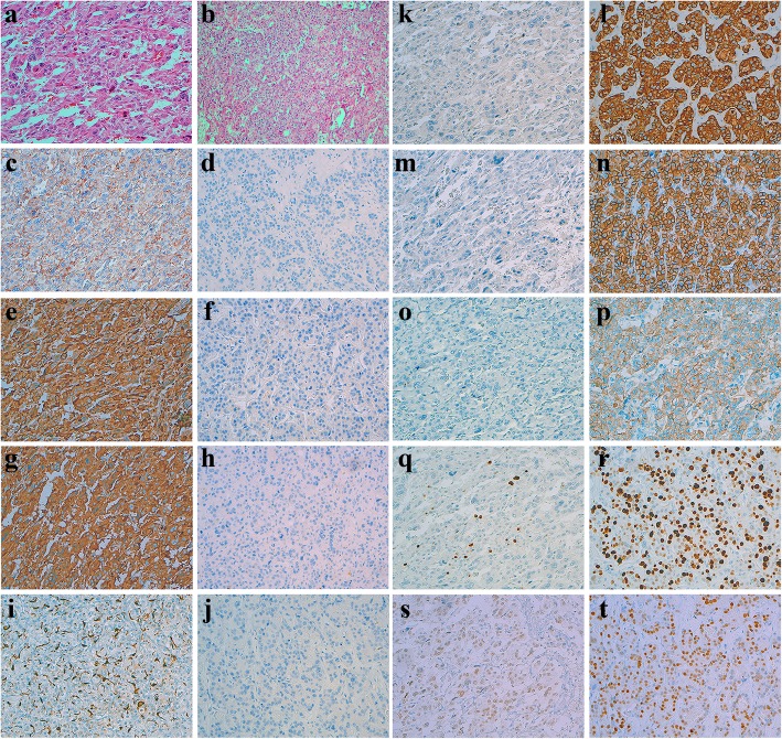
Case presentation: A 35-year-old woman was diagnosed with pheochromocytoma using computed tomography and ultrasound examinations. The tumor was surgically removed. Histological and immunohistochemical staining suggested that there were two components in the tumor: pheochromocytoma and metastatic cancer.

Conclusion: This is the second published case of pheochromocytoma with tumor-to-tumor metastasis from an invasive ductal carcinoma of the breast. Furthermore, we highlight the importance of awareness of tumor-to-tumor metastasis in pathological diagnosis.

Keywords: Breast carcinoma; Pheochromocytoma; Tumor-to-tumor metastasis.


Conflict of interest statement

The authors declare that they have no competing interests.

Figures

Fig. 1
Fig. 1
Computed tomography image showing a circular soft tissue density shadow in the right adrenal gland of the patient (a). The lesion in the arterial phase is markedly heterogeneous with a clear boundary after enhancement (b)
Fig. 2
Fig. 2
Ultrasound image showing a hypoechoic mass with a clear boundary in the right adrenal gland. The internal echo is heterogeneous (a). Color Doppler flow image showing a rich blood flow signal inside this mass (b)
Fig. 3
Fig. 3
The cut surface of the tumor is divided into two parts: whitish and pinkish-grey (a). Histological staining with hematoxylin and eosin shows that the cells of both parts are similar (b). The proliferative index (Ki67) of metastatic breast cancer is significantly higher than that of pheochromocytoma (c). Immunohistochemistry shows that pheochromocytoma cells are strongly positive for CgA, SYN and CD56 (d, e and f), and metastatic breast carcinoma cells are strongly positive for CK, HER-2 and membrane positive for E-cadherin (g, h and i)
Fig. 4
Fig. 4
Comparison of the two tumor components in morphology and IHC staining, × 200. Hematoxylin and eosin staining of two tumor cells (a Pheochromocytoma, b Metastatic breast carcinoma). CD56 is expressed on the membrane of pheochromocytoma cells (c Pheochromocytoma, d Metastatic breast carcinoma). CgA is diffusely expressed in pheochromocytoma (e Pheochromocytoma, f Metastatic breast carcinoma). SYN is also diffusely expressed in pheochromocytoma cells (g Pheochromocytoma, h Metastatic breast carcinoma). S100 is expressed in the sustentacular cells of pheochromocytoma (i Pheochromocytoma, j Metastatic breast carcinoma). CK is stongly positive in metastatic breast cancer cells (k Pheochromocytoma, l Metastatic breast carcinoma). HER-2 is stongly positive (3+) in metastatic breast cancer cells (m Pheochromocytoma, m Metastatic breast carcinoma). E-cadherin is membrane positive in metastatic breast cancer cells (o Pheochromocytoma, p Metastatic breast carcinoma). The proliferative index (Ki67) of pheochromocytoma is not more than 10% (q) and that of metastatic breast cancer is about 70% (r). GATA-3 is weakly positive in pheochromocytoma cells (s) and significantly positive in metastatic breast cancer cells (t)

References

    1. Erdogan H, Aydin M, Tasdemiroglu E. Tumor-to-tumor metastasis of the central nervous system. Turk Neurosurg. 2014;24:151–162. - PubMed
    1. Moody P, Murtagh K, Piduru S, Brem S, Murtagh R, Rojiani A. Tumor-to-tumor metastasis: pathology and neuroimaging considerations. Int J Clin Exp Pathol. 2012;5:367–373. - PMC - PubMed
    1. Sayegh E, Burch E, Henderson G, Oh T, Bloch O, Parsa A. Tumor-to-tumor metastasis: breast carcinoma to meningioma. J Clin Neurosci. 2015;22:268–274. doi: 10.1016/j.jocn.2014.07.002. - DOI - PubMed
    1. Wagnerova H, Lazurova I, Felsoci M. Adrenal metastases. Bratisl Lek Listy. 2013;114:237–240. - PubMed
    1. Seitz G, Schüder G. Neoplasm to neoplasm metastasis. Pheochromocytoma harboring a metastasis of breast cancer. Pathol Res Pract. 1987;182:228–232. doi: 10.1016/S0344-0338(87)80109-7. - DOI - PubMed

MeSH terms