

Identifying the molecular and cellular signature of cardiac dilation following myocardial infarction
- PMID: 31109452
- PMCID: PMC6530589
- DOI: 10.1016/j.bbadis.2018.09.023
Identifying the molecular and cellular signature of cardiac dilation following myocardial infarction
Abstract
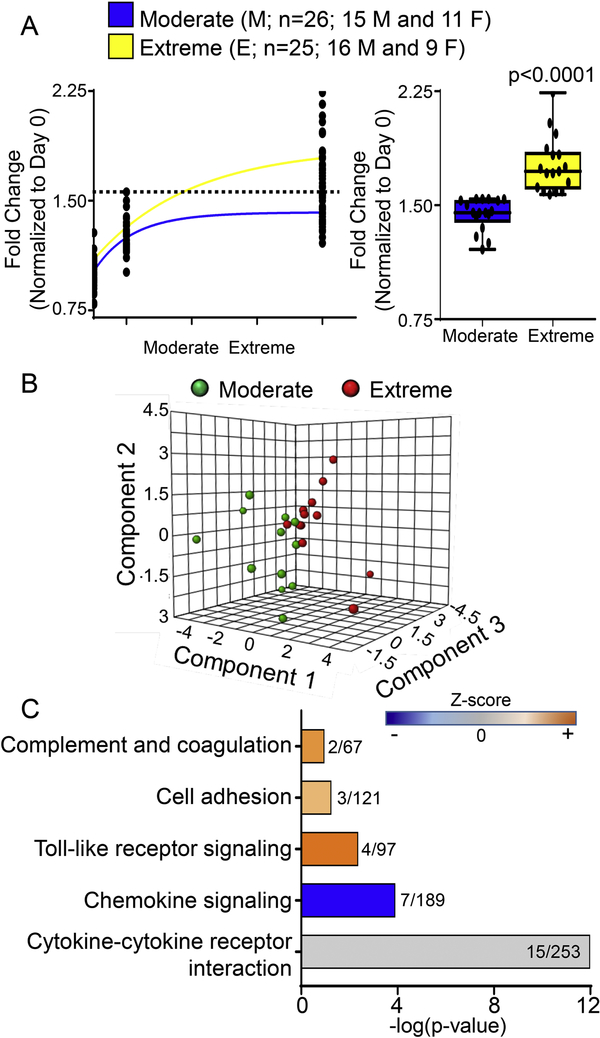
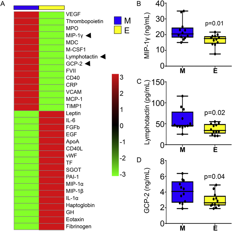
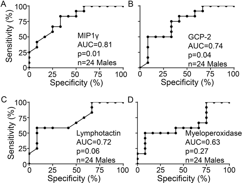
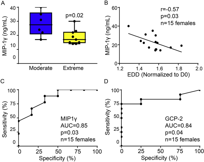
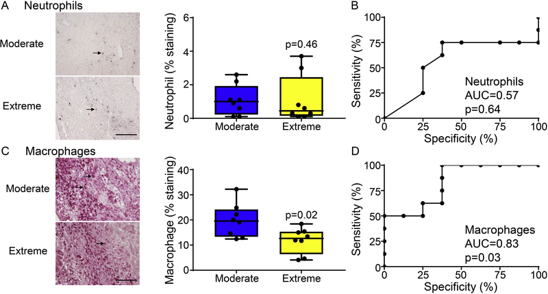
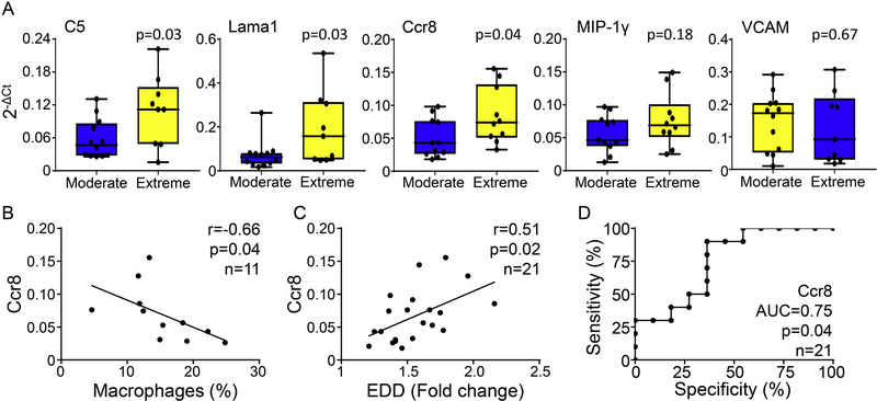
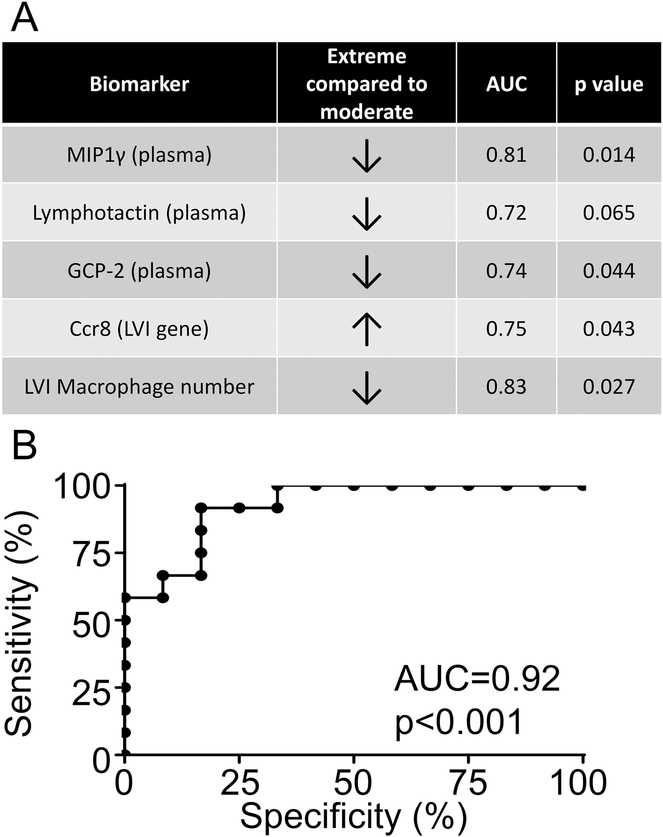
Establishing molecular and cellular indicators that reflect the extent of dilation of the left ventricle (LV) after myocardial infarction (MI) may improve diagnostic and prognostic capabilities. We queried the Mouse Heart Attack Research Tool (mHART) 1.0 for day 7 post-MI mice (age 3-9 months, untreated males and females) with serial echocardiographic data at days 0, 1, and 7 (n = 51). Mice were classified into two subgroups determined by a median fold change of 1.6 in end-diastolic dimensions (EDD) normalized to pre-MI values; n = 26 fell below (moderate; mean of 1.42 ± 0.01) and n = 25 fell above this cut-off (extreme; mean of 1.79 ± 0.01; p < 0.001 vs. moderate). Plasma proteomic profiling of 34 analytes measured at day 7 post-MI from male mice (n = 12 moderate and 12 extreme) were evaluated as the test dataset, and receiver operating curve (ROC) analysis was used to assess strength of biomarkers. Females (n = 6 moderate and 9 extreme) were used as the validation dataset. Both by t-test and characteristic (ROC) curve analysis, lower macrophage inflammatory protein-1 gamma (MIP-1γ), lymphotactin, and granulocyte chemotactic protein-2 (GCP-2) were identified as plasma indicators for dilation status (p < 0.05 for all). Macrophage numbers were decreased and complement C5, laminin 1, and Ccr8 gene levels were significantly higher in the LV infarcts of the extreme dilation group (p < 0.05 for all). A composite panel including plasma MIP-1γ, lymphotactin, and GCP-2, and LV infarct Ccr8 and macrophage numbers strongly mirrored LV dilation status (AUC = 0.92; p < 0.0001). Using the mHART 1.0 database, we determined that a failure to mount sufficient macrophage-mediated inflammation was indicative of exacerbated LV dilation.
Keywords: Big data; Cardiovascular disease; Inflammation; LV dilation; Macrophage; Myocardial infarction; Proteomics.
Published by Elsevier B.V.
Figures
Similar articles
-
Clinical aspects of left ventricular diastolic function assessed by Doppler echocardiography following acute myocardial infarction.Dan Med Bull. 2001 Nov;48(4):199-210. Dan Med Bull. 2001. PMID: 11767125 Review.
-
Dimethyl fumarate preserves left ventricular infarct integrity following myocardial infarction via modulation of cardiac macrophage and fibroblast oxidative metabolism.J Mol Cell Cardiol. 2021 Sep;158:38-48. doi: 10.1016/j.yjmcc.2021.05.008. Epub 2021 May 21. J Mol Cell Cardiol. 2021. PMID: 34023353 Free PMC article.
-
Exogenous CXCL4 infusion inhibits macrophage phagocytosis by limiting CD36 signalling to enhance post-myocardial infarction cardiac dilation and mortality.Cardiovasc Res. 2019 Feb 1;115(2):395-408. doi: 10.1093/cvr/cvy211. Cardiovasc Res. 2019. PMID: 30169632 Free PMC article.
-
Building a better infarct: Modulation of collagen cross-linking to increase infarct stiffness and reduce left ventricular dilation post-myocardial infarction.J Mol Cell Cardiol. 2015 Aug;85:229-39. doi: 10.1016/j.yjmcc.2015.06.006. Epub 2015 Jun 12. J Mol Cell Cardiol. 2015. PMID: 26080361 Free PMC article.
-
Knowledge gaps to understanding cardiac macrophage polarization following myocardial infarction.Biochim Biophys Acta. 2016 Dec;1862(12):2288-2292. doi: 10.1016/j.bbadis.2016.05.013. Epub 2016 May 27. Biochim Biophys Acta. 2016. PMID: 27240543 Free PMC article. Review.
Cited by
-
Harnessing the Plasma Proteome to Mirror Current and Predict Future Cardiac Remodeling After Myocardial Infarction.J Cardiovasc Transl Res. 2023 Feb;16(1):3-16. doi: 10.1007/s12265-022-10326-w. Epub 2022 Oct 5. J Cardiovasc Transl Res. 2023. PMID: 36197585 Free PMC article.
-
Identification of Independent and Communal Differentially Expressed Genes as Well as Potential Therapeutic Targets in Ischemic Heart Failure and Non-Ischemic Heart Failure.Pharmgenomics Pers Med. 2021 Jun 14;14:683-693. doi: 10.2147/PGPM.S313621. eCollection 2021. Pharmgenomics Pers Med. 2021. PMID: 34163213 Free PMC article.
-
Guidelines for in vivo mouse models of myocardial infarction.Am J Physiol Heart Circ Physiol. 2021 Dec 1;321(6):H1056-H1073. doi: 10.1152/ajpheart.00459.2021. Epub 2021 Oct 8. Am J Physiol Heart Circ Physiol. 2021. PMID: 34623181 Free PMC article. Review.
-
CircRNA ACAP2 Is Overexpressed in Myocardial Infarction and Promotes the Maturation of miR-532 to Induce the Apoptosis of Cardiomyocyte.J Cardiovasc Pharmacol. 2021 Jun 18;78(2):247-252. doi: 10.1097/FJC.0000000000001065. J Cardiovasc Pharmacol. 2021. PMID: 34139744 Free PMC article.
-
Cardiomyocytes induced from hiPSCs by well-defined compounds have therapeutic potential in heart failure by secreting PDGF-BB.Signal Transduct Target Ther. 2022 Jul 29;7(1):253. doi: 10.1038/s41392-022-01045-4. Signal Transduct Target Ther. 2022. PMID: 35902567 Free PMC article.
References
-
- DeLeon KY, Yabluchanskiy A, Winniford MD, Lange RA, Chilton RJ, Lindsey ML, Modifying Matrix Remodeling To Prevent Heart Failure, in: Li R, Weisel RD (Eds.) Cardiac Regeneration And Repair Volume I: Pathology And Therapies, Woodhead, 2013.
Publication types
MeSH terms
Substances
Grants and funding
LinkOut - more resources
Full Text Sources
Medical
Research Materials
Miscellaneous
