Review
. 2020 Feb 3;10(2):a034173.
doi: 10.1101/cshperspect.a034173.

Vagus Nerve Stimulation and the Cardiovascular System

Affiliations
Review

Vagus Nerve Stimulation and the Cardiovascular System

Michael J Capilupi et al. Cold Spring Harb Perspect Med. .

Abstract

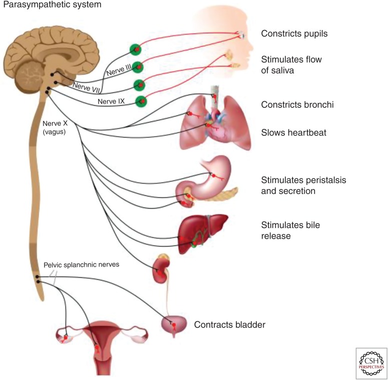
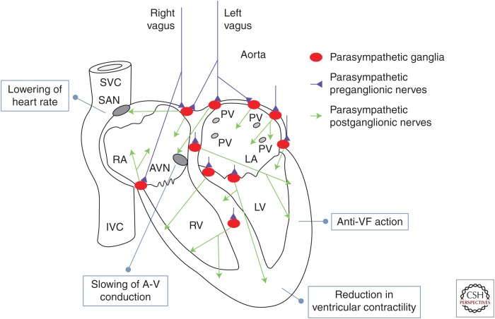
The vagus nerve plays an important role in maintaining physiological homeostasis, which includes reflex pathways that regulate cardiac function. The link between vagus nerve activity and the high-frequency component of heart rate variability (HRV) has been well established, correlating with vagal tone. Recently, vagus nerve stimulation (VNS) has been investigated as a therapeutic for a multitude of diseases, such as treatment-resistant epilepsy, rheumatoid arthritis, Crohn's disease, and asthma. Because of the vagus nerve's innervation of the heart, VNS has been identified as a potential therapy for cardiovascular disorders, such as cardiac arrest, acute myocardial infarction, and stroke. Here, we review the current state of preclinical and clinical studies, as well as the potential application of VNS in relation to the cardiovascular system.


Figures

Figure 1.
Figure 1.
Schematic representation of the efferent (motor) projections of the parasympathetic autonomic nervous system (ANS). Cranial nerves III, VII, and IX innervate the pupils, producing pupillary constriction, and the salivary glands, resulting in increase in the flow of saliva. The pelvic splachnic nerves innervate the bladder, with their activation resulting in bladder constriction and facilitation of urination, the lower intestine, facilitating defecation; they also innervate the male and female sexual organs. The main efferent conduit of the parasympathetic ANS, the motor vagus, starts in cells in two nuclei in the brainstem, the dorsal motor nucleus of the vagus, and the nucleus ambiguus. The axons of those cells project to a variety of organs, forming synapses with ganglionic cells in parasympathetic ganglia found at or very close to those organs. Postganglionic axons from ganglionic cells innervate the lungs (promoting bronchoconstriction), the heart (slowing of heart rate, reduction in cardiac contractility), the pulmonary and systemic vessels (vasodilation and reduction in blood pressure), stomach (promoting peristalsis and gastric secretion), liver and pancreas (complex metabolic actions), spleen (preganglionic axons interacting with the splenic nerve, promoting anti-inflammatory effects), and kidneys (affecting vascular tone and filtration rate).
Figure 2.
Figure 2.
Vagus nerve innervations in the heart. Shown in blue are the two cardiac vagal preganglionic nerves (left and right). They synapse on a number of parasympathetic ganglia, shown in red, located in the epicardium and in the atrial and ventricular septum. The parasympathetic ganglionic neurons project postganglionic axons, shown in green, to different parts of the heart; the anatomical distribution of those axons can help explain the physiological actions of the vagus in the heart. In more detail: sinoatrial node (SAN): vagus nerve activation resulting in lowering of heart rate (negative chronotropic action); atrioventricular node (AVN): slowing of A-V conduction (negative dromotropic action); walls of the right and left ventricles (RVs, LVs) and conduction system: reduction in ventricular contractility (negative inotropic action); increase in threshold for induction of ventricular fibrillation (VF). SVC, Superior vena cava; IVC, inferior vena cava; RA/LA, right and left atrium; PV, pulmonary vein. (Image modified from Coote 2013, with permission, from The Physiological Society © 2013.)

References

    1. Al-Khatib SM, Stevenson WG. 2018. Management of ventricular arrhythmias and sudden cardiac death risk related to ischemic and nonischemic cardiomyopathy. JAMA Cardiol 3: 541–542. 10.1001/jamacardio.2018.0016 - DOI - PubMed
    1. Annoni EM, Xie X, Lee SW, Libbus I, KenKnight BH, Osborn JW, Tolkacheva EG. 2015. Intermittent electrical stimulation of the right cervical vagus nerve in salt-sensitive hypertensive rats: Effects on blood pressure, arrhythmias, and ventricular electrophysiology. Physiol Rep 3: e12476 10.14814/phy2.12476 - DOI - PMC - PubMed
    1. Ay I, Sorensen AG, Ay H. 2011. Vagus nerve stimulation reduces infarct size in rat focal cerebral ischemia: An unlikely role for cerebral blood flow. Brain Res 1392: 110–115. 10.1016/j.brainres.2011.03.060 - DOI - PMC - PubMed
    1. Bailey P, Bremer F. 1938. A sensory cortical representation of the vagus nerve: with a note on the effects of low blood pressure on the cortical electrogram. J Neurophysiol 1: 405–412. 10.1152/jn.1938.1.5.405 - DOI
    1. Beaumont E, Southerland EM, Hardwick JC, Wright GL, Ryan S, Li Y, KenKnight BH, Armour JA, Ardell JL. 2015. Vagus nerve stimulation mitigates intrinsic cardiac neuronal and adverse myocyte remodeling postmyocardial infarction. Am J Physiol Heart Circ Physiol 309: H1198–H1206. 10.1152/ajpheart.00393.2015 - DOI - PMC - PubMed

LinkOut - more resources