. 2019 Jun 28;294(26):10300-10314.
doi: 10.1074/jbc.RA118.006302. Epub 2019 May 20.

Parkin recruitment to impaired mitochondria for nonselective ubiquitylation is facilitated by MITOL

Affiliations

Parkin recruitment to impaired mitochondria for nonselective ubiquitylation is facilitated by MITOL

Fumika Koyano et al. J Biol Chem. .

Abstract

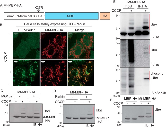
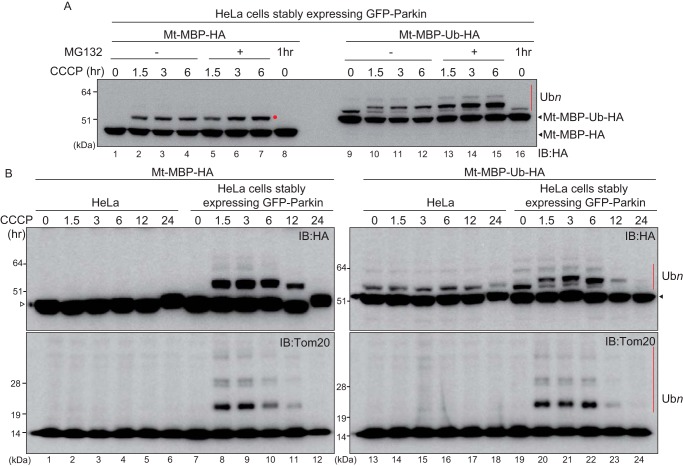
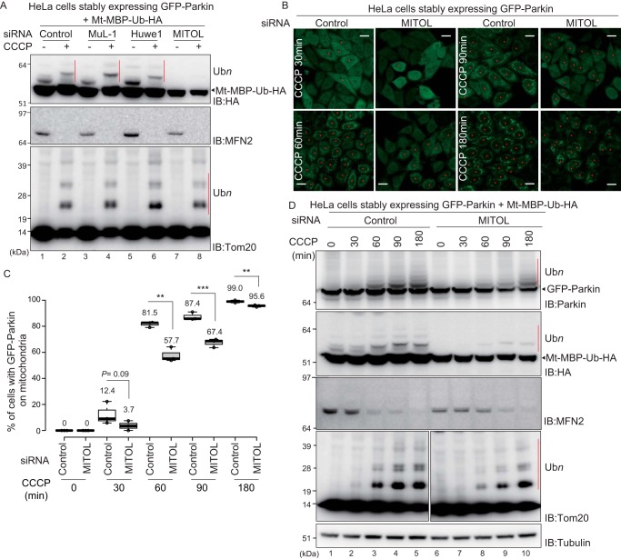
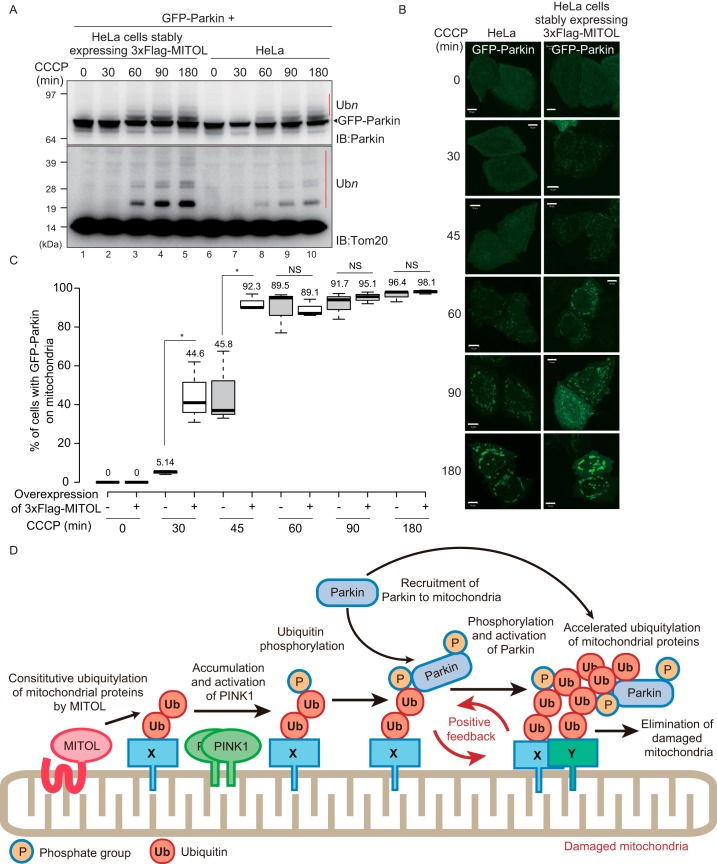
PINK1 (PARK6) and PARKIN (PARK2) are causal genes of recessive familial Parkinson's disease. Parkin is a ubiquitin ligase E3 that conjugates ubiquitin to impaired mitochondrial proteins for organelle degradation. PINK1, a Ser/Thr kinase that accumulates only on impaired mitochondria, phosphorylates two authentic substrates, the ubiquitin-like domain of Parkin and ubiquitin. Our group and others have revealed that both the subcellular localization and ligase activity of Parkin are regulated through interactions with phosphorylated ubiquitin. Once PINK1 localizes on impaired mitochondria, PINK1-catalyzed phosphoubiquitin recruits and activates Parkin. Parkin then supplies a ubiquitin chain to PINK1 for phosphorylation. The amplified ubiquitin functions as a signal for the sequestration and degradation of the damaged mitochondria. Although a bewildering variety of Parkin substrates have been reported, the basis for Parkin substrate specificity remains poorly understood. Moreover, the mechanism underlying initial activation and translocation of Parkin onto mitochondria remains unclear, because the presence of ubiquitin on impaired mitochondria is thought to be a prerequisite for the initial PINK1 phosphorylation process. Here, we show that artificial mitochondria-targeted proteins are ubiquitylated by Parkin, suggesting that substrate specificity of Parkin is not determined by its amino acid sequence. Moreover, recruitment and activation of Parkin are delayed following depletion of the mitochondrial E3, MITOL/March5. We propose a model in which the initial step in Parkin recruitment and activation requires protein ubiquitylation by MITOL/March5 with subsequent PINK1-mediated phosphorylation. Because PINK1 and Parkin amplify the ubiquitin signal via a positive feedback loop, the low substrate specificity of Parkin might facilitate this amplification process.

Keywords: Parkinson disease; mitophagy; parkin; ubiquitin; ubiquitin ligase.


Conflict of interest statement

The authors declare that they have no conflicts of interest with the contents of this article

Figures

Figure 1.
Figure 1.
Parkin-catalyzed ubiquitylation of mitochondria-localized GFP following CCCP treatment. A, schematic representation of Mt-GFP. GFP was fused with the mitochondrial anchoring sequence of Tom20 (i.e. the N-terminal 33 amino acids (a.a.) of Tom20). B, mitochondrial localization of Mt-GFP. HeLa cells stably expressing HA–Parkin were transfected with Mt-GFP, treated with 15 μm CCCP for 3 h, and then subjected to immunocytochemistry. The Mt-GFP signal co-localized with the mitochondrial marker Tom22 ± CCCP. Scale bars, 10 μm. C, immunoblot-based (IB) detection of Mt-GFP ubiquitylation. HeLa cells stably expressing HA–Parkin were transfected with Mt-GFP or MtK27R–GFP, in which Lys-27 of the Tom20 mitochondrial anchoring sequence was mutated to Arg to prevent ubiquitin conjugation to the Tom20 moiety. The cells were treated with 15 μm CCCP ± 10 μm MG132 for 3 h and then immunoblotted with an anti-GFP antibody. The red dots and bars indicate ubiquitylated Mt-GFP, and the black asterisk indicates cross-reacting bands. D, mitochondrial GFP rather than cytosolic GFP is specifically ubiquitylated in a Parkin-dependent manner. HeLa cells stably expressing HA–Parkin transfected with MtK27R–GFP or GFP, and intact HeLa cells transfected with MtK27R–GFP were treated with 15 μm CCCP ± 10 μm MG132 for 3 h and then immunoblotted with an anti-GFP antibody. The red dot and bar indicate ubiquitylated Mt-GFP, and the black asterisk indicates a cross-reacting band. E, ubiquitylation of MtK27R–GFP involves ubiquitin and phosphoubiquitin. HeLa cells stably expressing HA–Parkin transfected with MtK27R–GFP were treated with 15 μm CCCP + 10 μm MG132 for 3 h and were then immunoprecipitated (IP) with an anti-GFP antibody. The total cell lysate (input) and immunoprecipitated products were immunoblotted with the indicated antibodies. The red bars indicate ubiquitylation (Ubn) or phosphoubiquitylation (phospho-Ubn) of MtK27R–GFP.
Figure 2.
Figure 2.
Parkin-catalyzed ubiquitylation of mitochondria-localized MBP following CCCP treatment. A, schematic representation of Mt–MBP–HA. B, mitochondrial localization of Mt–MBP–HA. HeLa cells stably expressing GFP–Parkin were transfected with Mt–MBP–HA, treated with 15 μm CCCP for 3 h, and then subjected to immunocytochemistry. Mt–MBP–HA localized to mitochondria with GFP–Parkin following a decrease in mitochondrial membrane potential. Scale bars, 10 μm. C, HeLa cells stably expressing GFP–Parkin transfected with Mt–MBP–HA were treated with 15 μm CCCP ± 10 μm MG132 for 3 h and then immunoblotted (IB) with an anti-HA antibody. The proteasomal inhibitor MG132 did not enhance ubiquitylation of Mt–MBP–HA. D, intact HeLa cells or HeLa cells stably expressing GFP–Parkin were transfected with Mt–MBP–HA and then immunoblotted with an anti-HA antibody. Mt–MBP–HA ubiquitylation depended on Parkin. E, ubiquitylation of Mt–MBP–HA involves ubiquitin and phosphoubiquitin. HeLa cells stably expressing GFP–Parkin transfected with Mt–MBP–HA were treated with 15 μm CCCP for 3 h and then immunoprecipitated (IP) with an anti-HA antibody. Total cell lysate (input) and the immunoprecipitated products were immunoblotted with the indicated antibodies. The red bars indicate ubiquitylation (Ubn) or phosphoubiquitylation (phospho-Ubn) of Mt–MBP–HA. F, Parkin ubiquitylates mitochondria-localized MBP-HA but not cytosolic MBP-HA. HeLa cells stably expressing GFP–Parkin transfected with Mt–MBP–HA or MBP-HA were treated with 15 μm CCCP for 3 h and were then immunoblotted with an anti-HA antibody. The red bar indicates Parkin-mediated ubiquitylation of Mt–MBP–HA. a.a., amino acids.
Figure 3.
Figure 3.
Ubiquitin fusion accelerates Parkin-catalyzed ubiquitylation of mitochondria-localized MBP. A, schematic representation of Mt–MBP–HA and Mt–MBP–Ub–HA. Gly-76 of the ubiquitin moiety was mutated to Val to avoid cleavage by DUB. B, HeLa cells stably expressing GFP–Parkin were transfected with Mt–MBP–HA or Mt–MBP–Ub–HA, treated with 15 μm CCCP + 10 μm MG132 for 3 h, and then immunoblotted (IB) with an anti-HA antibody. Mt–MBP–Ub–HA ubiquitylation (indicated by the red bar) was more efficient than that of Mt–MBP–HA. C, HeLa cells stably expressing GFP–Parkin were transfected with Mt–MBP–Ub–HA or Mt–MBP–Ub(S65A)–HA, treated with 15 μm CCCP + 10 μm MG132 for 3 h, and then immunoprecipitated (IP) with an anti-HA antibody. Immunoprecipitated products were immunoblotted with the indicated antibodies. The red bars indicate ubiquitylation (Ubn) or phosphoubiquitylation (phospho-Ubn) of Mt–MBP–Ub–HA and Mt–MBP–Ub(S65A)–HA. Phosphorylation of the ubiquitin moiety enhanced Parkin-catalyzed ubiquitylation of Mt–MBP–Ub–HA. D, HeLa cells stably expressing GFP–Parkin were transfected with Mt–MBP–Ub–HA or Mt–MBP–Ub(S65A)–HA, treated with 15 μm CCCP + 10 μm MG132 for the indicated times (min), and then immunoblotted with an anti-HA antibody. A phosphorylation-deficient S65A mutation in the ubiquitin moiety delayed Parkin-catalyzed ubiquitylation of Mt–MBP–Ub–HA. E, intact HeLa cells or HeLa cells stably expressing GFP–Parkin were transfected with Mt–MBP–HA or Mt–MBP–Ub–HA, treated with 15 μm CCCP + 10 μm MG132 for 3 h, and then immunoblotted with an anti-HA antibody. The red bar indicates ubiquitylation (Ubn) of Mt–MBP–Ub–HA. F, HeLa cells stably expressing GFP–Parkin were transfected with Mt–MBP–Ub–HA, Mt–MBP–Ub(K48R)–HA, or Mt-MBP-Ub(K63)-HA and then treated with 10 μm MG132 ± 15 μm CCCP for 3 h. Cell lysates were immunoblotted with an anti-HA antibody. H indicates higher molecular weight ubiquitylation that depends on both Parkin and CCCP; L indicates lower molecular weight Parkin-independent ubiquitylation. G, Parkin-mediated ubiquitylation of Mt–MBP–3HA and Mt–MBP–Ub–3HA reconstituted in vitro. Isolated mitochondria from CCCP-treated HeLa cells stably expressing Mt–MBP–3HA or Mt–MBP–Ub–3HA were mixed with recombinant ubiquitin, E1, E2 (UbcH7), and Parkin in the presence of ATP, MgCl2, and TCEP for 30 min in vitro. Ubiquitylation was analyzed by immunoblotting using anti-HA, anti-Tom20, and anti-MTCO2 antibodies. The red bars indicate ubiquitylation (Ubn) of Mt–MBP–3HA and Mt–MBP–Ub–3HA. a.a., amino acids.
Figure 4.
Figure 4.
Ubiquitylation of mitochondria-localized MBP and mitochondria-localized MBP-ubiquitin over time. A, HeLa cells stably expressing GFP–Parkin were transfected with Mt–MBP–HA or Mt–MBP–Ub–HA and then treated with 15 μm CCCP ± 10 μm MG132 for the indicated times. Cell lysates were then immunoblotted (IB) with an anti-HA antibody. Unlike ubiquitylated Mt–MBP–HA (lanes 1–7), MG132 treatment promoted accumulation of ubiquitylated Mt–MBP–Ub–HA (lanes 9–15). The red dot and red bar indicate ubiquitylation of Mt–MBP–HA and Mt–MBP–Ub–HA, respectively. B, intact HeLa cells or HeLa cells stably expressing GFP–Parkin were transfected with Mt–MBP–HA or Mt–MBP–Ub–HA and then treated with 15 μm CCCP for the indicated times. Cell lysates were immunoblotted with an anti-HA antibody or an anti-Tom20 antibody. The white triangle by the left panel indicates Mt–MBP–HA, and the black triangle by the right panel indicates Mt–MBP–Ub–HA. The ubiquitylation patterns of both Mt–MBP–HA and Mt–MBP–Ub–HA were comparable with the ubiquitylation pattern of the endogenous substrate, Tom20. The red bars indicate ubiquitylation (Ubn) of Mt–MBP–Ub–HA and Tom20.
Figure 5.
Figure 5.
Knockdown of MITOL attenuates Parkin recruitment to depolarized mitochondria and Parkin-catalyzed ubiquitylation. A, HeLa cells stably expressing GFP–Parkin were simultaneously transfected with Mt–MBP–Ub–HA and siRNAs against three different mitochondrial E3s. The cells were incubated for 2 days and then treated with 15 μm CCCP for 3 h. Cell lysates were then immunoblotted (IB) with the indicated antibodies. The red bars indicate Parkin-catalyzed ubiquitylation (Ubn) of Mt–MBP–Ub–HA and Tom20. MITOL knockdown attenuated the ubiquitylation of Mt–MBP–Ub–HA. B, HeLa cells stably expressing GFP–Parkin were transfected with a control siRNA or siRNA for MITOL, followed by CCCP treatment (15 μm) for the indicated times, and then the subcellular localization of GFP–Parkin was observed. The red asterisks indicate cells in which GFP–Parkin was recruited to mitochondria. Scale bars, 20 μm. C, statistical analysis of the subcellular localization of GFP–Parkin following 15 μm CCCP treatment for the indicated times in cells treated with control or MITOL siRNA. The percentage of cells with the indicated GFP–Parkin localization was calculated using > 100 cells. The numbers in the box-and-whisker plot are mean values across three independent experiments. Statistical significance was calculated using a one-tailed Student's t test. **, p < 0.01; ***, p < 0.001. D, HeLa cells stably expressing GFP–Parkin were simultaneously transfected with Mt–MBP–Ub–HA and control siRNA or siRNA for MITOL. The cells were incubated for 2 days and then treated with 15 μm CCCP for the indicated times. The cell lysates were then immunoblotted with the indicated antibodies. The red bars indicate autoubiquitylation of GFP–Parkin or Parkin-catalyzed ubiquitylation of Mt–MBP–Ub–HA and Tom20.
Figure 6.
Figure 6.
MITOL knockout attenuates Parkin recruitment to depolarized mitochondria and Parkin-catalyzed ubiquitylation. A, MITOL knockout HCT116 cells were generated using the CRISPR/Cas9 system. Intact HCT116 cells and MITOL knockout HCT116 cells stably expressing GFP–Parkin were treated with 15 μm CCCP for 60 min, and then the subcellular localization of GFP–Parkin was observed. Scale bars, 20 μm. B, statistical analysis of GFP–Parkin mitochondrial localization following 15 μm CCCP treatment for the indicated times in WT or MITOL knockout HCT116 cells. The percentage of cells with Parkin-positive mitochondria were determined using >100 cells. The numbers in the box-and-whisker plot are mean values across three independent experiments. Statistical significance was calculated using a one-tailed Student's t test. ***, p < 0.001. NS, not significant. C, MITOL depletion in MITOL knockout HCT116 cells was verified by immunoblotting (IB) with an anti-MITOL antibody. D, WT or MITOL knockout HCT116 cells stably expressing GFP–Parkin were treated with 15 μm CCCP for the indicated times and then immunoblotted with the indicated antibodies. The red bars indicate Parkin-catalyzed ubiquitylation (Ubn) of the endogenous substrates HKI, Tom20, and MitoNEET/CISD1.
Figure 7.
Figure 7.
Overexpression of MITOL enhances Parkin recruitment to depolarized mitochondria and Parkin-catalyzed ubiquitylation. A, intact HeLa cells or HeLa cells stably expressing 3×FLAG–MITOL were transfected with GFP–Parkin, treated with 15 μm CCCP for the indicated times, and then immunoblotted (IB) with anti-Parkin and anti-Tom20 antibodies. The red bars (Ubn) indicate autoubiquitylation of GFP–Parkin or Parkin-catalyzed ubiquitylation of Tom20. B, subcellular localization of transiently transfected GFP–Parkin in intact HeLa cells or HeLa cells stably expressing 3×FLAG–MITOL following 15 μm CCCP treatment for the indicated times. Scale bars, 10 μm. C, statistical analysis of the subcellular localization of GFP–Parkin following 15 μm CCCP treatment for the indicated times in intact HeLa cells or HeLa cells stably expressing 3×FLAG–MITOL. The percentage of cells with the mitochondria GFP–Parkin localization was calculated using >100 cells. Numbers in the box-and-whisker plot are mean values across three independent experiments. Statistical significance was calculated using a one-tailed Student's t test. *, p < 0.05. B and C show that overexpression of MITOL accelerates the recruitment of GFP–Parkin onto impaired mitochondria. D, schematic model. At steady-state conditions, MITOL ubiquitylates the endogenous mitochondrial substrate. When the mitochondrial membrane potential decreases, this MITOL-catalyzed constitutive ubiquitylation is used as a “seed” ubiquitin for subsequent PINK1/Parkin-dependent ubiquitylation, thereby generating a positive feedback cycle that amplifies ubiquitylation. Because Parkin lacks substrate specificity, the amplified ubiquitylation of proteins on damaged mitochondria can proceed efficiently. X, MITOL substrate; Y, any type of mitochondrial protein as Parkin lacks substrate specificity; NS, not significant.

References

    1. Schapira A. H. (2008) Mitochondria in the aetiology and pathogenesis of Parkinson's disease. Lancet Neurol. 7, 97–109 10.1016/S1474-4422(07)70327-7 - DOI - PubMed
    1. Tanner C. M., Kamel F., Ross G. W., Hoppin J. A., Goldman S. M., Korell M., Marras C., Bhudhikanok G. S., Kasten M., Chade A. R., Comyns K., Richards M. B., Meng C., Priestley B., Fernandez H. H., et al. (2011) Rotenone, paraquat, and Parkinson's disease. Environ. Health Perspect. 119, 866–872 10.1289/ehp.1002839 - DOI - PMC - PubMed
    1. Uversky V. N. (2004) Neurotoxicant-induced animal models of Parkinson's disease: understanding the role of rotenone, maneb and paraquat in neurodegeneration. Cell Tissue Res. 318, 225–241 10.1007/s00441-004-0937-z - DOI - PubMed
    1. Valente E. M., Abou-Sleiman P. M., Caputo V., Muqit M. M., Harvey K., Gispert S., Ali Z., Del Turco D., Bentivoglio A. R., Healy D. G., Albanese A., Nussbaum R., González-Maldonado R., Deller T., Salvi S., et al. (2004) Hereditary early-onset Parkinson's disease caused by mutations in PINK1. Science 304, 1158–1160 10.1126/science.1096284 - DOI - PubMed
    1. Kitada T., Asakawa S., Hattori N., Matsumine H., Yamamura Y., Minoshima S., Yokochi M., Mizuno Y., and Shimizu N. (1998) Mutations in the parkin gene cause autosomal recessive juvenile parkinsonism. Nature 392, 605–608 10.1038/33416 - DOI - PubMed

Publication types

MeSH terms

LinkOut - more resources