Wunderlich's syndrome in pregnancy: a shocking triad
- PMID: 31110067
- PMCID: PMC6536169
- DOI: 10.1136/bcr-2019-229219
Wunderlich's syndrome in pregnancy: a shocking triad
Abstract
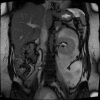
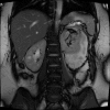
Wunderlich's syndrome, non-traumatic renal haemorrhage into the subscapular and perinephric space, in pregnancy, is a very rare clinical entity. We describe a case of Wunderlich's syndrome in a 29-year-old gravida 5 para 4 who presented to our emergency department with sudden onset severe left flank pain. On assessment, she was clinically shocked-hypotensive, tachycardic and perfused poorly peripherally. Ultrasound of the abdomen and pelvis and subsequent MRI of the left kidney revealed a large hypervascular exophytic lesion arising from the left renal pole-appearance consistent with an angiomyolipoma. This specific presentation is clinically characterised as Lenk's triad-acute flank pain, flank mass and hypovolaemic shock. The patient was adequately resuscitated and interventional radiological embolisation of the mass was performed. She went on to have an uneventful pregnancy and delivered vaginally after induction at 38 weeks of gestation.
Keywords: obstetrics and gynaecology; urology.
© BMJ Publishing Group Limited 2019. Re-use permitted under CC BY-NC. No commercial re-use. See rights and permissions. Published by BMJ.
Conflict of interest statement
Competing interests: None declared.
Figures


References
-
- Polkey HJ. Spontaneous nontraumatic perirenal and renal hematomas. Arch Surg 1933;26:196 10.1001/archsurg.1933.01170020030002 - DOI
-
- Illescas Molina T, Montalvo Montes J, Contreras Cecilia E, et al. . tuberous sclerosis and pregnancy. Ginecol Obstet Mex 2009;77:380–6. - PubMed
-
- EAU. EAU Guidelines. 2018. http://uroweb.org/guideline/renal-cell-carcinoma/
Publication types
MeSH terms
LinkOut - more resources
Full Text Sources
Medical
