. 2019 Aug 15;25(16):4933-4944.
doi: 10.1158/1078-0432.CCR-19-0183. Epub 2019 May 20.

A Phase I Dose-Escalation Trial of BN-CV301, a Recombinant Poxviral Vaccine Targeting MUC1 and CEA with Costimulatory Molecules

Affiliations

A Phase I Dose-Escalation Trial of BN-CV301, a Recombinant Poxviral Vaccine Targeting MUC1 and CEA with Costimulatory Molecules

Margaret E Gatti-Mays et al. Clin Cancer Res. .

Abstract

Purpose: BN-CV301 is a poxviral-based vaccine comprised of recombinant (rec.) modified vaccinia Ankara (MVA-BN-CV301; prime) and rec. fowlpox (FPV-CV301; boost). Like its predecessor PANVAC, BN-CV301 contains transgenes encoding tumor-associated antigens MUC1 and CEA as well as costimulatory molecules (B7.1, ICAM-1, and LFA-3). PANVAC was reengineered to make it safer and more antigenic.

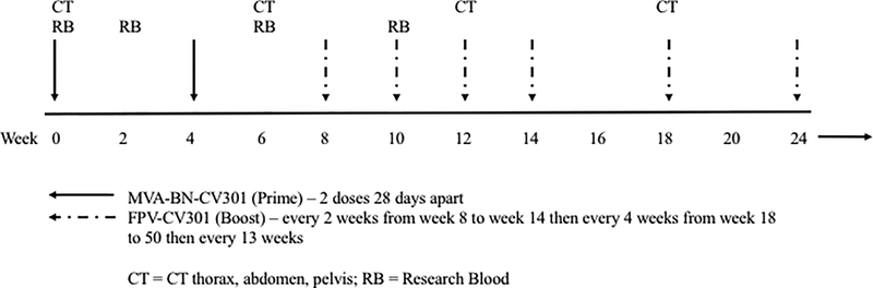
Patients and methods: This open-label, 3+3 design, dose-escalation trial evaluated three dose levels (DL) of MVA-BN-CV301: one, two, or four subcutaneous injections of 4 × 10⁸ infectious units (Inf.U)/0.5 mL on weeks 0 and 4. All patients received FPV-CV301 subcutaneously at 1 × 10⁹ Inf.U/0.5 mL every 2 weeks for 4 doses, then every 4 weeks. Clinical and immune responses were evaluated.

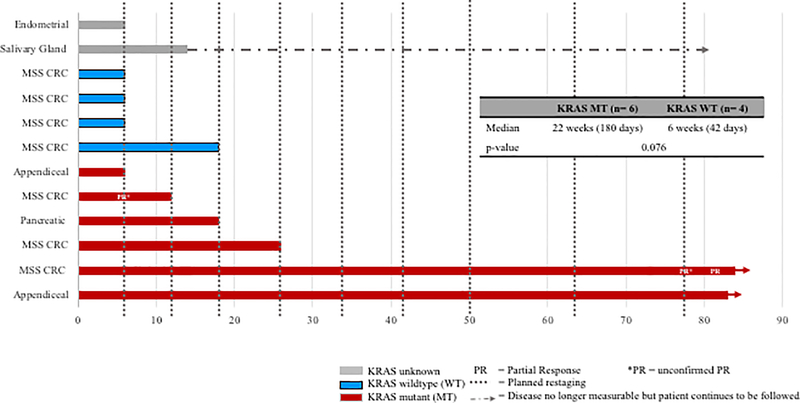
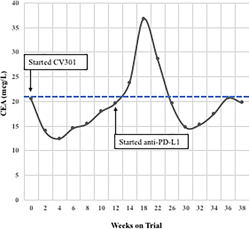
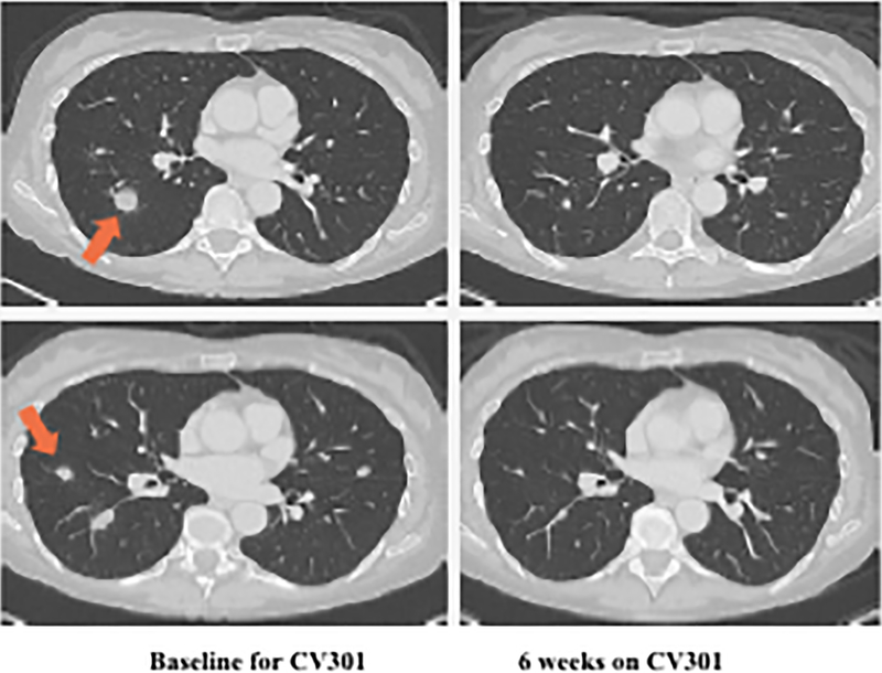
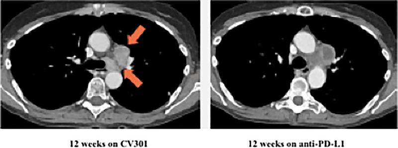
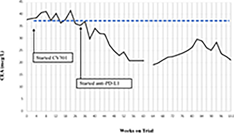
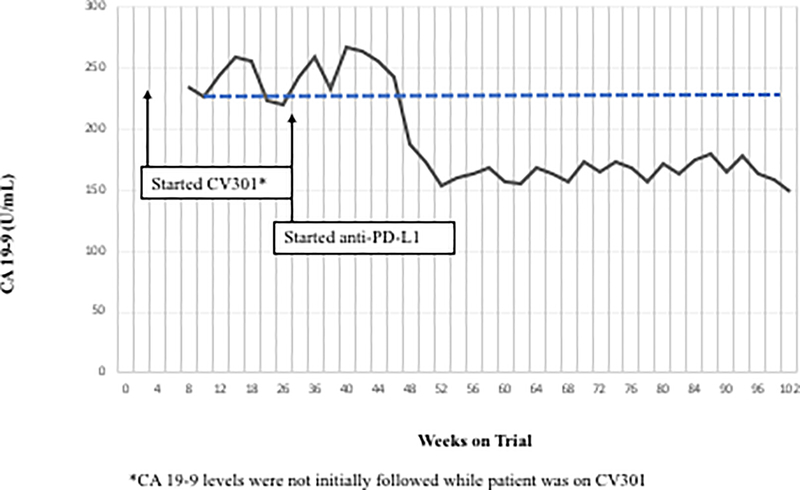
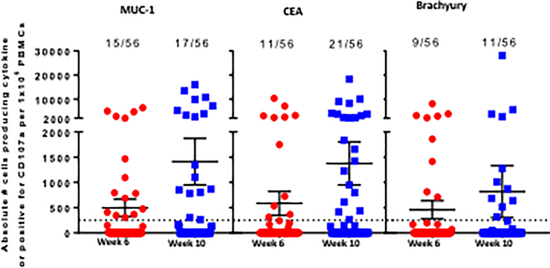
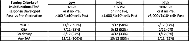
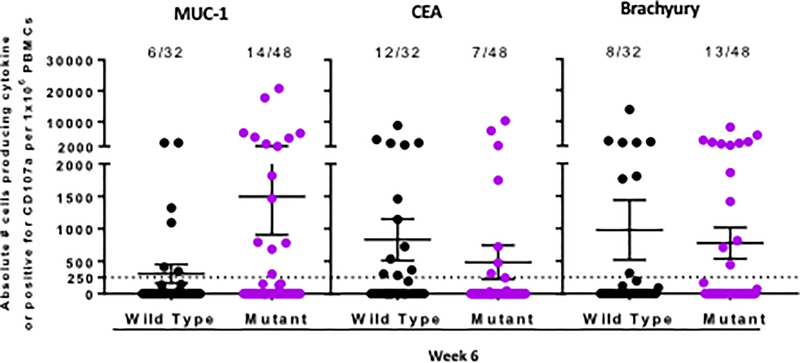
Results: There were no dose-limiting toxicities. Twelve patients enrolled on trial [dose level (DL) 1 = 3, DL2 = 3, DL3 = 6). Most side effects were seen with the prime doses and lessened with subsequent boosters. All treatment-related adverse events were temporary, self-limiting, grade 1/2, and included injection-site reactions and flu-like symptoms. Antigen-specific T cells to MUC1 and CEA, as well as to a cascade antigen, brachyury, were generated in most patients. Single-agent BN-CV301 produced a confirmed partial response (PR) in 1 patient and prolonged stable disease (SD) in multiple patients, most notably in KRAS-mutant gastrointestinal tumors. Furthermore, 2 patients with KRAS-mutant colorectal cancer had prolonged SD when treated with an anti-PD-L1 antibody following BN-CV301.

Conclusions: The BN-CV301 vaccine can be safely administered to patients with advanced cancer. Further studies of the vaccine in combination with other agents are planned.See related commentary by Repáraz et al., p. 4871.


Conflict of interest statement

Disclosure of Potential Conflicts of Interest:

M. Gatti-Mays, J. Strauss , R. Donahue, C. Palena, J. Del Rivero, J. Redman, R. Madan, J. Marté, L. Cordes, E. Lamping, J. Schlom and J. Gulley declare they have no potential conflicts to disclose. A. Orpia and A. Burmeister are employees of Leidos Biomedical Research. E. Wagner is an employee of Bavarian Nordic GmbH. C. Pico Navarro and C. Heery are employees of Bavarian Nordic.

Figures

Figure 1.
Figure 1.. BN-Phase I schema and clinical outcomes.
(A) BN-CV301 Phase I study schema. (B) Progression-free survival on BN-CV301 for individual patients by KRAS status. Post-hoc KRAS analysis performed with an exact two-tailed log-rank test suggests there is a trend towards a difference by KRAS status but is limited by sample size.
Figure 2.
Figure 2.
Two patients with KRAS mutation, MSS mCRC had a ≥ 35% decrease in tumor markers associated with prolonged stable disease after treatment with BN-CV301 followed by an anti-PD-L1 antibody. Patient #1: A 62-year-old female with KRAS MT, MSS CRC with progressive disease despite 6 prior regimens (DL=1). She had an initial decrease in CEA (A) and an unconfirmed partial response at the first restaging (6 weeks; B) followed by growth of a non-target lesion (new mediastinal adenopathy) at the 12-week restaging. The patient then enrolled on an anti-PD-L1 trial and experienced a subsequent decrease in CEA as well as a radiographic response (C) to treatment with necrosis of mediastinal adenopathy and decreasing tumor markers at week 12 of treatment with the anti-PD-L1 antibody. This patient had stable disease for 43 weeks while on an anti-PD-L1 antibody. Patient #2: A 54-year-old female with KRAS MT, MSS CRC with progressive disease despite 8 prior regimens (DL=1). While on BN-CV301 trial, CEA and tumor burden were stable but the patient eventually developed progressive disease at 26 weeks. Patient was then enrolled on an anti-PD-L1 trial and experienced a subsequent decrease in tumor markers (D and E). Radiographically the patient has continued stable disease on the anti-PD-L1 antibody ongoing at 71 weeks. Prior trials have found a median progression-free survival of 10 weeks (2.2 months) in patients with MSS, mCRC who receive an anti-PD-L1 antibody. Blue dotted lines represent baseline tumor markers.
Figure 2.
Figure 2.
Two patients with KRAS mutation, MSS mCRC had a ≥ 35% decrease in tumor markers associated with prolonged stable disease after treatment with BN-CV301 followed by an anti-PD-L1 antibody. Patient #1: A 62-year-old female with KRAS MT, MSS CRC with progressive disease despite 6 prior regimens (DL=1). She had an initial decrease in CEA (A) and an unconfirmed partial response at the first restaging (6 weeks; B) followed by growth of a non-target lesion (new mediastinal adenopathy) at the 12-week restaging. The patient then enrolled on an anti-PD-L1 trial and experienced a subsequent decrease in CEA as well as a radiographic response (C) to treatment with necrosis of mediastinal adenopathy and decreasing tumor markers at week 12 of treatment with the anti-PD-L1 antibody. This patient had stable disease for 43 weeks while on an anti-PD-L1 antibody. Patient #2: A 54-year-old female with KRAS MT, MSS CRC with progressive disease despite 8 prior regimens (DL=1). While on BN-CV301 trial, CEA and tumor burden were stable but the patient eventually developed progressive disease at 26 weeks. Patient was then enrolled on an anti-PD-L1 trial and experienced a subsequent decrease in tumor markers (D and E). Radiographically the patient has continued stable disease on the anti-PD-L1 antibody ongoing at 71 weeks. Prior trials have found a median progression-free survival of 10 weeks (2.2 months) in patients with MSS, mCRC who receive an anti-PD-L1 antibody. Blue dotted lines represent baseline tumor markers.
Figure 3.
Figure 3.
Magnitude and breadth of combined antigen-specific CD4+ and CD8+ T-cell responses post- (vs pre) BN-CV301 (MVA-BN-CV301/FPV-CV301) vaccine. (A) Seven patients were tested and TAA responses compared both at 6 weeks (2 weeks after the 2nd MVA-BN-CV301 prime, red) and 10 weeks (2 weeks after the 1st FPV-CV301 boost, blue). The absolute number of CD4+ and/or CD8+ T-cells producing IFNγ, TNF, or IL-2, or positive for the degranulation marker CD107a per 1 × 10⁶ PBMC plated at the start of the stimulation assay was calculated. Any background signal (obtained with the HLA peptide pool) and any signal obtained prior to vaccination was subtracted ([post-TAA – post-HLA] – [pre-TAA – pre-HLA]). Each point indicates the magnitude of a cytokine/CD107a measure, with 8 measures assessed per patient (CD8+IFNγ+, CD8+TNF+, CD8+IL-2+, CD8+CD107a+, CD4+IFNγ+, CD4+TNF+, CD4+IL-2+, CD4+CD107a+). Frequency of positive measures (>250 CD4+ and CD8+ T-cells producing cytokine and/or positive for CD107a) is indicated. (B) Polyfunctional TAA responses (CD4+ and CD8+ T cells expressing 2 or more of the following: IFNγ, TNF, IL-2, or CD107a) were measured before and after any time point post-vaccination in all 12 patients. The frequency of patients developing a low, mid, or high magnitude of multifunctional TAA-specific T-cells after vaccination at any time point post- vs pre- is indicated. (C) TAA responses in 4 patients with known wild type KRAS (Black) and six patients with known/presumed KRAS mutations (Purple) were compared at 6 weeks (2 weeks after the 2nd MVA Prime). The absolute number of CD4+ or CD8+ T-cells producing IFNg, TNF, or IL-2, or positive for the degranulation marker CD107a per 1×10⁶ PBMC plated at the start of the stimulation assay was calculated. Each point indicates the magnitude of a cytokine/CD107a measure, with 8 measures assessed per patient. Frequency of positive measures (>250 CD4+ and CD8+ T-cells producing cytokine and/or positive for CD107a) is indicated.
Figure 3.
Figure 3.
Magnitude and breadth of combined antigen-specific CD4+ and CD8+ T-cell responses post- (vs pre) BN-CV301 (MVA-BN-CV301/FPV-CV301) vaccine. (A) Seven patients were tested and TAA responses compared both at 6 weeks (2 weeks after the 2nd MVA-BN-CV301 prime, red) and 10 weeks (2 weeks after the 1st FPV-CV301 boost, blue). The absolute number of CD4+ and/or CD8+ T-cells producing IFNγ, TNF, or IL-2, or positive for the degranulation marker CD107a per 1 × 10⁶ PBMC plated at the start of the stimulation assay was calculated. Any background signal (obtained with the HLA peptide pool) and any signal obtained prior to vaccination was subtracted ([post-TAA – post-HLA] – [pre-TAA – pre-HLA]). Each point indicates the magnitude of a cytokine/CD107a measure, with 8 measures assessed per patient (CD8+IFNγ+, CD8+TNF+, CD8+IL-2+, CD8+CD107a+, CD4+IFNγ+, CD4+TNF+, CD4+IL-2+, CD4+CD107a+). Frequency of positive measures (>250 CD4+ and CD8+ T-cells producing cytokine and/or positive for CD107a) is indicated. (B) Polyfunctional TAA responses (CD4+ and CD8+ T cells expressing 2 or more of the following: IFNγ, TNF, IL-2, or CD107a) were measured before and after any time point post-vaccination in all 12 patients. The frequency of patients developing a low, mid, or high magnitude of multifunctional TAA-specific T-cells after vaccination at any time point post- vs pre- is indicated. (C) TAA responses in 4 patients with known wild type KRAS (Black) and six patients with known/presumed KRAS mutations (Purple) were compared at 6 weeks (2 weeks after the 2nd MVA Prime). The absolute number of CD4+ or CD8+ T-cells producing IFNg, TNF, or IL-2, or positive for the degranulation marker CD107a per 1×10⁶ PBMC plated at the start of the stimulation assay was calculated. Each point indicates the magnitude of a cytokine/CD107a measure, with 8 measures assessed per patient. Frequency of positive measures (>250 CD4+ and CD8+ T-cells producing cytokine and/or positive for CD107a) is indicated.

Comment in

  • When Cancer Vaccines Go Viral.
    Repáraz D, Llopiz D, Sarobe P. Repáraz D, et al. Clin Cancer Res. 2019 Aug 15;25(16):4871-4873. doi: 10.1158/1078-0432.CCR-19-1652. Epub 2019 Jun 21. Clin Cancer Res. 2019. PMID: 31227502

References

    1. Rajabi H, Kufe D. MUC1-C oncoprotein integrates a program of EMT, epigenetic reprogramming and immune evasion in human carcinomas. Biochim Biophys Acta 2017;1868(1):117–22 doi 10.1016/j.bbcan.2017.03.003. - DOI - PMC - PubMed
    1. Kharbanda A, Rajabi H, Jin C, Alam M, Wong KK, Kufe D. MUC1-C confers EMT and KRAS independence in mutant KRAS lung cancer cells. Oncotarget 2014;5(19):8893–905 doi 10.18632/oncotarget.2360. - DOI - PMC - PubMed
    1. Bouillez A, Adeegbe D, Jin C, Hu X, Tagde A, Alam M, et al. MUC1-C promotes the suppressive immune microenvironment in non-small cell lung cancer. OncoImmunology 2017;6(9):e1338998 doi 10.1080/2162402X.2017.1338998. - DOI - PMC - PubMed
    1. Zeng Y, Zhang Q, Zhang Y, Lu M, Liu Y, Zheng T, et al. MUC1 predicts colorectal cancer metastasis: a systematic review and meta-analysis of case controlled studies. PLoS One 2015;10(9):e0138049 doi 10.1371/journal.pone.0138049. - DOI - PMC - PubMed
    1. Kharbanda A, Rajabi H, Jin C, Raina D, Kufe D. Oncogenic MUC1-C promotes tamoxifen resistance in human breast cancer. Mol Cancer Res 2013;11(7):714–23 doi 10.1158/1541-7786.MCR-12-0668. - DOI - PMC - PubMed