. 2019 Aug;17(8):1652-1664.
doi: 10.1158/1541-7786.MCR-19-0144. Epub 2019 May 20.

Identification of TP53RK-Binding Protein (TPRKB) Dependency in TP53-Deficient Cancers

Affiliations

Identification of TP53RK-Binding Protein (TPRKB) Dependency in TP53-Deficient Cancers

Moloy T Goswami et al. Mol Cancer Res. 2019 Aug.

Abstract

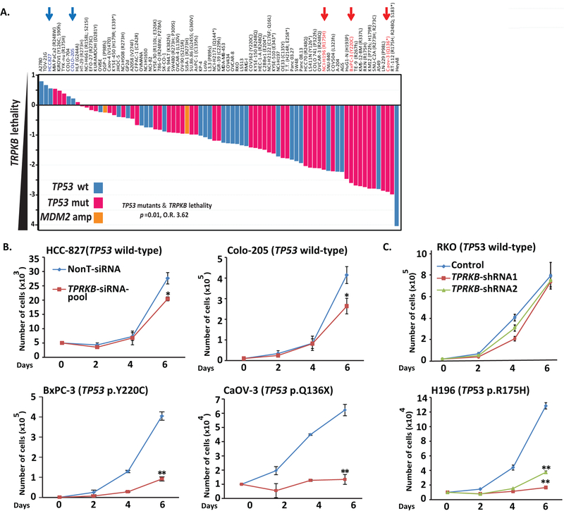
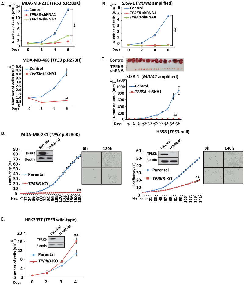
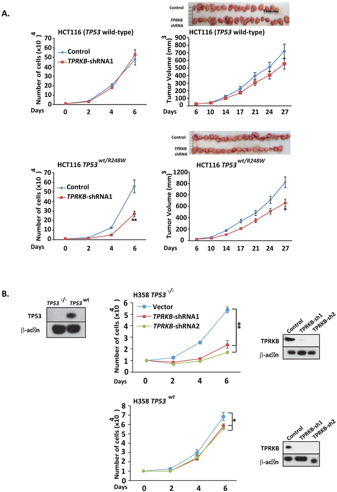
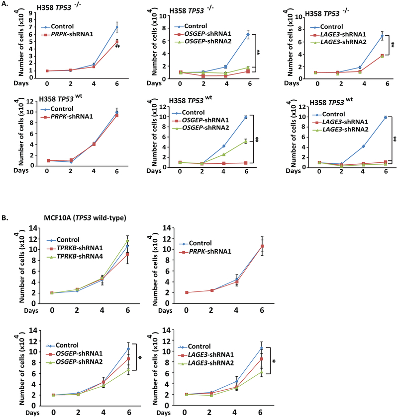
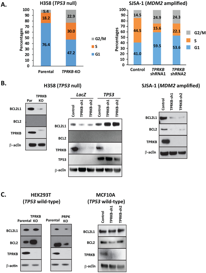
Tumor protein 53 (TP53; p53) is the most frequently altered gene in human cancer. Identification of vulnerabilities imposed by TP53 alterations may enable effective therapeutic approaches. Through analyzing short hairpin RNA (shRNA) screening data, we identified TP53RK-Binding Protein (TPRKB), a poorly characterized member of the tRNA-modifying EKC/KEOPS complex, as the most significant vulnerability in TP53-mutated cancer cell lines. In vitro and in vivo, across multiple benign-immortalized and cancer cell lines, we confirmed that TPRKB knockdown in TP53-deficient cells significantly inhibited proliferation, with minimal effect in TP53 wild-type cells. TP53 reintroduction into TP53-null cells resulted in loss of TPRKB sensitivity, confirming the importance of TP53 status in this context. In addition, cell lines with mutant TP53 or amplified MDM2 (E3-ubiquitin ligase for TP53) also showed high sensitivity to TPRKB knockdown, consistent with TPRKB dependence in a wide array of TP53-altered cancers. Depletion of other EKC/KEOPS complex members exhibited TP53-independent effects, supporting complex-independent functions of TPRKB. Finally, we found that TP53 indirectly mediates TPRKB degradation, which was rescued by coexpression of PRPK, an interacting member of the EKC/KEOPS complex, or proteasome inhibition. Together, these results identify a unique and specific requirement of TPRKB in a variety of TP53-deficient cancers. IMPLICATIONS: Cancer cells with genomic alterations in TP53 are dependent on TPRKB.


Figures

Figure 1.
Figure 1.. Identification and validation of TPRKB vulnerability in cancer cell lines with TP53 alterations.
A) TPRKB was the only gene identified in the Project Achilles genome-wide shRNA database as showing significant enrichment for dependency in TP53 altered cell lines (two-sided Fisher’s exact test odds ratio (O.R.) and p-value are shown for original (2015) TP53 annotation status; O.R. = 2.6 and p=0.06 for TP53 status and TPRKB dependency [> or < 1.4] for 2019 comprehensive cell line encyclopaedia [CCLE] TP53 annotation status). TPRKB dependency (fold-change in shRNA abundance versus control transfected cells) for cancer cell lines from Project Achilles is plotted, with cell lines ordered by increasing TPRKB dependency. The color of the bars indicates mutational status from 2019 CCLE annotation: blue bars indicate TP53 wild-type cells, red bars indicate TP53 hotspot/deleterious mutants, and orange bars indicate MDM2-amplifications. Blue and red arrows indicate cell lines with wild-type and mutant TP53, respectively, that were chosen for validation experiments. B) Differential effects of pooled siRNA against TPRKB (or scrambled control) on cell proliferation were confirmed in TP53 wt (Colo-205 and HCC-827) and TP53 mut cell lines (BxPC-3 and CaOV-3). C) As in B, but using independent shRNAs against TPRKB (or scrambled control) in RKO (TP53 wt) and H196 (TP53 mut). Confirmation of TPRKB knockdown is shown in Figures S1. All experiments utilized triplicate samples, with the average and standard error plotted. * indicate p-values < 0.05 and ** indicate p-values <0.01.
Figure 2.
Figure 2.. Various classes of TP53 perturbation result in marked TPRKB dependent proliferation.
Cancer cell lines with A) hotspot TP53 mutations (MDA-MB-231 and MDA-MB-469) or B) MDM2 amplification (SJSA-1; see Figure S2 for additional cell lines) were assessed for TPRKB dependent proliferation using shRNA. C) As in B, except using in vivo mouse xenografts with tumor volume plotted and tumors at sacrifice shown. D) CRISPR-Cas9 mediated TPRKB knockout in TP53 mut MDA-MB-231 and H358 (TP53 deep deletion) cells confirmed results from siRNA/shRNA. TPRKB knockout was confirmed by Western blotting, and % confluency was plotted with representative phase contrast photomicrographs at the indicated time points shown. E) Minimal proliferation inhibition was observed upon TPRKB knockout by CRISPR-Cas9 in benign immortalized TP53 wt cells (HEK293T). All experiments utilized triplicate samples, with the average and standard error plotted. * indicate p-values < 0.05 and ** indicate p-values <0.01.
Figure 3 –
Figure 3 –. Isogenic cells show wild-type TP53 presence rescues proliferation defects from TPRKB knockdown
A) TPRKB was stably knocked down in isogenic HCT116 (TP53 wild-type) and HCT116 TP53WT/R248W cells. The stable clones were assayed for cell proliferation and tumor formation in nude mice. B) TPRKB knockdown by shRNA was performed in H358 cells stably expressing LacZ control (left) or TP53 (right) and proliferation was monitored. Inset Western blot panels confirm TP53 over-expression from lentiviral transduction and TPRKB knockdown. All experiments utilized triplicate samples, with the average and standard error plotted. * indicate p-values < 0.05 and ** indicate p-values <0.01.
Figure 4-
Figure 4-. Knockdown of other EKC/KEOPS complex members does not produce the same TP53-dependent effects as TPRKB.
A) TPRKB is a member of the EKC/KEOPS complex, which includes the canonical proteins PRPK, OSGEP and LAGE3. To investigate the TP53 dependent effects of other EKC/KEOPS complex members on proliferation, PRPK, OSGEP or LAGE3 were knocked down using shRNA in H358 TP53−/− LacZ and H358 TP53WT cells as in Figure 3. B) Proliferation of benign immortalized MCF10A (TP53 wt) cells was similarly assessed after stable knockdown of the indicated EKC/KEOPS complex member by shRNA. All experiments utilized triplicate samples, with the average and standard error plotted. * indicate p-values < 0.05 and ** indicate p-values <0.01.
Figure 5-
Figure 5-. Loss of TPRKB leads to cell cycle arrest and reduced anti-apoptotic protein expression in TP53-deficient cells.
A) Serum stimulated synchronized H358 (parental) or CRISPR-Cas9 generated H358-TPRKB knockout (KO) cells and SJSA-1 control or TPRKB knockdown cells were assessed for cell cycle analysis by flow cytometry. The proportion of cells in G1, S, and G2/M is plotted. B) Anti-apoptotic protein (BCL2L1 and BCL2) expression was determined by Western blotting in H358 control, H358 TPRKB-KO, H358 TP53−/− LacZ, and H358 TP53WT and SJSA-1 control and TPRKB shRNA knockdown cells. C) Additional TP53 wild-type cell lines were assayed for BCL2 and BCL2L1 expression: HEK293T cells with TPRKB-KO or PRPK-KO and MCF10A control or TPRKB knockdown cells.
Figure 6.
Figure 6.. TP53 degrades TPRKB through the proteasome, while PRPK expression stabilizes TPRKB.
A) Parental HEK293T and HEK293T with CRISPR introduced FLAG epitope into the endogenous TPRKB locus (HEK293T-TPRKB-Flag) were used for co-immunoprecipitation. After Flag pulldown, samples were tested for endogenous TPRKB-Flag interaction with PRPK or TP53 B) Increasing amounts of exogenous TP53-V5 in HEK293T cells were used to observe effects of TP53 expression on TPRKB-HA protein levels by Western Blot. C) The impact of TP53 protein level on TPRKB expression was assessed through exogenous expression of TP53 in H358 cells and, conversely, knockdown of endogenous TP53 in HEK293T. D) Co-expression of PRPK-Flag and knockout of PRPK in HEK293T cells was used to determine PRPK effects on TPRKB protein levels. E) Using TPRKB knockout cells, we looked at PRPK expression by Western blot.
Figure 7-
Figure 7-. Model of TPRKB dependency in TP53-deficient cells.
A) Interactions between PRPK and TPRKB lead to TPRKB stabilization, while the presence of TP53 leads to reduced TPRKB levels. Additionally, TP53 is able to mediate TPRKB degradation through the proteasome, despite the fact that TPRKB and TP53 do not directly interact. Influences of these proteins may be key in phenotypes we witness. B) In cells that maintain wild-type TP53, loss of TPRKB does not have a profound effect on cell viability. In cells in which TP53 is perturbed, either through loss of wild-type protein, mutation of TP53, or disturbance of TP53 in some other manner (i.e. MDM2 amplification), TPRKB depletion leads to a reduction in cell proliferation.

References

    1. Vogelstein B, Lane D, Levine AJ. Surfing the p53 network. Nature 2000;408:307–10 - PubMed
    1. Bieging KT, Mello SS, Attardi LD. Unravelling mechanisms of p53-mediated tumour suppression. Nature reviews Cancer 2014;14:359–70 - PMC - PubMed
    1. Kandoth C, McLellan MD, Vandin F, Ye K, Niu B, Lu C, et al. Mutational landscape and significance across 12 major cancer types. Nature 2013;502:333–9 - PMC - PubMed
    1. Soussi T, Wiman KG. TP53: an oncogene in disguise. Cell death and differentiation 2015;22:1239–49 - PMC - PubMed
    1. Gurpinar E, Vousden KH. Hitting cancers’ weak spots: vulnerabilities imposed by p53 mutation. Trends in cell biology 2015;25:486–95 - PubMed

Publication types

MeSH terms

Substances