Case Reports
. 2019 Mar 30;7(5):930-934.
doi: 10.1002/ccr3.2100. eCollection 2019 May.

A case of adenomyoepithelioma with myoepithelial carcinoma of the breast

Affiliations
Case Reports

A case of adenomyoepithelioma with myoepithelial carcinoma of the breast

Ryota Ito et al. Clin Case Rep. .

Abstract

Adenomyoepithelioma with myoepithelial carcinoma of the breast is rare and diagnosed with histology and immunohistochemistry. We present a case of malignant transformation over 10 years, with ultrasonographic findings, highlighting the importance of an early excisional biopsy. Conservative surgery and radiation therapy were performed. There was no recurrence for 2 years.

Keywords: adenomyoepithelioma; breast cancer; myoepithelial carcinoma.


Conflict of interest statement

None declared.

Figures

Figure 1
Figure 1
Ultrasonography of a lump in the left breast. Ultrasonography (A) at the initial visit shows a flat and well‐defined hypoechoic lesion measuring 0.8 cm (BI‐RADS category 2), (B) 8 y after the initial visit shows a homogenous hypoechoic lesion measuring 1.5 cm with posterior acoustic shadow, slightly taller than wide ratio and lobulation (BI‐RADS category 5), (C) 9 y after the initial visit shows a more lobulated hypoechoic lesion measuring 1.7 cm (BI‐RADS category 5), and (D) 10 y after the initial visit shows a solid irregular lobulated heterogeneous hypoechoic lesion with a partially unclear border measuring 2.5 cm (BI‐RADS category 5)
Figure 2
Figure 2
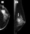
Preoperative left mammography. Shown is a high‐density, well‐demarcated lesion with round macrocalcification and no spicules in the outer, middle area of the left breast (white arrow; BI‐RADS category 3). CC, craniocaudal view; MLO, mediolateral oblique view
Figure 3
Figure 3
Characteristics of the tumor. A, Macroscopically, the tumor is solid and grayish with focal yellowish parts and cystic degeneration. B, Microscopically, the right side of the figure shows the AME component, while the left side shows the myoepithelial carcinoma component. No separation is seen between the two components. Black asterisk indicates necrosis (×2). C, The AME component shows biphasic proliferation of both ductal epithelium and myoepithelium (×20), and D, the myoepithelial carcinoma component shows monophasic proliferation. The cytoplasm is pale, and the nuclei are pleomorphic and heterogeneous in size. Black arrows indicate mitosis (×40) (B‐D: hematoxylin and eosin stain)
Figure 4
Figure 4
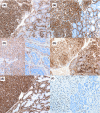
Immunohistochemical analysis. Myoepithelial cells of the AME (right side) and myoepithelial carcinoma (left side) components are positive for (A) HHF35 (×20), (B) α‐SMA (×20), (C) calponin (×20), (D) S‐100 (×20), (E) CD10 (×20), and (F) p63 (×20)

References

    1. Santhosh R, Padu K, Singh Th B, Sharma MB, Singh TS. Myoepithelial carcinoma of the breast. J Clin Diagn Res. 2013;7:1191‐1193. - PMC - PubMed
    1. Liang YF, Zeng JC, Ruan JB, et al. Malignant myoepithelioma of the breast: a case report and review of literature. Int J Clin Exp Pathol. 2014;7:2654‐2657. - PMC - PubMed
    1. Lakhani SR, International Agency for Research on Cancer , World Health Organization . World Health Organization classification of tumours In: Lakhani SR, Ellis IO, Schnitt SJ, et al. eds. WHO Classification of Tumours of the Breast, 4th edn Lyon: International Agency for Research on Cancer; 2012:120‐121.
    1. Korolczuk A, Amarowicz M, Bak K, Korobowicz E, Koncewicz T. Adenomyoepithelioma of the breast with late pulmonary metastases ‐ case report and review of the literature. J Cardiothorac Surg. 2016;11:121. - PMC - PubMed
    1. Moritz AW, Wiedenhoefer JF, Profit AP, Jagirdar J. Breast adenomyoepithelioma and adenomyoepithelioma with carcinoma (malignant adenomyoepithelioma) with associated breast malignancies: a case series emphasizing histologic, radiologic, and clinical correlation. Breast. 2016;29:132‐139. - PubMed

Publication types

LinkOut - more resources