

Correlation Between Corneal Topographic, Densitometry, and Biomechanical Parameters in Keratoconus Eyes
- PMID: 31110913
- PMCID: PMC6504131
- DOI: 10.1167/tvst.8.3.12
Correlation Between Corneal Topographic, Densitometry, and Biomechanical Parameters in Keratoconus Eyes
Abstract
Purpose: To investigate the correlation between corneal densitometry, corneal topographic parameters, and corneal biomechanical properties in keratoconus.
Methods: A total of 76 eyes of 76 keratoconus patients were enrolled in this cross-sectional study. Corneal densitometry and topography were measured using Pentacam HR. Corneal biomechanical properties were measured using CorVis ST.
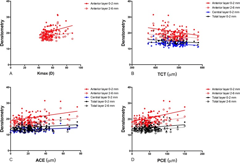
Results: The corneal densitometry values of the anterior 0 to 2 and 2 to 6 mm layers significantly correlated with the maximum keratometry values (R = 0.373, P = 0.001 and R = 0.276, P = 0.016, respectively), thinnest corneal thickness values (R = -0.331, P = 0.003 and R = -0.234, P = 0.042, respectively), anterior corneal elevation (R = 0.392, P < 0.001 and R = 0.323, P = 0.004, respectively), and posterior corneal elevation (R = 0.450, P < 0.001 and R = 0.367, P = 0.001, respectively). The stiffness parameter-applanation time 1 (SP-A1) significantly correlated with the corneal densitometry values for the anterior 0 to 2 mm (R = -0.397, P < 0.001), anterior 2 to 6 mm (R = -0.331, P = 0.004), central 0 to 2 mm (R = -0.306, P = 0.007), central 2 to 6 mm (R = -0.228, P = 0.048), posterior 2 to 6 mm (R = -0.243, P = 0.035), total 0 to 2 mm (R = -0.291, P = 0.011), and total 2 to 6 mm (R = -0.295, P = 0.010) layers.
Conclusions: The corneal densitometry values correlated with the severity of keratoconus and the SP-A1 values.
Translational relevance: Corneal densitometry values may serve as markers to predict the severity of keratoconus.
Keywords: CorVis ST; Pentacam HR; biomechanics; corneal structure; keratoconus.
Figures


Similar articles
-
Keratectasia severity staging and progression assessment based on the biomechanical E-staging.Eye Vis (Lond). 2024 Jul 1;11(1):24. doi: 10.1186/s40662-024-00392-3. Eye Vis (Lond). 2024. PMID: 38946004 Free PMC article. Review.
-
Relationship Among Corneal Stiffness, Thickness, and Biomechanical Parameters Measured by Corvis ST, Pentacam and ORA in Keratoconus.Front Physiol. 2019 Jun 13;10:740. doi: 10.3389/fphys.2019.00740. eCollection 2019. Front Physiol. 2019. PMID: 31263429 Free PMC article.
-
[Influence factors and differences of posterior corneal elevation measured by Pentacam system combined with Corvis ST].Zhonghua Yan Ke Za Zhi. 2020 Feb 11;56(2):110-117. doi: 10.3760/cma.j.issn.0412-4081.2020.02.006. Zhonghua Yan Ke Za Zhi. 2020. PMID: 32074821 Chinese.
-
Relationship between corneal biomechanical parameters and corneal sublayer thickness measured by Corvis ST and UHR-OCT in keratoconus and normal eyes.Eye Vis (Lond). 2021 Jan 8;8(1):2. doi: 10.1186/s40662-020-00225-z. Eye Vis (Lond). 2021. PMID: 33419485 Free PMC article.
-
Identification of genetic variants in five chinese families with keratoconus: Pathogenicity analysis and characteristics of parental corneal topography.Front Genet. 2022 Oct 6;13:978684. doi: 10.3389/fgene.2022.978684. eCollection 2022. Front Genet. 2022. PMID: 36276932 Free PMC article.
Cited by
-
Comparison of the quantitative contrast sensitivity function between early keratoconus and normal eyes.BMC Ophthalmol. 2024 Oct 18;24(1):458. doi: 10.1186/s12886-024-03695-0. BMC Ophthalmol. 2024. PMID: 39425133 Free PMC article.
-
Impacts and Correlations on Corneal Biomechanics, Corneal Optical Density and Intraocular Pressure after Cataract Surgery.Diagnostics (Basel). 2024 Jul 18;14(14):1557. doi: 10.3390/diagnostics14141557. Diagnostics (Basel). 2024. PMID: 39061693 Free PMC article.
-
Keratectasia severity staging and progression assessment based on the biomechanical E-staging.Eye Vis (Lond). 2024 Jul 1;11(1):24. doi: 10.1186/s40662-024-00392-3. Eye Vis (Lond). 2024. PMID: 38946004 Free PMC article. Review.
-
Comparison of bilateral differential characteristics of corneal biomechanics between keratoconus and normal eyes.Front Bioeng Biotechnol. 2023 Jun 1;11:1163223. doi: 10.3389/fbioe.2023.1163223. eCollection 2023. Front Bioeng Biotechnol. 2023. PMID: 37324412 Free PMC article.
-
Investigation of How Corneal Densitometry Artefacts Affect the Imaging of Normal and Keratoconic Corneas.Bioengineering (Basel). 2024 Feb 1;11(2):148. doi: 10.3390/bioengineering11020148. Bioengineering (Basel). 2024. PMID: 38391634 Free PMC article.
References
-
- de Sanctis U, Loiacono C, Richiardi L, Turco D, Mutani B, Grignolo FM. Sensitivity and specificity of posterior corneal elevation measured by Pentacam in discriminating keratoconus/subclinical keratoconus. Ophthalmology. 2008;115:1534–1539. - PubMed
-
- Swartz T, Marten L, Wang M. Measuring the cornea: the latest developments in corneal topography. Curr Opin Ophthalmol. 2007;18:325–333. - PubMed
-
- Shen Y, Chen Z, Knorz MC, Li M, Zhao J, Zhou X. Comparison of corneal deformation parameters after SMILE, LASEK, and femtosecond laser-assisted LASIK. J Refract Surg. 2014;30:310318. - PubMed
LinkOut - more resources
Full Text Sources
