

The crown-root morphology of central incisors in different skeletal malocclusions assessed with cone-beam computed tomography
- PMID: 31111270
- PMCID: PMC6527728
- DOI: 10.1186/s40510-019-0272-2
The crown-root morphology of central incisors in different skeletal malocclusions assessed with cone-beam computed tomography
Abstract
Background: To determine the discrepancy of crown-root morphology of central incisors among different types of skeletal malocclusion using cone-beam computed tomography (CBCT) and to provide guidance for proper torque expression of anterior teeth and prevention of alveolar fenestration and dehiscence.
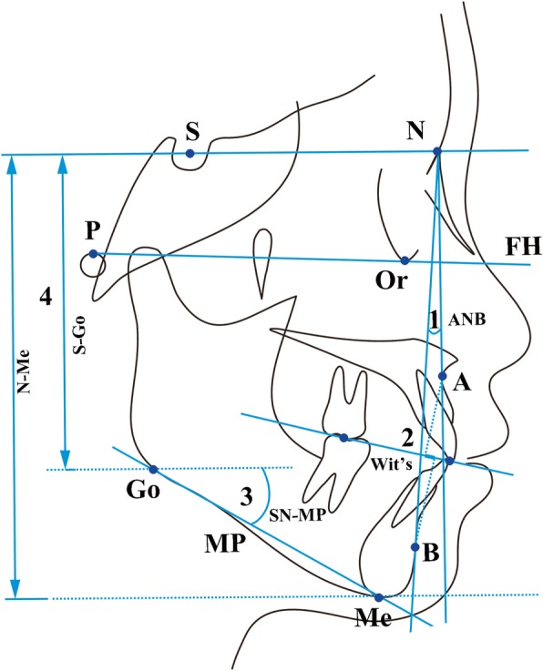
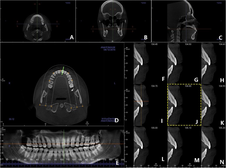
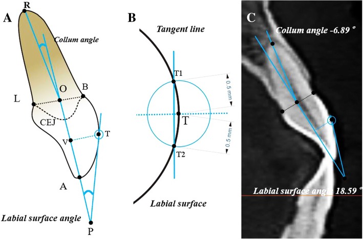
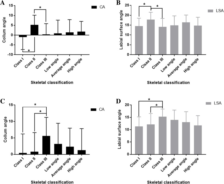
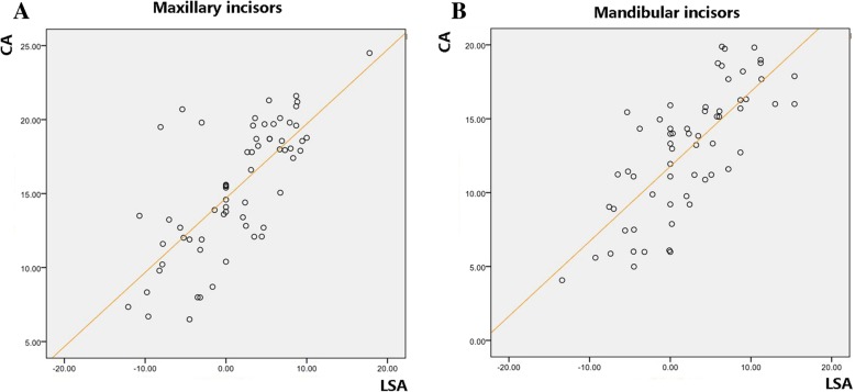
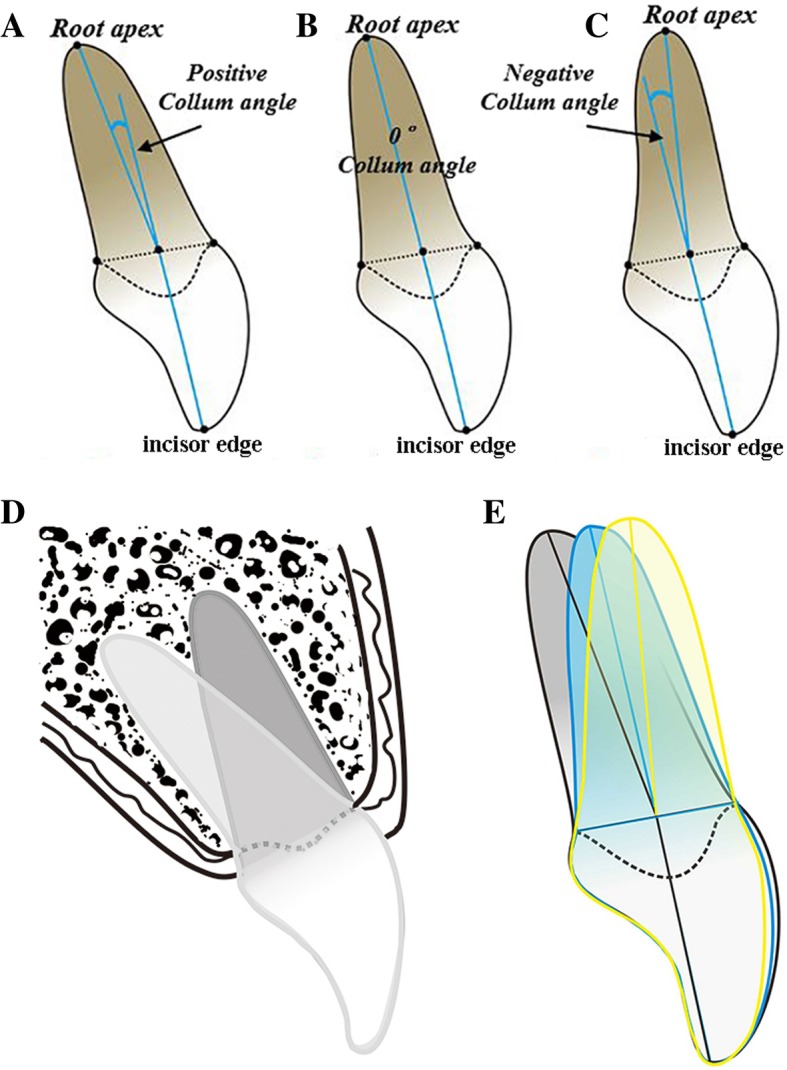
Methods: In this retrospective study, a total of 108 CBCT images were obtained (ranging from 18.0 to 30.0 years, mean age 25.8 years). Patients were grouped according to routine sagittal and vertical skeletal malocclusion classification criteria. The patients in sagittal groups were all average vertical patterns, with Class I comprised 24 patients-14 females and 10 males; Class II comprised 20 patients-13 females and 7 males; and Class III comprised 22 subjects-13 females and 9 males. The patients in vertical groups were all skeletal Class I malocclusions, with low angle comprised 21 patients-12 females and 9 males; average angle comprised 24 patients; and high angle comprised 21 patients-11 females and 10 males. All the CBCT data were imported into Invivo 5.4 software to obtain a middle labio-lingual section of right central incisors. Auto CAD 2007 software was applied to measure the crown-root angulation (Collum angle), and the angle formed by a tangent to the central of the labial surface of the crown and the long axis of the crown (labial surface angle). One-way analysis of variance (ANOVA) and Scheffe's test were used for statistical comparisons at the P < 0.05 level, and the Pearson correlation analysis was applied to investigate the association between the two measurements.
Results: The values of Collum angle and labial surface angle in maxillary incisor of Class II and mandibular incisor of Class III were significantly greater than other types of sagittal skeletal malocclusions (P < 0.05); no significant difference was detected among vertical skeletal malocclusions. Notably, there was also a significant positive correlation between the two measurements.
Conclusions: The maxillary incisor in patients with sagittal skeletal Class II malocclusion and mandibular incisor with Class III malocclusion present remarkable crown-root angulation and correspondingly considerable labial surface curvature. Equivalent deviation during bracket bonding may cause greater torque expression error and increase the risk of alveolar fenestration and dehiscence.
Keywords: Collum angle; Cone-beam CT; Crown-root morphology; Labial surface angle; Skeletal malocclusion.
Conflict of interest statement
The authors declare that they have no competing interests.
Figures
References
-
- Kong WD, Ke JY, Hu XQ, Zhang W, Li SS, Feng Y. Applications of cone-beam computed tomography to assess the effects of labial crown morphologies and Collum angles on torque for maxillary anterior teeth. Am J Orthod Dentofacial Orthop. 2016;150:789–795. doi: 10.1016/j.ajodo.2016.03.029. - DOI - PubMed
-
- Heravi F, Salari S, Tanbakuchi B, Loh S, Amiri M. Effects of crown-root angle on stress distribution in the maxillary central incisors’ PDL during application of intrusive and retraction forces: a three-dimensional finite element analysis. Prog Orthod. 2013;14:26. doi: 10.1186/2196-1042-14-26. - DOI - PMC - PubMed
-
- Shen YW, Hsu JT, Wang YH, Huang HL, Fuh LJ. The Collum angle of the maxillary central incisors in patients with different types of malocclusion. J Dent Sci. 2012;7:72–76. doi: 10.1016/j.jds.2012.01.010. - DOI
MeSH terms
LinkOut - more resources
Full Text Sources
Other Literature Sources
Miscellaneous
