. 2019 Jul;23(7):4829-4838.
doi: 10.1111/jcmm.14406. Epub 2019 May 21.

Dihydromyricetin protects HUVECs of oxidative damage induced by sodium nitroprusside through activating PI3K/Akt/FoxO3a signalling pathway

Affiliations

Dihydromyricetin protects HUVECs of oxidative damage induced by sodium nitroprusside through activating PI3K/Akt/FoxO3a signalling pathway

Xiaoying Zhang et al. J Cell Mol Med. 2019 Jul.

Abstract

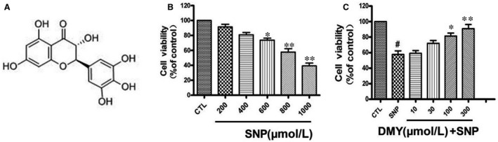
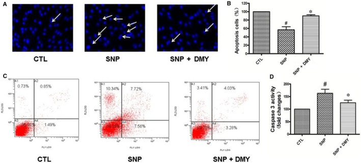
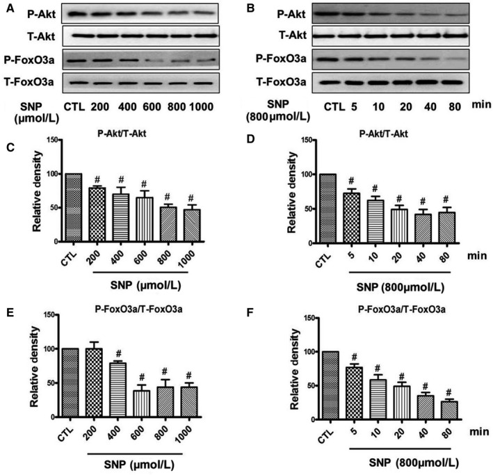
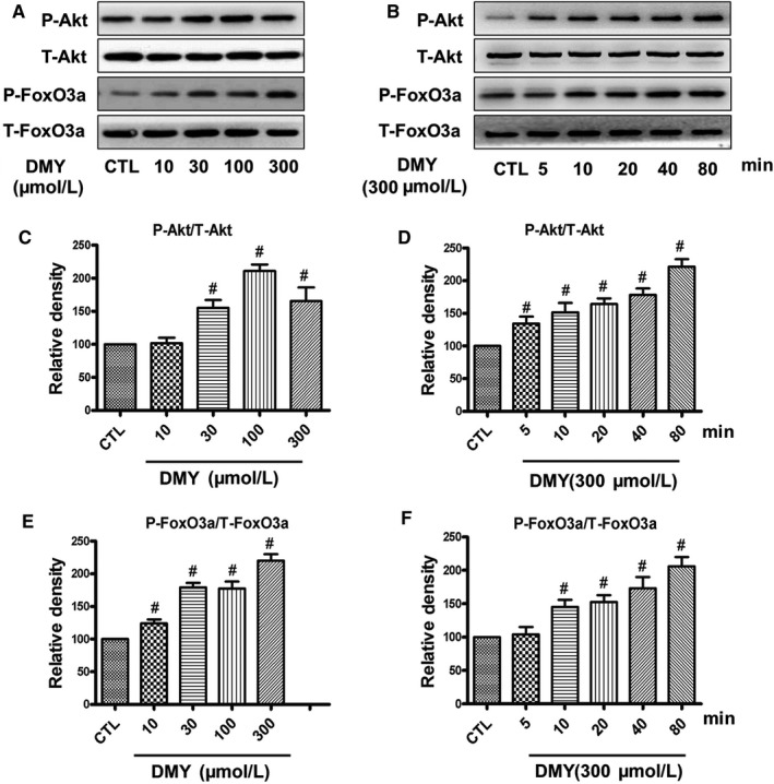
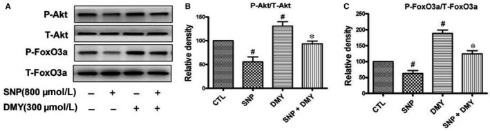
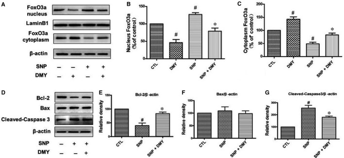
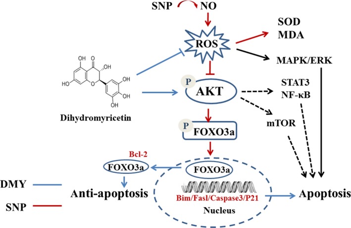
The damage of vascular endothelial cells induced by oxidative stress plays an important role in the pathogenesis of atherosclerosis. Dihydromyricetin (DMY) is considered as a natural antioxidant. However, the mechanism of DMY on endothelial cell injury induced by oxidative stress remains unclear. In this study, we found that DMY could reduce the oxidative damage of HUVECs induced by sodium nitroprusside (SNP), HUVECs pre-treated with DMY suppressed SNP-induced apoptosis by reduced ROS overproduction of intracellular, decreased MDA level and elevated the superoxide dismutase activity. Meanwhile, we found that DMY could promote the expression of phosphorylated FoxO3a and Akt, and affect the nuclear localization of FoxO3a, when treated with the PI3K inhibitor LY294002, the effect of DMY was blocked. These data suggest that DMY protects HUVECs from oxidative stress by activating PI3K/Akt/FoxO3a signalling pathway. Therefore, DMY may have great therapeutic potential as a new drug for atherosclerosis.

Keywords: apoptosis; atherosclerosis; dihydromyricetin; oxidative stress; sodium nitroprusside.


Conflict of interest statement

The authors declare no conflict of interest.

Figures

Figure 1
Figure 1
Dihydromyricetin attenuated the decrease in cell viability induced by sodium nitroprusside (SNP) in HUVECs. (A) The structure of Dihydromyricetin. (B) Cells were treated with SNP (200‐1000 μmol/L) for 24 h and cell viability was measured using the MTT assay. (C) Cells were pre‐treated with Dihydromyricetin at indicated concentrations for 2 h and then incubated with or without 800 μmol/L SNP for a further 24 h. Results are shown as the mean ± SD, # P < 0.05 vs CTL group;*P < 0.05, **P < 0.01 vs SNP group
Figure 2
Figure 2
The protective effect of Dihydromyricetin on sodium nitroprusside (SNP)‐induced apoptosis in the HUVECs. Cells were pre‐treated with Dihydromyricetin and then were incubated with or without 800 μmol/L SNP for 24 h. The apoptosis of HUVECs cells was detected by Hoechst staining (A) and Flow cytometry (C), the number of apoptotic nuclei with condensed chromatin was counted from the photomicrographs and presented as a percentage of the total number of nuclei (B); And the activity of Caspase 3 was measured by Caspase‐3/CPP32 Fluorometric Assay Kit (D). Results are shown as the mean ± SD, # P < 0.05 vs CTL group, *P < 0.05 vs SNP group
Figure 3
Figure 3
Effect of Dihydromyricetin on sodium nitroprusside (SNP)‐induced oxidative stress in the HUVECs. HUVECs were cultured in 24‐well plates and treated with SNP with or without DMY for 24 h, (A) the cells were incubated with 10 μmol/L dihydroethidium (DHE) for 30 min at 37°C, Fluorescence intensity was detected using inversion fluorescence microscope, (B) and the relative change was processed with the ImageJ analysis. (C, D) The relative level of MDA and SOD was measured by MDA and SOD detection kit respectively. Results are shown as the mean ± SD, # P < 0.05, ## P < 0.01 vs CTL group; *P < 0.05 vs SNP group
Figure 4
Figure 4
Effects of sodium nitroprusside (SNP) on the phosphorylation of Akt and FoxO3a in HUVECs. HUVECs were treated with different concentrations of SNP for 80 min (A) or treated with 800 µmol/L SNP for various times (B), the phosphorylation of Akt and FoxO3a were analysed by Western blotting. (C‐F) Densitometric analysis of the immunoblot was expressed as a percentage of control. Results are shown as the mean ± SD, # P < 0.05 vs CTL group
Figure 5
Figure 5
Effects of dihydromyricetin (DMY) on inhibitory effect of sodium nitroprusside (SNP) on the phosphorylation of Akt and FoxO3a in HUVECs. HUVECs were treated with 800 µmol/L SNP with or without DMY for 80 min, (A) phosphorylation of Akt and FoxO3a was analysed by Western blotting. (B, C) Densitometric analysis of the immunoblot was expressed as a percentage of control. Results are shown as the mean ± SD, # P < 0.05 vs CTL group; *P < 0.05 vs SNP group
Figure 6
Figure 6
Effects of dihydromyricetin (DMY) on the phosphorylation of Akt and FoxO3a in HUVECs. HUVECs were treated with different concentrations of DMY for 80 min (A) or treated with 300 µmol/L DMY for various times (B), the phosphorylation of Akt and FoxO3a were analysed by Western blotting. (C‐F) Densitometric analysis of the immunoblot was expressed as a percentage of control. Results are shown as the mean ± SD, # P < 0.05 vs CTL group
Figure 7
Figure 7
Effects of dihydromyricetin (DMY) and sodium nitroprusside (SNP) in the FoxO3a nuclear translocation and the expression of apoptosis‐related protein. HUVECs were treated with DMY or SNP or SNP + DMY, the nuclear and cytosolic protein of FoxO3a (A) and the level of Bcl‐2, Bax and cleaved‐caspase3 (D) were analysed immunoblotting. (B‐C, E‐G) Relative levels of the nuclear and cytosolic protein of FoxO3a and Bcl‐2, Bax and cleaved‐caspase3 expression in each sample were determined by densitometry of the blots, densitometric analysis of the immunoblot was expressed as a percentage of control. Results are shown as the mean ± SD, # P < 0.05 vs CTL group, *P < 0.05 vs SNP group
Figure 8
Figure 8
Effects of LY294002 on the phosphorylation of Akt and FoxO3a induced by dihydromyricetin (DMY) and sodium nitroprusside (SNP) in HUVECs. HUVECs pre‐treated with LY294002 were treated with DMY or SNP. The phosphorylation of Akt and FoxO3a was analysed by immunoblotting. (B, C) Relative levels of p‐Akt versus total Akt and p‐FoxO3a versus total FoxO3a in each sample was determined by blot densitometry, densitometric analysis of the immunoblotting was expressed as a percentage of control. Data are shown as the mean ± SD, # P < 0.05 vs CTL group, *P < 0.05 vs SNP group, & P < 0.05 vs SNP + DMY group
Figure 9
Figure 9
The possibly mechanisms underlying dihydromyricetin (DMY) against sodium nitroprusside (SNP) in the HUVECs. DMY treatment activates PI3K/AKT/ FoxO3a pathway and subsequently drives FoxO3a out of nuclei, which inhibits apoptosis. SNP, a nitric oxide donor, creates oxidase stress and subsequently down‐regulates PI3K/AKT/FoxO3a pathway. Additionally, DMY may modulate others signal pathway such as mTOR, STAT3/NF‐κB to antiapoptosis in the HUVECs

Similar articles

Cited by

References

    1. Mozaffarian . Heart disease and stroke statistics‐2015 update: A report from the American heart association. Circulation. 2015; 131: E535. - PubMed
    1. Bentzon JF, Otsuka F, Virmani R, Falk E. Mechanisms of plaque formation and rupture. Circ Res. 2014;114:1852‐1866. - PubMed
    1. Weintraub NL. Understanding abdominal aortic aneurysm. New Engl J Med. 2009;361:1114‐1116. - PMC - PubMed
    1. Dernbach E, Urbich C, Brandes RP, Hofmann WK, Zeiher AM, Dimmeler S. Antioxidative stress‐associated genes in circulating progenitor cells: evidence for enhanced resistance against oxidative stress. Blood. 2004;104:3591‐3597. - PubMed
    1. Higashi Y, Noma K, Yoshizumi M, Kihara Y. Endothelial function and oxidative stress in cardiovascular diseases. Circ J. 2009;73:411‐418. - PubMed

Publication types

MeSH terms