. 2019 May;24(5):1-12.
doi: 10.1117/1.JBO.24.5.056002.

Intraoperative photoacoustic screening of breast cancer: a new perspective on malignancy visualization and surgical guidance

Affiliations

Intraoperative photoacoustic screening of breast cancer: a new perspective on malignancy visualization and surgical guidance

Ivan Kosik et al. J Biomed Opt. 2019 May.

Abstract

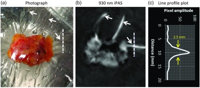
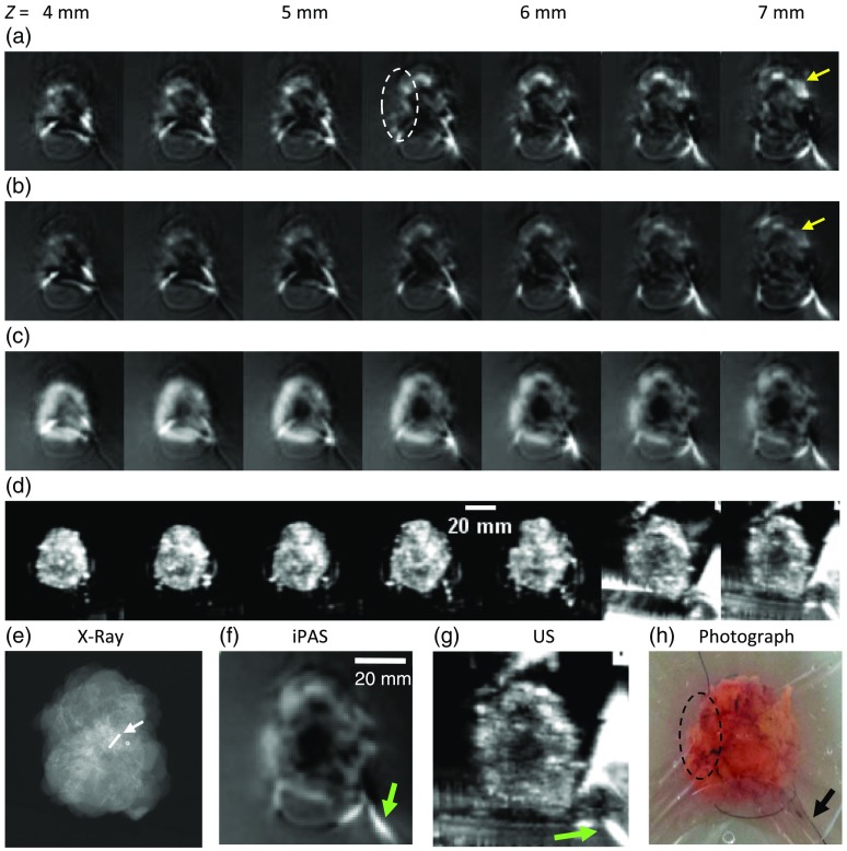
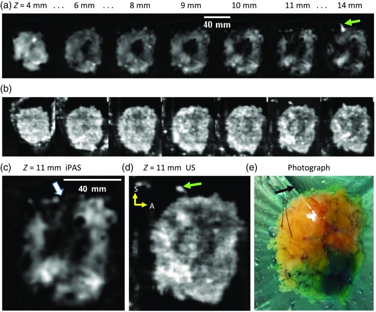
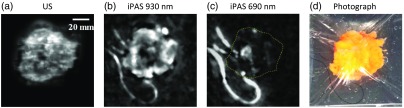
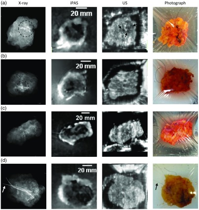
High re-excision rates in breast-conserving surgery call for a new intraoperative approach to the lumpectomy margin evaluation problem. The unique intraoperative imaging system, presented here, demonstrated the capability of photoacoustic tomography (PAT) to deliver optical sensitivity and specificity, along with over 2-cm imaging depth, in a clinical setting. The system enabled the evaluation of tumor extent, shape, morphology, and position within lumpectomy specimens measuring up to 11 cm in diameter. The investigation included all major breast cancer-related lesions, such as invasive ductal carcinoma (IDC), multifocal IDC, ductal carcinoma in situ and combinations of these variants. Coregistration with established ultrasound (US) technology, as well as comparison to specimen radiography, validated the performance of PAT, which appeared to facilitate better tumor visualization. Contrary to expected PA contrast mechanisms, PAT images of hemoglobin distribution correlated poorly with US-determined tumor location, while hypointense regions in lipid-weighted PAT images were in better agreement with US.

Keywords: breast cancer; photoacoustic; surgical guidance; three-dimensional; tomography.


Figures

Fig. 1
Fig. 1
(a) Schematic representation of the major components of the iPAS system for intraoperative lumpectomy margin evaluation during BCS. (b) A labeled photograph of the intraoperative system within surgical suite, consisting of US system on the left and iPAS system on the right. The front panel of the iPAS system is removed to facilitate convenient viewing of internal components. (c) A close-up photograph of the arc-shaped 24-channel PA transducer array (arrow) mounted on the effector of the Epson SCARA robot. (d) The PA array from below, along with scale bar indicating dimensions and the location of the conventional 6.6-MHz linear US transducer array (yellow arrow). (d) The positioning of the individual transducer elements on the array chassis (black arrows). (e) The custom-built lumpectomy specimen holder which contains a freshly excised compressively restrained lumpectomy specimen.
Fig. 2
Fig. 2
(a) Selected 690-nm iPAS z-slices showing a graphite/agar gel phantom reconstructed using spatial domain stitching, resulting in unwanted scan grid artifacts in the final composite image. (b) The same scan data but with stitching implemented in the gradient domain, resulting in complete or near complete elimination of the grid artifacts. The larger images in (c) and (d) allow for an easier appreciation of the result, while the photograph in (e) provides an effective gold standard reference.
Fig. 3
Fig. 3
The iPAS system resolution evaluation via line profile analysis of surgical suture image. The black sutures, indicated by white arrows in the photograph of panel (a), are clearly visualized on the 930-nm iPAS scan result of (b). The suture, indicated by an orthogonal 20-mm dashed line in the photo and iPAS image, is used to estimate the in-plane resolution of the imaging system. FWHM analysis indicates a resolution of 2.5  mm.
Fig. 4
Fig. 4
Image stacks of lumpectomy specimen containing an 18-mm diameter, grade II IDC, acquired using the iPAS system at the indicated Z-depths below the illuminated surface. (a) and (b) iPAS results for 690- and 800-nm scans, respectively, illustrating the small difference between targeting deoxyhemoglobin (690 nm) and total hemoglobin (800 nm). The increased intensity of the 690-nm scan results, compared to 800 nm (yellow arrows), likely indicates the presence of mostly deoxygenated blood in the excised specimen. Nonetheless, both wavelengths fail to unambiguously differentiate the malignancy region, best indicated in the near 7-mm-deep slice of the 6.6-MHz US scan shown in (d) and (g), as well as in the x-ray image found in (e). On the other hand, the iPAS slices in (c) and (f), acquired using lipid-weighted 930-nm scans, clearly show a 20-mm diameter centrally located hypointense area corresponding to the x-ray and US findings.
Fig. 5
Fig. 5
(a) The 930-nm iPAS slices corresponding to the indicated depth below the illumination surface, showing a grade II IDC along with a grade II DCIS-containing specimen, found by postoperative pathology to have a positive margin. (b) Coregistered slices acquired using a 6.6-MHz conventional US scanner. (c) and (d) A larger view of the slice near Z=11  mm for iPAS and US, respectively. The iPAS slice in (c) clearly shows intrusion of the specimen edge by the hypointense extension (white arrow). For reference, a photograph is included in (e), which, along with the 14-mm iPAS and US slices, shows the superior orientation suture (black and green arrows). The sutures facilitate the orientation of the specimen with superior (S) at the top and anterior (A) to the right (yellow arrows).
Fig. 6
Fig. 6
The iPAS assessment of lumpectomy specimen belonging to an 88-year-old patient confirmed by pathology to contain a 15-mm grade I IDC along with grade II DCIS. Interestingly, in this case, the hypointense tumor areas found in the US and 930 nm iPAS images of (a) and (b) correspond well to the hyperintense area in the 690 nm iPAS image of (c). This is likely caused by the simultaneous presence of increased vascularity along with decreased lipid content of the tumor region. For easier comparison the dashed yellow line in the 690-nm iPAS image indicates the outer perimeter of the specimen which is difficult to see due to the low hemoglobin content of the healthy margin. For reference, a photograph is included in (d).
Fig. 7
Fig. 7
Montage with slides of commonly encountered BCS lesions, acquired using the iPAS lumpectomy evaluation system. Note that these results may not be representative of all cases. The first column shows intraoperative transmission x-ray images. The second column depicts the corresponding 930-nm iPAS slices, which are coregistered to the US slices shown in the third column. Finally, the last column shows specimen photographs, which, along with the x-ray images, are not coregistered. The specimen in (a) contained a pathologically confirmed 17-mm IDC with concordant findings on iPAS and x-ray (dashed circle), and to a lesser degree, US. (b) A specimen which contained a lesion composed of an 8-mm IDC with an extension of DCIS. (c) A lumpectomy specimen found by pathology to contain two adjacent foci of IDC, measuring 16 mm and 15 mm each, along with high-grade DCIS. Last, the lumpectomy found in (d) is confirmed to be a pure DCIS lesion measuring at least 18 mm in diameter and is removed using wire-guided excision. The hooked localization wires can be appreciated in the photograph and on the x-ray image (arrows). The iPAS results appear to be in agreement as they show hypointense areas corresponding to the lesion location in the x-ray.

References

    1. Siegel R. L., Miller K. D., Jemal A., “Cancer statistics,” CA Cancer J Clin. 65(1), 5–29 (2015). 10.3322/caac.21254 - DOI - PubMed
    1. Ananthakrishnan P., Balci F. L., Crowe J. P., “Optimizing surgical margins in breast conservation,” Int. J. Surg. Oncol. 2012, 585670 (2012). 10.1155/2012/585670 - DOI - PMC - PubMed
    1. Lagios M. D., Silverstein M. J., “Ductal carcinoma in situ: recent history and areas of controversy,” Breast J. 21(1), 21–26 (2015). 10.1111/tbj.12359 - DOI - PubMed
    1. Silverstein M. J., Lagios M. D., “Treatment selection for patients with ductal carcinoma in situ (DCIS) of the Breast Using the University of Southern California/Van Nuys (USC/VNPI) Prognostic Index,” Breast J. 21(2), 127–132 (2015). 10.1111/tbj.12368 - DOI - PubMed
    1. Waljee J. F., et al. , “Predictors of re-excision among women undergoing breast-conserving surgery for cancer,” Ann. Surg. Oncol. 15(5), 1297–1303 (2008). 10.1245/s10434-007-9777-x - DOI - PubMed

Publication types

MeSH terms

Grants and funding