. 2019 Mar;13(2):143-159.
doi: 10.1080/17435390.2018.1530392.

Lung deposition patterns of MWCNT vary with degree of carboxylation

Affiliations

Lung deposition patterns of MWCNT vary with degree of carboxylation

Andrij Holian et al. Nanotoxicology. 2019 Mar.

Abstract

Functionalization of multi-walled carbon nanotubes (MWCNT) is known to affect the biological response (e.g. toxicity, inflammation) in vitro and in vivo. However, the reasons for these changes in vivo are not well described. This study examined the degree of MWCNT functionalization with regard to in vivo mouse lung distribution, particle retention, and resulting pathology. A commercially available MWCNT (source MWCNT) was functionalized (f-MWCNT) by systematically varying the degree of carboxylation on the particle's surface. Following a pilot study using seven variants, two f-MWCNT variants were chosen and for lung pathology and particle distribution using oropharyngeal aspiration administration of MWCNT in Balb/c mice. Particle distribution in the lung was examined at 7 and 28 days post-instillation by bright-field microscopy, CytoViva hyperspectral dark-field imaging, and Stimulated Raman Scattering (SRS) microscopy. Examination of the lung tissue by bright-field microscopy showed some acute inflammation for all MWCNT that was highest with source MWCNT. Hyperspectral imaging and SRS were employed to assess the changes in particle deposition and retention. Highly functionalized MWCNT had a higher lung burden and were more disperse. They also appeared to be associated more with epithelial cells compared to the source and less functionalized MWCNT that were mostly interacting with alveolar macrophages (AM). These results showing a slightly reduced pathology despite the extended deposition have implications for the engineering of safer MWCNT and may establish a practical use as a targeted delivery system.

Keywords: MWCNT; Stimulated Raman Scatter; carboxylation; functionalization; macrophage.


Conflict of interest statement

Competing Interests

The authors declare no conflicts of interest.

Figures

Figure 1:
Figure 1:
Spectra used in SFF hyperspectral imaging (Cytoviva). A) Background tissue intensity (green) verses non-tissue background (yellow). B) Particle signal (red) verses background tissue intensity (green) and non-tissue background (yellow). C) Raman spectra for tissue background.
Figure 2:
Figure 2:
TEM images of A) source MWCNT, B) minimal f-MWCNT38.4, and C) maximal f-MWCNT14.7 demonstrating little visible difference (20 nm diameters in all instances).
Figure 3:
Figure 3:
Relative lung pathology 7 and 28 days post-particle exposure contrasting the source MWCNT with minimally and maximally carboxylated MWCNT. Pathology scores range from 0 to 4, with 0 indicating no pathology and 4 being the worst possible indication of disease. Data expressed as median scores ± range. Asterisks ** indicate statistical significance at P < 0.01, or * P < 0.05 compared to the corresponding DM control.
Figure 4:
Figure 4:
Representative H&E bright-field photomicrographs of Balb/c mouse lung sections exposed to various MWCNT for 7 days post-particle exposure. Dark areas indicate MWCNT deposition. A) Dispersion media control for 7 days. B) The source MWCNT for 7 days with particle isolated primarily in the alveolar macrophages. C) Particle f-MWCNT38.4 (5 min) for 7 days isolated exclusively in numerous alveolar macrophages. D) Particle f-MWCNT14.7 (120 min). Magnification at 1000x for all images and micrometer scale bar = microns.
Figure 5:
Figure 5:
Representative H&E bright-field photomicrographs of Balb/c mouse lung sections exposed to various MWCNT for 28 days post-particle exposure. Dark areas indicate MWCNT deposition. A) Dispersion media control for 28 days. B) The source MWCNT for 28 days C) Particle f-MWCNT38.4 (5 min) for 28 days. D) Particle f-CNT14.7 (120 min) for 28 days. Magnification at 1000x for all images and micrometer scale bar = microns.
Figure 6:
Figure 6:
Representative CytoViva hyperspectral dark-field photomicrographs (SFF) of Balb/c mouse lung sections exposed to various MWCNT for 7 days post-particle exposure. Bright green areas indicate MWCNT deposition. A) Dispersion media control for 7 days. B) The source MWCNT for 7 days with particle that appears to be isolated primarily in alveolar macrophages within the alveolar spaces of the lung section. C) Particle f-MWCNT38.4 (5 min) for 7 days that appears to be isolated exclusively in numerous alveolar macrophages in a lung airway. D) Particle f-MWCNT14.7 (120 min) for 7 days dispersing throughout the epithelial tissue matrix in addition to alveolar macrophage uptake. This particle deposition is not limited to particular lung areas, but freely disperses throughout the lung tissue. Magnification at 400x (40x objective) for all images and micrometer scale bar = 20 microns.
Figure 7:
Figure 7:
Representative CytoViva hyperspectral dark-field photomicrographs (SFF) of Balb/c mouse lung sections exposed to various MWCNT for 28 days post-particle exposure. Bright green areas indicate MWCNT deposition. A) Dispersion media control for 28 days. B) The source MWCNT for 28 days C) Particle f-MWCNT38.4 (5 min) for 28 days. D) Particle f-MWCNT14.7 (120 min) for 28 days. Magnification at 400x (40x objective) for all images and micrometer scale bar = 20 microns.
Figure 8:
Figure 8:
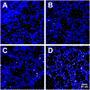
Particle deposition in lung sections assessed by stimulated Raman scattering after 7-day exposure. Representative regions of merged tissue (blue) and MWCNT (white) channels. A) Dispersion media control for 7 days. B) The source MWCNT for 7 days. C) Particle f-MWCNT38.4 (5 min) for 7 days. D) Particle f-MWCNT14.7 (120 min) for 7 days. Magnification equal for all images; scale bar = 50 μm.
Figure 9:
Figure 9:
Particle deposition in lung sections assessed by stimulated Raman scattering after 28-day exposure. Representative regions of merged tissue (blue) and MWCNT (white) channels. A) Dispersion media control for 28 days. B) The source MWCNT for 28 days. C) Particle f-MWCNT38.4 (5 min) for 28 days. D) Particle f-MWCNT14.7 (120 min) for 28 days. Magnification equal for all images; scale bar = 50 μm.
Figure 10:
Figure 10:
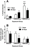
Particle quantification from stimulated Raman scattering for 7- and 28-day MWCNT lung exposures. A) Mean ± SEM percent particle burden (pink) in the lung sections (blue). B) Mean ± SEM particle cluster size in the lung tissue expressed as microns squared. Asterisks indicate statistical significance at *** P < 0.001 or at * P > 0.05 compared to corresponding control particle (no COOH MWCNT). Daggers ††† indicate significance at P < 0.001 or † P < 0.05 compared to corresponding f-MWCNT. Symbol § indicates significance at P < 0.05 compared to day-7 for the same particle. Sample size was 3 mice per exposure.

Similar articles

Cited by

References

    1. Allegri M, Perivoliotis DK, Bianchi MG, Chiu M, Pagliaro A, Koklioti MA, Trompeta AFA, Bergamaschi E, Bussolati O & Charitidis CA, 2016. Toxicity determinants of multi-walled carbon nanotubes: The relationship between functionalization and agglomeration. Toxicology Reports, 3, 230–243. - PMC - PubMed
    1. Bai W, Wu Z, Mitra S & Brown JM, 2016. Effects of multiwalled carbon nanotube surface modification and purification on bovine serum albumin binding and biological responses. J Nanomater, 2016. - PMC - PubMed
    1. Borm PJ & Driscoll K, 1996. Particles, inflammation and respiratory tract carcinogenesis. Toxicol Lett, 88, 109–13. - PubMed
    1. Bunderson-Schelvan M, Hamilton R, Trout K, Jessop F, Gulumian M & Holian A, 2016. Approaching a Unified Theory for Particle-Induced Inflammation In Otsuki T, Yoshioka Y & Holian A (eds.) Biological Effects of Fibrous and Particulate Substances. Springer; Japan, 51–76.
    1. Castranova V, Schulte PA & Zumwalde RD, 2013. Occupational nanosafety considerations for carbon nanotubes and carbon nanofibers. Acc Chem Res, 46, 642–9. - PMC - PubMed

Publication types

MeSH terms

Substances