

Chronic Spinal Cord Injury Reduces Gastrin-Releasing Peptide in the Spinal Ejaculation Generator in Male Rats
- PMID: 31111794
- PMCID: PMC6892439
- DOI: 10.1089/neu.2019.6509
Chronic Spinal Cord Injury Reduces Gastrin-Releasing Peptide in the Spinal Ejaculation Generator in Male Rats
Abstract
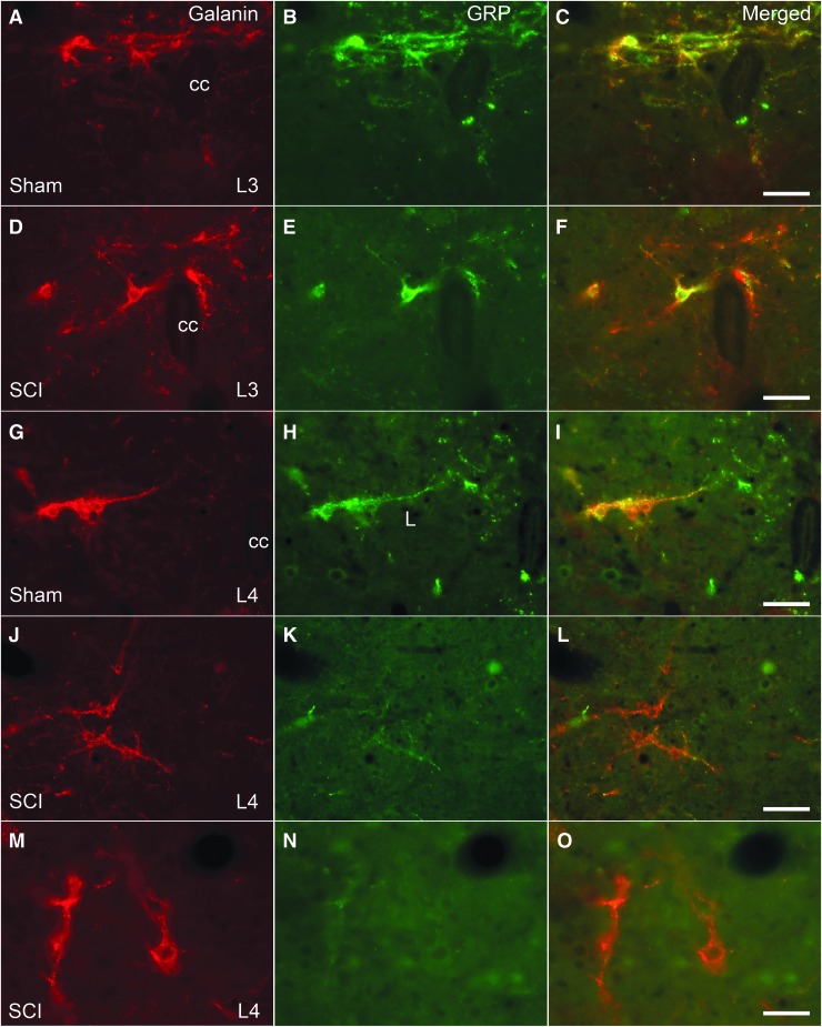
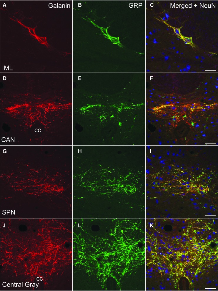
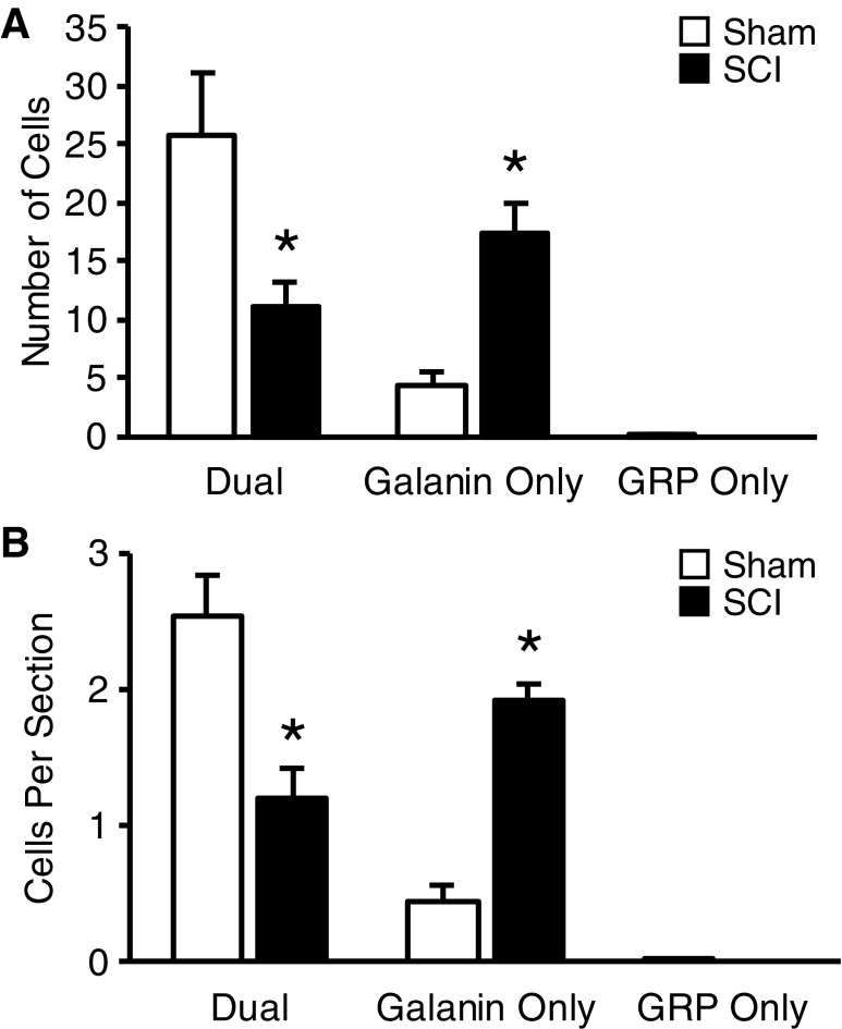
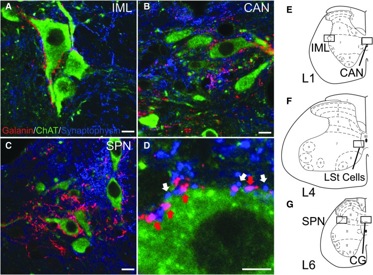
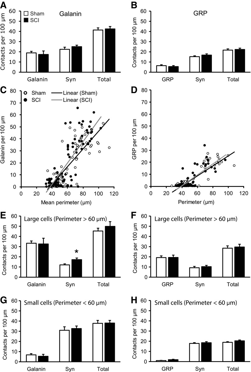
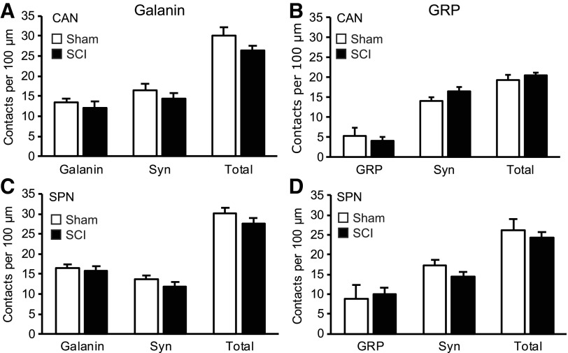
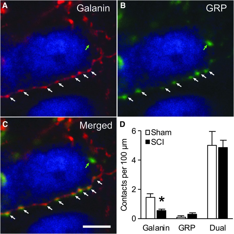
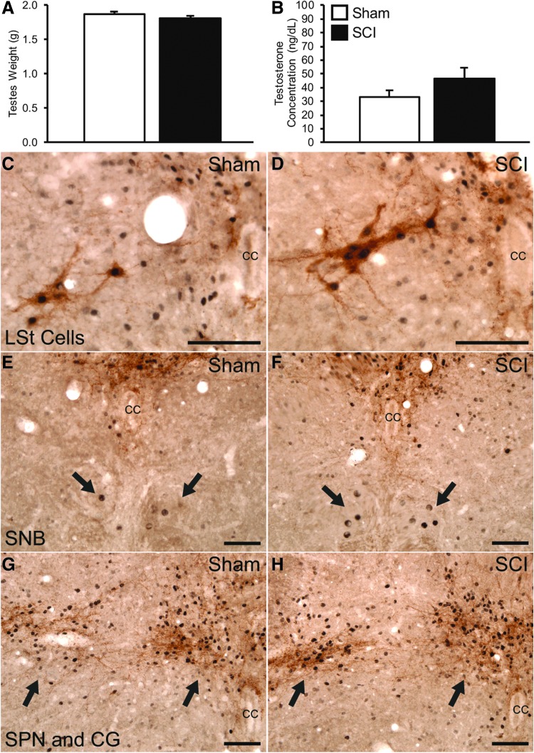
Spinal cord injury (SCI) causes sexual dysfunction, including anejaculation in men. Likewise, chronic mid-thoracic contusion injury impairs ejaculatory reflexes in male rats. Ejaculation is controlled by a spinal ejaculation generator (SEG) comprised of a population of lumbar spinothalamic (LSt) neurons. LSt neurons co-express four neuropeptides, including gastrin-releasing peptide (GRP) and galanin and control ejaculation via release of these peptides in lumbar and sacral autonomic and motor nuclei. Here, we tested the hypothesis that contusion injury causes a disruption of the neuropeptides that are expressed in LSt cell bodies and axon terminals, thereby causing ejaculatory dysfunction. Male Sprague Dawley rats received contusion or sham surgery at spinal levels T6-7. Five to six weeks later, animals were perfused and spinal cords were immunoprocessed for galanin and GRP. Results showed that numbers of cells immunoreactive for galanin were not altered by SCI, suggesting that LSt cells are not ablated by SCI. In contrast, GRP immunoreactivity was decreased in LSt cells following SCI, evidenced by fewer GRP and galanin/GRP dual labeled cells. However, SCI did not affect efferent connections of LSt, cells as axon terminals containing galanin or GRP in contact with autonomic cells were not reduced following SCI. Finally, no changes in testosterone plasma levels or androgen receptor expression were noted after SCI. In conclusion, chronic contusion injury decreased immunoreactivity for GRP in LSt cell soma, but did not affect LSt neurons per se or LSt connections within the SEG. Since GRP is essential for triggering ejaculation, such loss may contribute to ejaculatory dysfunction following SCI.
Keywords: anejaculation; contusion injury; lumbar spinal cord; sexual dysfunction; urogenital.
Conflict of interest statement
No competing financial interests exist.
Figures
Similar articles
-
Spinal Cord Injury Causes Reduction of Galanin and Gastrin Releasing Peptide mRNA Expression in the Spinal Ejaculation Generator of Male Rats.Front Neurol. 2021 Jun 22;12:670536. doi: 10.3389/fneur.2021.670536. eCollection 2021. Front Neurol. 2021. PMID: 34239493 Free PMC article.
-
Chronic Contusion Spinal Cord Injury Impairs Ejaculatory Reflexes in Male Rats: Partial Recovery by Systemic Infusions of Dopamine D3 Receptor Agonist 7OHDPAT.J Neurotrauma. 2016 May 15;33(10):943-53. doi: 10.1089/neu.2015.4232. Epub 2016 Jan 15. J Neurotrauma. 2016. PMID: 26437577
-
Activation of gastrin-releasing peptide receptors in the lumbosacral spinal cord is required for ejaculation in male rats.J Sex Med. 2012 May;9(5):1303-18. doi: 10.1111/j.1743-6109.2012.02688.x. Epub 2012 Mar 16. J Sex Med. 2012. PMID: 22429708
-
[Spinal gastrin-releasing peptide system mediates sexual function of males: advances in studies].Zhonghua Nan Ke Xue. 2014 Jun;20(6):554-7. Zhonghua Nan Ke Xue. 2014. PMID: 25029865 Review. Chinese.
-
Descending pathways modulating the spinal circuitry for ejaculation: effects of chronic spinal cord injury.Prog Brain Res. 2006;152:415-26. doi: 10.1016/S0079-6123(05)52028-4. Prog Brain Res. 2006. PMID: 16198717 Review.
Cited by
-
Sexual Experience Induces the Expression of Gastrin-Releasing Peptide and Oxytocin Receptors in the Spinal Ejaculation Generator in Rats.Int J Mol Sci. 2021 Sep 26;22(19):10362. doi: 10.3390/ijms221910362. Int J Mol Sci. 2021. PMID: 34638701 Free PMC article.
-
Spinal Cord Injury Causes Reduction of Galanin and Gastrin Releasing Peptide mRNA Expression in the Spinal Ejaculation Generator of Male Rats.Front Neurol. 2021 Jun 22;12:670536. doi: 10.3389/fneur.2021.670536. eCollection 2021. Front Neurol. 2021. PMID: 34239493 Free PMC article.
References
-
- Anderson K.D., Borisoff J.F., Johnson R.D., Stiens S.A., and Elliott S.L. (2007). The impact of spinal cord injury on sexual function: concerns of the general population. Spinal Cord 45, 328–337 - PubMed
-
- Anderson K.D., Borisoff J.F., Johnson R.D., Stiens S.A., and Elliott S.L. (2007). Long-term effects of spinal cord injury on sexual function in men: implications for neuroplasticity. Spinal Cord 45, 338–348 - PubMed
-
- Brackett N.L., Ferrell S.M., Aballa T.C., Amador M.J., Padron O.F., Sonksen J., and Lynne C.M. (1998). An analysis of 653 trials of penile vibratory stimulation in men with spinal cord injury. J. Urol. 159, 1931–1934 - PubMed
-
- Brackett N.L., Ibrahim E., Iremashvili V., Aballa T.C., and Lynne C.M. (2010). Treatment for ejaculatory dysfunction in men with spinal cord injury: an 18-year single center experience. J. Urol. 183, 2304–2308 - PubMed
-
- Brackett N.L., Lynne C.M., Ibrahim E., Ohl D.A., and Sonksen J. (2010). Treatment of infertility in men with spinal cord injury. Nat. Rev. Urol. 7, 162–172 - PubMed
Publication types
MeSH terms
Substances
Grants and funding
LinkOut - more resources
Full Text Sources
Medical
