Review
. 2019 May;7(3):10.1128/microbiolspec.gpp3-0059-2018.
doi: 10.1128/microbiolspec.GPP3-0059-2018.

The Bacteriophages of Streptococcus pyogenes

Affiliations
Review

The Bacteriophages of Streptococcus pyogenes

W Michael McShan et al. Microbiol Spectr. 2019 May.

Abstract

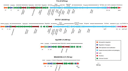
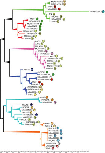
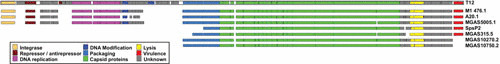
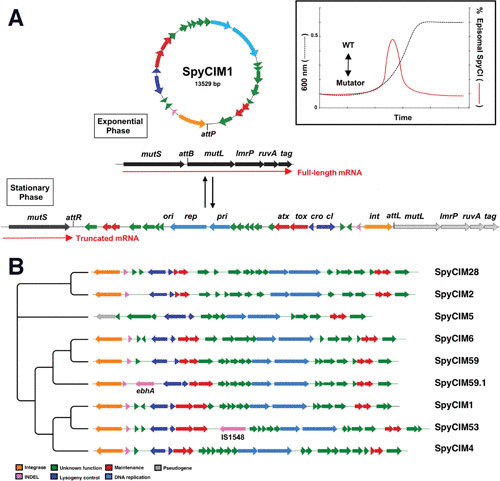
The bacteriophages of Streptococcus pyogenes (group A streptococcus) play a key role in population shaping, genetic transfer, and virulence of this bacterial pathogen. Lytic phages like A25 can alter population distributions through elimination of susceptible serotypes but also serve as key mediators for genetic transfer of virulence genes and antibiotic resistance via generalized transduction. The sequencing of multiple S. pyogenes genomes has uncovered a large and diverse population of endogenous prophages that are vectors for toxins and other virulence factors and occupy multiple attachment sites in the bacterial genomes. Some of these sites for integration appear to have the potential to alter the bacterial phenotype through gene disruption. Remarkably, the phage-like chromosomal islands (SpyCI), which share many characteristics with endogenous prophages, have evolved to mediate a growth-dependent mutator phenotype while acting as global transcriptional regulators. The diverse population of prophages appears to share a large pool of genetic modules that promotes novel combinations that may help disseminate virulence factors to different subpopulations of S. pyogenes. The study of the bacteriophages of this pathogen, both lytic and lysogenic, will continue to be an important endeavor for our understanding of how S. pyogenes continues to be a significant cause of human disease.


Figures

FIGURE 1
FIGURE 1
The genome of bacteriophage A25 reveals an escape from lysogeny. The 33,900-bp generalized transducing phage A25 is shown. The portion of the chromosome included in the shaded box is the high homology region that contains the remnant lysogeny module and other genes that A25 shares with prophages from genome strains MGAS10270 (M2), MGAS315 (M3), MGAS10570 (M4), and STAB902 (M4). Unlike these complete lysogens, A25 only has the operator and antirepressor from the lysogeny module (shown in expanded view below the map), apparently having lost the integrase and repressor for lysogeny some time in the past. This A25 expanded region is compared to the homologous region from genome prophage MGAS10270.2, which contains these elements as well as the upstream genes including the cI-like repressor. Promoters are shown as directional arrows. Introduction of the MGAS10270.2 repressor into an A25-sensitive S. pyogenes strain results in its conversion to a high level of A25 resistance (32). The genome follows a typical modular arrangement, with the predicted function for A25 genes indicated by color: regulation, dark red; DNA replication, pink; encode endonucleases, dark blue; genome packaging, light blue; structural, green; and lysis, yellow. The figure is redrawn from McCullor et al. (32).
FIGURE 2
FIGURE 2
Prophage attachment sites in the S. pyogenes genome. The locations of the genome prophages are shown as a generalized chromosome backbone based on the SF370 M1 genome; each diamond represents a genome prophage identified at that site. The M type of the host for each prophage is indicated by the number within the diamond, and the circled letter is the identifier linked to Table 2 for attB gene identification, the integration target within that gene (5′ or 3′), and associated prophage virulence genes. The rRNA operons are indicated as green blocks, and the hypervariable regions containing virulence genes associated with emm or prtF are hatched. The origin of replication is indicated (OriC).
FIGURE 3
FIGURE 3
Integration of prophage SF370.1 may provide an alternative promoter for dipeptidase Spy0713. In strains lacking an integrated prophage at this site, the native promoter for dipeptidase Spy0713 is downstream of the uncharacterized gene Spy0654; the predicted sequence is shown above. Integration of phage SF370.1 into Spy0713 separates this gene from that of the native promoter, and a predicted promoter encoded by the prophage is now positioned in front of the dipeptidase ORF. This phage-encoded promoter is preceded also by a canonical CinA box (56), which is not part of the native promoter. Transcription of prophage virulence genes speC and spd1 is from the opposite strand and should not influence transcription of Spy0713. Promoter predictions were done using the online tool at http://www.fruitfly.org/seq_tools/promoter.html (110).
FIGURE 4
FIGURE 4
Morphology of streptococcal lysogenic phages. Prophages SF370.1 (A) and T12 (B) release typical Siphoviridae virions following induction. The SF370.1 head is about 55 nm across and the tail is 168 nm in length in this micrograph. In this image, the tail fibers that contain hyaluronate lyase (hyaluronidase) are visible. The T12 capsid has similar dimensions, with the head being about 66 nm and the tail length 196 nm. Electron micrographs provided by W.M. McShan and S.V. Nguyen.
FIGURE 5
FIGURE 5
The genetic structure of streptococcal prophages and phage-like chromosomal islands. The prophages found in the genomes of S. pyogenes follow a typical lambdoid pattern in their organization with genetic modules for lysogeny, DNA replication, regulation, head morphogenesis, head-tail joining, tails and tail fibers, lysis, and virulence.
FIGURE 6
FIGURE 6
Phylogenetic relationships of S. pyogenes prophages. An unrooted phylogenetic tree was created by DNA alignment of the genome prophages. Prophages MGAS10394.2, HKU16.1, NZ131.1, m46.1, and MGAS10394.4 were so dissimilar from the other prophages that each occupied an independent branch; consequently, for clarity, they are not shown on the tree. The alignment organized the remaining prophages into six major branches, and the encircled letter identifier by each prophage refers to its associated attachment site (attB) described in Table 2; each identifier is colored to facilitate viewing. The groups are defined by shared modules for structural genes (Table 3). The tree was created using the software packages Clustal-omega and TreeGraph 2 (111, 112).
FIGURE 7
FIGURE 7
Shared genetic modules of the T12-related prophage family. The top line is the simplified genetic map of bacteriophage T12 colored by gene or genetic module for the integrase, repressor-antirepressor, DNA replication, DNA modification, DNA packaging, capsid proteins, lysis, and virulence (speA). Regions of unknown or uncertain function are colored gray. Beneath T12 is shown the genetic maps of the other genome prophages that share the extended region dedicated to packaging, capsid proteins, and lysis. DNA regions that are divergent from T12 are not shown. The figure illustrates that a structural gene module can be associated with divergent attachment sites or virulence genes. The alignment was derived from the phylogenetic tree presented in Fig. 5.
FIGURE 8
FIGURE 8
Identity matrix of genome prophages grouped by M type. The identity matrix presents the Clustal-omega DNA alignment from Fig. 5 as the percentage identity between genome prophages, which are grouped by the M type of their host streptococcus. The numbers within each cell represent the identity rounded to the nearest whole number, and the cell colors show the range into which each identity falls by increasing percentages of 10.
FIGURE 9
FIGURE 9
(A) SpyCIM1 regulation of the MMR operon through dynamic site-specific excision and integration. The MMR operon of S. pyogenes groups the genes encoding DNA MMR (mutS and mutL), multidrug efflux (lmrP), Holliday-junction resolvase (ruvA), and base excision repair glycosylase (tag). The orientation of this chromosomal region is shown here from the lagging strand to emphasize the MMR operon transcription. During exponential phase, SpyCIM1 excises from the chromosome, circularizes, and replicates as an episome, restoring transcription of the entire DNA MMR operon (WT). Excision and mobilization occur early in logarithmic growth in response to as yet unknown cellular signals (insert; adapted from Scott et al. [53]). As logarithmic growth continues, SpyCIM1 reintegrates into mutL at attB, and by the time the culture reaches the stationary phase, the integration process has completed, again blocking transcription of the MMR operon. WT, wild-type phenotype associated with unimpeded expression of the MMR operon. Reproduced from Frontiers in Microbiology (52) under the Creative Commons Attribution License (CC-BY 4.0). (B) The phylogenetic tree of the SpyCI DNA sequences is presented. The tree was created with TreeGraph 2 (111) using previously analyzed data (52).

References

    1. Twort FW. 1915. An investigation on the nature of ultra-microscopic viruses. Lancet 186:1241–1243 10.1016/S0140-6736(01)20383-3. - DOI - PMC - PubMed
    1. Cantacuzene J, Boncieu O. 1926. Modifications subies pare des streptococques d’origine non-scarlatineuse qu contact des produits scarlatineux filtres. C R Acad Sci 182:1185.
    1. Frobisher M, Brown JH. 1927. Transmissible toxicogenicity of streptococci. Bull Johns Hopkins Hosp 41:167–173.
    1. Evans AC. 1933. Inactivation of antistreptococcus bacteriophage by animal fluids. Public Health Rep 48:411–426 10.2307/4580757. - DOI
    1. Evans AC. 1934. Streptococcus bacteriophage: a study of four serological types. Public Health Rep 49:1386–1401 10.2307/4581377. - DOI

LinkOut - more resources