. 2019 May 21;5(13):e96002.
doi: 10.1172/jci.insight.96002.

Tie2 regulates endocardial sprouting and myocardial trabeculation

Affiliations

Tie2 regulates endocardial sprouting and myocardial trabeculation

Xianghu Qu et al. JCI Insight. .

Abstract

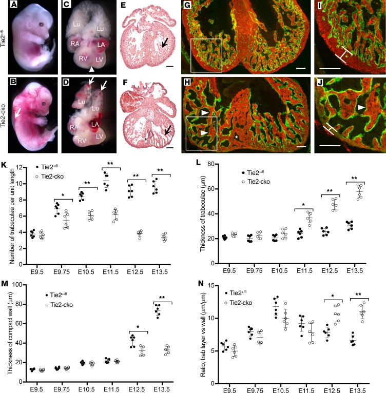
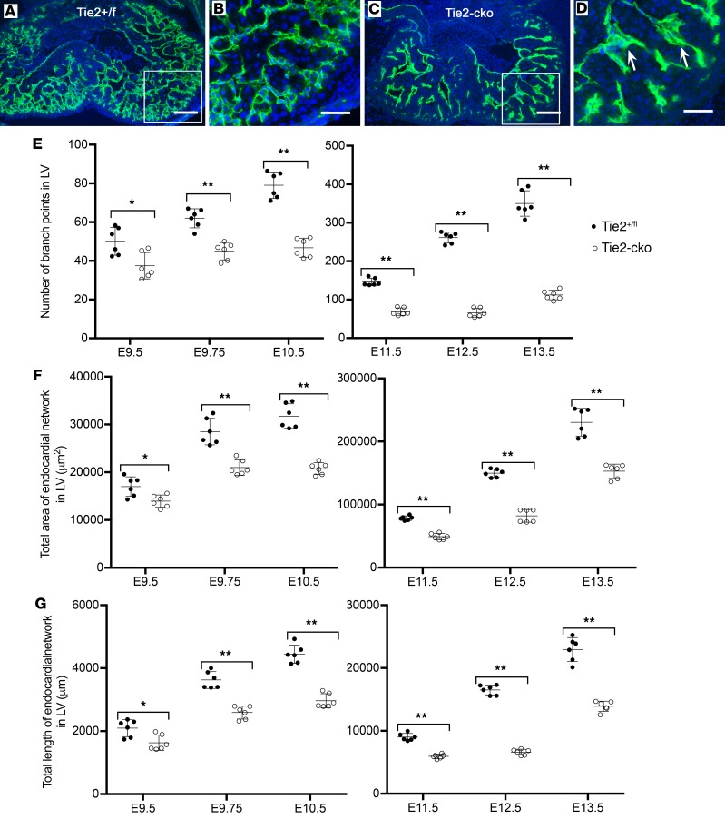
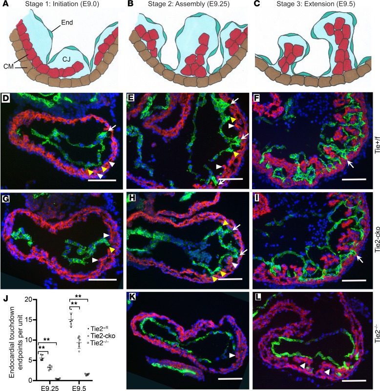
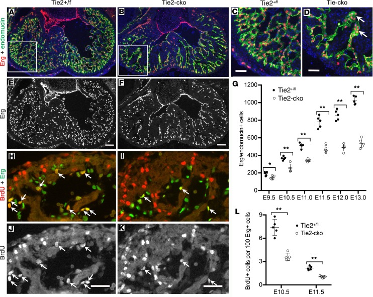
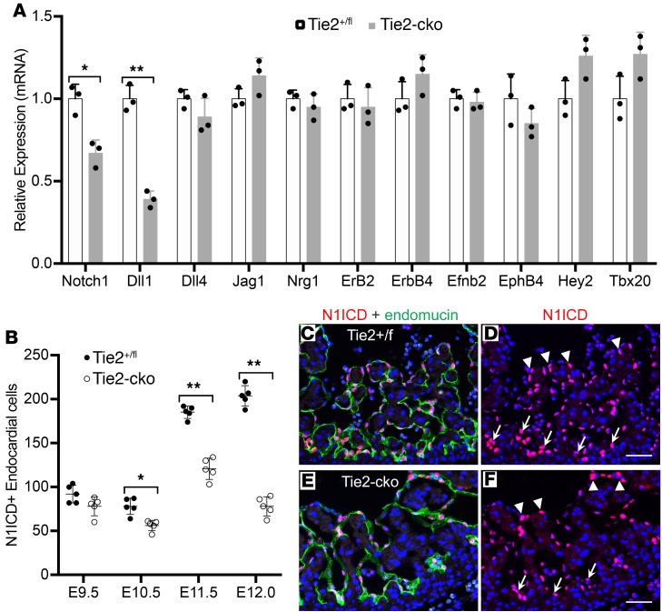
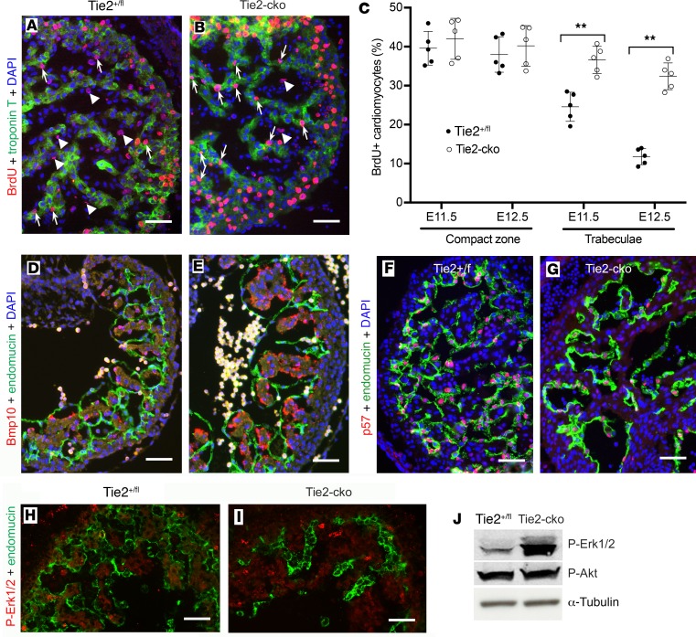
The ang1-Tie2 pathway is required for normal vascular development, but its molecular effectors are not well-defined during cardiac ontogeny. Here we show that endocardial specific attenuation of Tie2 results in mid-gestation lethality due to heart defects associated with a hyperplastic but simplified trabecular meshwork (fewer but thicker trabeculae). Reduced proliferation and production of endocardial cells (ECs) following endocardial loss of Tie2 results in decreased endocardial sprouting required for trabecular assembly and extension. The hyperplastic trabeculae result from enhanced proliferation of trabecular cardiomyocyte (CMs), which is associated with upregulation of Bmp10, increased retinoic acid (RA) signaling, and Erk1/2 hyperphosphorylation in the myocardium. Intriguingly, myocardial phenotypes in Tie2-cko hearts could be partially rescued by inhibiting in utero RA signaling with pan-retinoic acid receptor antagonist BMS493. These findings reveal two complimentary functions of endocardial Tie2 during ventricular chamber formation: ensuring normal trabeculation by supporting EC proliferation and sprouting, and preventing hypertrabeculation via suppression of RA signaling in trabecular CMs.

Keywords: Cardiology; Cardiovascular disease; Development; Embryonic development; Mouse models.


Conflict of interest statement

Conflict of interest: The authors have declared that no conflict of interest exists.

Figures

Figure 1
Figure 1. Endocardial-specific deletion of Tie2 results in abnormal trabeculation.
(AD) Gross images of Tie2+/fl (A and C) and Tie2-cko (B and D) embryos, lungs, and hearts at E13.5, showing the perivascular hemorrhage, edema (arrow), and pulmonary congestion (arrows) in the mutant embryos. Arrowhead indicates formation of the ventricular groove in the control heart (C), which is usually not obvious in the mutants (D). (E and F) H&E–stained heart sections of E13.5 Tie2+/fl (E) and Tie2-cko (F) embryos. Arrows indicate trabeculae. (G and H) Tie2+/fl and Tie2-cko ventricular sections were stained with troponin T (myocardial marker) and endomucin (endocardial marker) antibodies. Compared with the control (Tie2+/fl), the mutant heart had fewer but thicker trabeculae (arrowheads), thinner compact myocardium, and ventricular septation defects (*). The boxed regions are enlarged in I and J. The width of the ventricular compact wall is indicated. LA, left atrium; Lu, lung; LV, left ventricle; RA, right atrium; RV, right ventricle. Scale bars: I and J, 50 μm; others, 100 μm. A representative of more than 10 images was chosen for each panel. (KN) Quantification of trabecular myocardium complexity (trabeculae density, trabeculae thickness, and ratio of trabecular layer and compact zone) and compact myocardium thickness from E9.5–E13.5 (n = 6 per group). Data are expressed as mean ± SEM. *P < 0.05; **P < 0.01, 2-way ANOVA.
Figure 2
Figure 2. Endocardial-specific deletion of Tie2 results in abnormal endocardial network.
(A and B) Tie2+/fl and Tie2-cko heart sections at E13.0 were immunostained with anti-endomucin antibody (green), showing simplified endocardial networks but thickened endocardial sproutings (arrows) in mutant ventricles. The boxed regions are enlarged in C and D. Nuclei are counterstained with DAPI (blue). Scale bars: A and B, 100 μm; C and D, 50 μm. (CE) Quantification of endocardial network complexity (branch points, total area and total length of endocardial network) of Tie2-cko embryos and their littermates at E9.5–E13.5 (n = 6 per group). *P < 0.05; **P < 0.01, 2-way ANOVA.
Figure 3
Figure 3. Endocardial loss of Tie2 impairs endocardial sprouting and trabecular assembly and extension.
(AC) The 3 basic stages of trabeculation (initiation, assembly, and extension) and their corresponding sections of Tie2+/fl (DF), Tie2-cko (GI), and Tie2–/– (K and L) embryos dual immunostained with troponin T (red) and endomucin (green) antibodies. Nuclei are counterstained with DAPI (blue). CJ, cardiac jelly; CM, cardiomyocytes; End, endocardium. At stage 1 (initiation, around E9.0), as the inner layer CMs delaminate into the lumen and form sheet-like protrusions (myocardial lamina, white arrowheads), the endocardium sends out sproutings to penetrate the thick cardiac jelly and make direct touchdown (arrows) with the outer layer of the myocardium in the control embryo. In Tie2-cko, however, the endocardial sproutings have not yet reached the outer layer of the myocardium (yellow arrowheads). At stage 2 (assembly, E9.25), as endocardial sproutings progress laterally beneath the myocardial lamina, they later assemble to isolated short trabecular clusters. At stage 3 (extension, E9.5), finger-like, long trabecular structures are formed by extension. More endocardial touchdown endpoints were detected in the control than in Tie2-cko and Tie2–/– ventricles. Although the myocardial wall in Tie2–/– ventricles (K and L) was able to thicken to become multicellular and form sheet-like protrusions (myocardial lamina, white arrowheads), assembly and extension of trabeculae were impaired. Scale bars: 100 μm. A representative of more than 8 images was chosen for each panel. (J) Quantification of endocardial touchdown endpoints in control, Tie2-cko, and Tie2–/– embryos at E9.25 and E9.5 (n = 6 per group). **P < 0.01, 1-way ANOVA.
Figure 4
Figure 4. Endocardial attenuation of Tie2 results in decreased EC number and proliferation.
(AF) Dual immunostaining of Tie2+/fl (A and C) and Tie2-cko (B and D) heart sections for Erg (red) and endomucin (green) at E13.0 reveals a significant decrease in Erg-positive EC number in the mutant ventricles following endocardial loss of Tie2. (C and D) Boxed regions in A and B are enlarged, and aggregations of ECs in dilated sproutings (arrow) are often observed in Tie2-cko endocardium. Nuclei are counterstained with DAPI (blue). (G) Quantification of total Erg/endomucin–positive cells indicated a gradual loss of ECs in the mutant endocardium, significant from E9.5. (HK) Dual immunostaining of control (H and J) and Tie2-cko (I and K) heart sections for BrdU (red) and Erg (green) at E10.5. The cells positive for both BrdU and Erg represent proliferating ECs (arrows), which are decreased in the mutant ventricles. (L) Quantification of BrdU/Erg–positive cells as a percentage of total Erg-positive cells in endocardium indicated that Tie2-cko displayed lower proliferation rates of ECs at E10.5 and at E11.5. Scale bars: C and D, 100 μm; others, 50 μm. A representative of more than 10 images was chosen for each panel (n ≥ 5 per group). *P < 0.05; **P < 0.01, 1-way ANOVA.
Figure 5
Figure 5. Endocardial attenuation of Tie2 results in decreased Notch signaling.
(A) qPCR analysis of genes associated with Notch signaling pathway detected reduced expression of Notch1 and Dll1 in E11.5 Tie2-cko hearts, although there was no significant change in expression of other components or downstream targets. (B) Quantification of N1ICD/endomucin double-immunostained hearts revealed that N1ICD+ ECs in Tie2-cko left ventricles were significantly reduced at E10.5, E11.5, and E12.0 when compared with the controls. (CF) Dual immunostaining of control and Tie2-cko heart sections at E11.5 for NICD1 (red) and endomucin (green). In comparison with the control, the reduction of N1ICD staining in the mutant endocardium is more significant at the base of the forming trabeculae (arrows) than in distal endocardium (arrowheads). Scale bars: 50 μm. A representative of more than 10 images was chosen for each panel; n = 3 (A) or 5 (B) per group. *P < 0.05; **P < 0.01, Student’s t test (A) and 2-way ANOVA (B).
Figure 6
Figure 6. Endocardial attenuation of Tie2 results in enhanced CM proliferation.
(A and B) BrdU pulse labeling and coimmunostaining for BrdU (red) and troponin T (green) showing more BrdU-positive CMs (arrows) but fewer BrdU-positive ECs (arrowheads) in the trabecular zone of Tie2-cko embryos at E11.5 than those in the control group. (C) Quantification of BrdU-positive nuclei as a percentage of total nuclei in myocardium indicated that Tie2-cko displayed higher proliferation rates of CMs in the trabecular zone at E11.5 and E12.5 (n = 5 per group). **P < 0.01, 2-way ANOVA. (D and E) Compared with the control littermates (D), Bmp10 expression (red, in situ hybridization) in the trabecular CMs of Tie2-cko embryos (E) at E10.5 was enhanced. Endocardium was labeled with endomucin antibody (green). (F and G) Dual immunostaining of control and Tie2-cko heart sections for the cell cycle inhibitor p57 (red) and endomucin (green) at E11.5 showing significantly reduced p57 expression in the mutant endocardium. (H and I) Dual immunostaining of control and Tie2-cko heart sections for p-Erk1/2 (red) and CD31 (green) at E11.5 showing Erk1/2 hyperphosphorylation in the mutant trabecular myocardium. (J) Western blot analysis of E12.5 control and Tie2-cko hearts confirming significantly enhanced phosphorylation of Erk1/2 in the mutants. α-Tubulin was used as a loading control. Scale bars: 50 μm. For the studies in AI, more than 5 embryos per genotype collected from at least 3 independent litters were analyzed. A representative of more than 10 images was chosen for each panel. For the studies in J, 58 embryonic hearts per genotype harvested from 23 independent litters were analyzed, and data are expressed as a representative of 3 independent experiments.
Figure 7
Figure 7. Myocardial phenotypes in Tie2-cko hearts were partially rescued by inhibiting in utero RA signaling with BMS493 treatment.
(A) qPCR analysis of genes associated with the RA signaling pathway in E11.5 hearts revealed elevated RA signaling following endocardial loss of Tie2. (B and C) Immunostaining of control and Tie2-cko heart sections at E9.5 for Crabp2 showing elevated Crabp2 expression in mutant CMs. (DF) Gross images of in utero BMS493 (pan-RA receptor antagonist)-treated Tie2+/fl (D), Tie2-cko/R (E, rescued group) and Tie2-cko/P (F, poorly rescued group) embryos at E14.0. Note that dorsal edema (arrow) was common in all mutant embryos, but pericardial effusions (arrowhead) were usually not obvious in the rescued group. (GI) Heart sections of BMS493-treated Tie2+/fl (G), Tie2-cko/R (H), and Tie2-cko/P (I) embryos at E14.0 were stained with troponin T (red) and endomucin (green) antibodies. (JL) Quantification of trabecular myocardium complexity (trabeculae density and thickness) and compact myocardium thickness from E11.5–E14.0. The trabeculae in the Tie2-cko rescued group were still thicker than those of BMS493-treated controls but much thinner than those of the Tie2-cko poorly rescued embryos, which were similar to the untreated Tie2-cko embryos. However, the defects on the density of trabeculae and thickness of compact wall and simplification of endocardium in the Tie2-cko rescued group were not significantly improved. (M and N) BrdU pulse labeling and immunostaining showed a little more BrdU-positive (red) CMs, stained with troponin T (green) in the trabecular zone of BMS493-treated Tie2-cko embryos (N) at E11.5 than those in the treated controls (M). (O) Quantification of BrdU-positive nuclei as a percentage of total nuclei in myocardium indicated that BMS493-treated Tie2-cko embryos displayed slightly higher proliferation rates of CMs in the trabecular zone at E11.5. Scale bars: GI, 100 μm; others, 50 μm. A representative of more than 10 images was chosen for each panel; n = 3 (A) or 6 (JL and O) per group. *P < 0.05; **P < 0.01, Student’s t test (A) and 2-way ANOVA (JL and O).

References

    1. Sedmera D, Pexieder T, Vuillemin M, Thompson RP, Anderson RH. Developmental patterning of the myocardium. Anat Rec. 2000;258(4):319–337. doi: 10.1002/(SICI)1097-0185(20000401)258:4<319::AID-AR1>3.0.CO;2-O. - DOI - PubMed
    1. Captur G, et al. Morphogenesis of myocardial trabeculae in the mouse embryo. J Anat. 2016;229(2):314–325. doi: 10.1111/joa.12465. - DOI - PMC - PubMed
    1. Samsa LA, Yang B, Liu J. Embryonic cardiac chamber maturation: trabeculation, conduction, and cardiomyocyte proliferation. Am J Med Genet C Semin Med Genet. 2013;163C(3):157–168. - PMC - PubMed
    1. Zhang W, Chen H, Qu X, Chang CP, Shou W. Molecular mechanism of ventricular trabeculation/compaction and the pathogenesis of the left ventricular noncompaction cardiomyopathy (LVNC) Am J Med Genet C Semin Med Genet. 2013;163C(3):144–156. - PMC - PubMed
    1. Grego-Bessa J, et al. Notch signaling is essential for ventricular chamber development. Dev Cell. 2007;12(3):415–429. doi: 10.1016/j.devcel.2006.12.011. - DOI - PMC - PubMed

Publication types

MeSH terms