. 2019 May 21;14(5):e0216811.
doi: 10.1371/journal.pone.0216811. eCollection 2019.

Three-dimensional label-free imaging throughout adipocyte differentiation by stimulated Raman microscopy

Affiliations

Three-dimensional label-free imaging throughout adipocyte differentiation by stimulated Raman microscopy

Maria Antonietta Ferrara et al. PLoS One. .

Abstract

Lipid droplets are lipid-storage organelles with a key role in lipid accumulation pathologies such as diabetes, obesity and atherosclerosis. Despite their important functions many aspects of lipid droplets biology are still unknown. This is partially due to the current use of exogenous labels to monitor their formation and remodelling by invasive imaging methods. Here, we apply stimulated Raman scattering microscopy to acquire images with high spatial resolution along with resolving capabilities of lipids and proteins and three-dimensional sectioning. Our images and data analysis demonstrate an increase in the number of large (>15μm2) lipid droplets in human adipocyte cells during differentiation process. In addition, spatially-resolved maps of lipids and proteins inside cells and three dimensional reconstructions of lipids at the initial and final steps of adipocyte differentiation are reported, too.


Conflict of interest statement

The authors declare that no competing interests exist.

Figures

Fig 1
Fig 1. Fluorescence imaging.
a) Representative confocal microscopy images of 3T3-L1 cells Undifferentiated or differentiated with cultured adipogenic media, fixed at day 5, day 10 and day 15 of the differentiation process, fixed and labeled with LipidTox Red (red), Oil Red O (red) or BODIPY 493/503 (green) fluorescent dyes. Cell nuclei were labelled with DAPI (blue). Scale bar, 10 μm. b) Quantification of LDs size (as area ranges, μm2) in 3T3-L1 cells treated, fixed and labeled as in (a) (Experimental data are reported in S1 File). c) Western blotting with anti-PPARγ and anti-FABP4 antibodies (as indicated) of total cell lysate (20 μg/lane) from 3T3-L1 cells treated as in (a). Actin is shown for the internal protein levels. Molecular weight standards (kDa) are indicated on the right of each panel. Of note, PPARγ is separated on 10% SDS-PAGE while FABP4 is separated on 15% SDS-PAGE (S2 Fig).
Fig 2
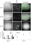
Fig 2. Subcellular localization of lipid droplets in adipocyte cells by SRS imaging.
a) Representative transmission microscopy images (first column) and SRS microscopy images acquired at 2,845 cm-1 (second column) of adipocyte cells, fixed at day 5, day 10 and day 15 of the differentiation process. Merged images are reported in the last. All scale bars are 5 μm. b) Quantification of LDs size (as area ranges, μm2) in adipocytes at day 5, day 10 and day 15 of the differentiation process (as indicated); experimental data are reported in S2 File. c) Quantification of major and minor axis (μm) of the fitted ellipsis in which LDs are arranged in adypocytes fixed at day 5, day 10 and day 15 of the differentiation process (as indicated); experimental data are reported in S3 File.
Fig 3
Fig 3. Representative single focal plane of Z stack SRS image of adipocytes fixed at day 5 of the differentiation process.
Images acquired with a 2.5x optical zoom at: a) 2,845 cm-1 (lipids contribution); b) 2,940 cm-1 (proteins contribution, retrieved image). c) Lipids (green) and proteins (red) contributions in cells acquired as in a-b. Scale bars, 5 μm.
Fig 4
Fig 4. Three-dimensional reconstruction of lipids, proteins and their simultaneous distribution in adipocytes fixed at days 5 of the differentiation process.
a) three-dimensional Z stack (13 frames, 1 μm/step) reconstruction of SRS images at 2,845 cm-1; b) three-dimensional reconstruction of SRS images at 2,940 cm-1; c) three-dimensional reconstruction of simultaneous lipids and proteins distribution. All the images are acquired with a 2.5x optical zoom.
Fig 5
Fig 5. SRS z-stack for 3D volume reconstruction of lipids.
a) Single representative z-sections (16 frames, 1 μm /step) of SRS images at 2,845 cm-1 of adipocyte fixed at day 15 of the differentiation process. b) three-dimensional reconstruction of lipids.

References

    1. Farese RV & Walther TC. Lipid Droplets Finally Get a Little R-E-S-P-E-C-T. Cell 2009; 139: 855–86. 10.1016/j.cell.2009.11.005 - DOI - PMC - PubMed
    1. Straub BK, Herpel E, Singer S, Zimbelmann R, Breuhahn K, Macher-Goeppinger S, et al. Lipid droplet-associated PAT-proteins show frequent and differential expression in neoplastic steatogenesis. Mod Pathol. 2010; 23: 480–92. 10.1038/modpathol.2009.191 - DOI - PubMed
    1. Tirinato L, Pagliari F, Limongi T, Marini M, Falqui A, Seco J, et al. An Overview of Lipid Droplets in Cancer and Cancer Stem Cells. Stem Cells Int. 2017; 1656053 10.1155/2017/1656053 - DOI - PMC - PubMed
    1. Yang L, Ding Y, Chen Y, Zhang S, Huo C, Wang Y, et al. The proteomics of lipid droplets: structure, dynamics, and functions of the organelle conserved from bacteria to humans. J Lipid Res. 2012; 53(7): 1245–53. 10.1194/jlr.R024117 - DOI - PMC - PubMed
    1. Thiam AR, Farese RV, Walther TC. The biophysics and cell biology of lipid droplets. Nat Rev Mol Cell Biol. 2013; 14(12): 775–86. 10.1038/nrm3699 - DOI - PMC - PubMed

Publication types