. 2019 May 21;10(1):146.
doi: 10.1186/s13287-019-1223-z.

IGF-1 overexpression improves mesenchymal stem cell survival and promotes neurological recovery after spinal cord injury

Affiliations

IGF-1 overexpression improves mesenchymal stem cell survival and promotes neurological recovery after spinal cord injury

Kyan James Allahdadi et al. Stem Cell Res Ther. .

Abstract

Background: Survival and therapeutic actions of bone marrow-derived mesenchymal stem cells (BMMSCs) can be limited by the hostile microenvironment present during acute spinal cord injury (SCI). Here, we investigated whether BMMSCs overexpressing insulin-like growth factor 1 (IGF-1), a cytokine involved in neural development and injury repair, improved the therapeutic effects of BMMSCs in SCI.

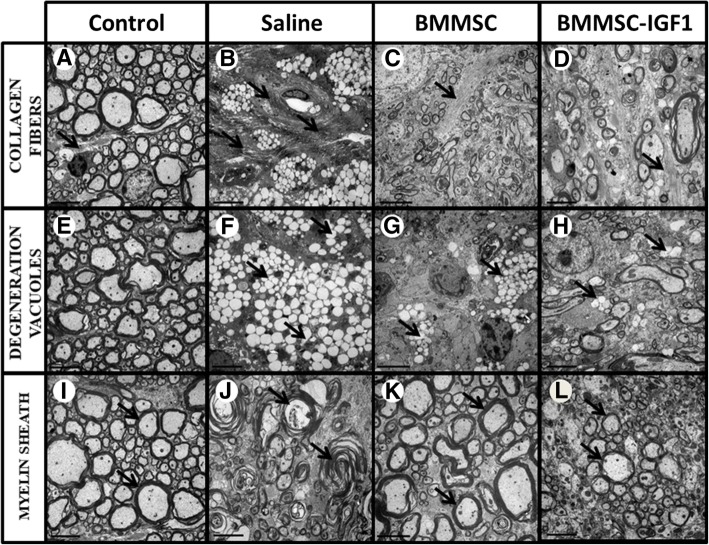
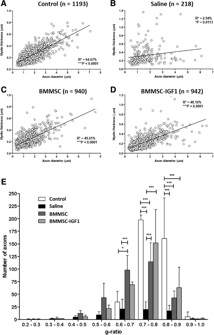
Methods: Using a SCI contusion model in C57Bl/6 mice, we transplanted IGF-1 overexpressing or wild-type BMMSCs into the lesion site following SCI and evaluated cell survival, proliferation, immunomodulation, oxidative stress, myelination, and functional outcomes.

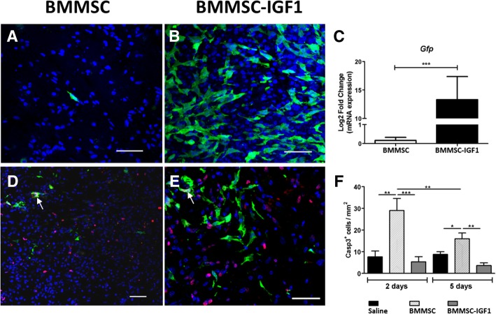
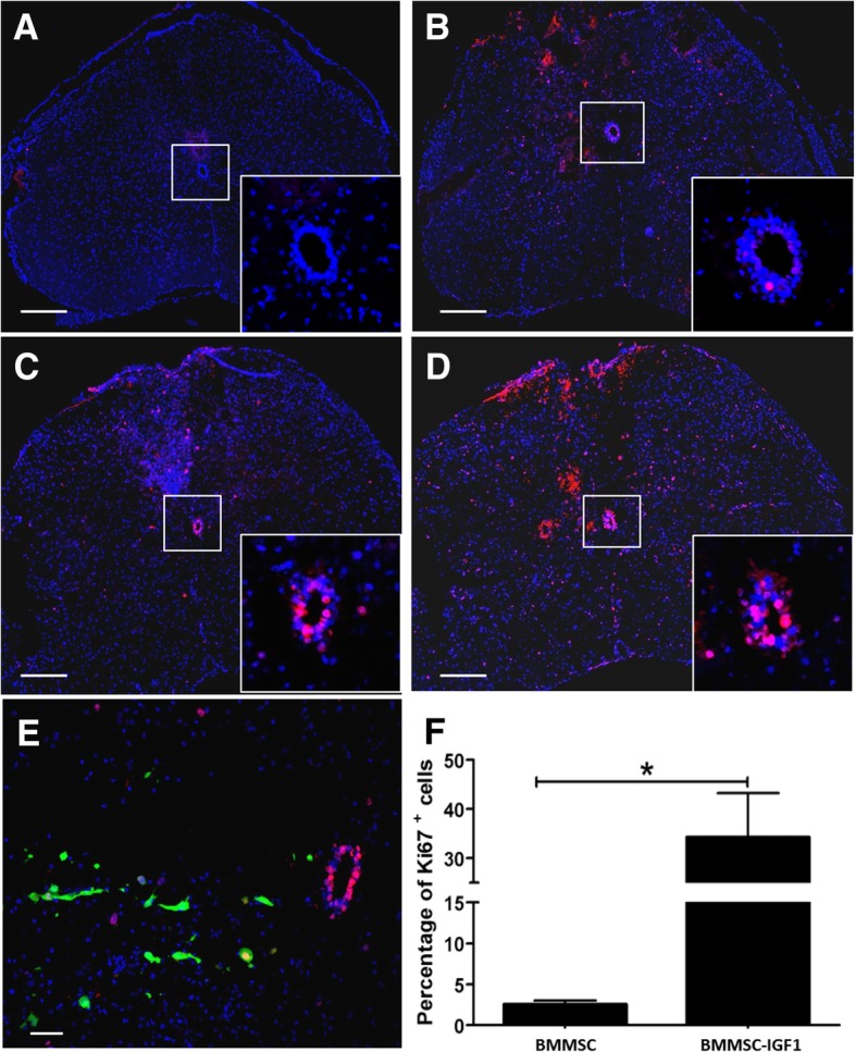
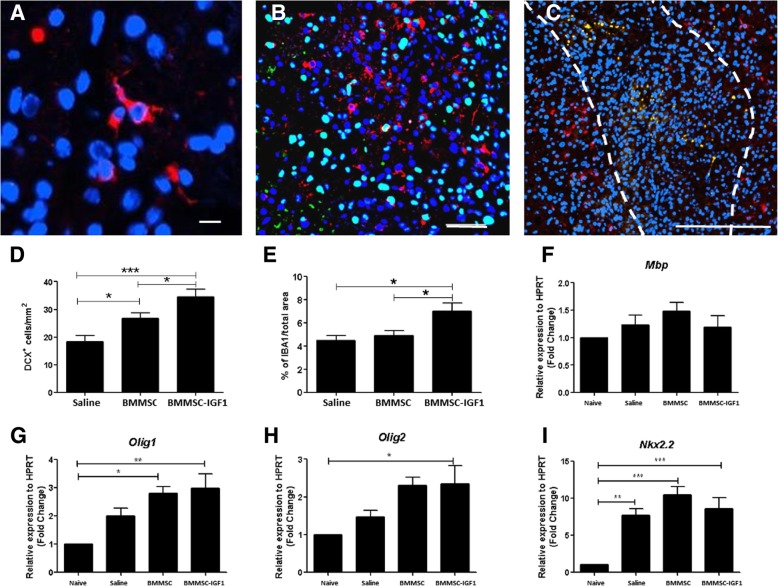
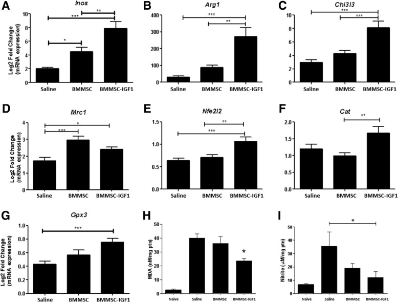
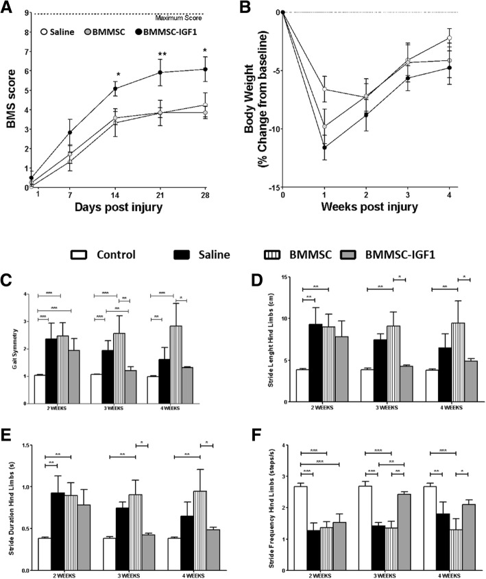
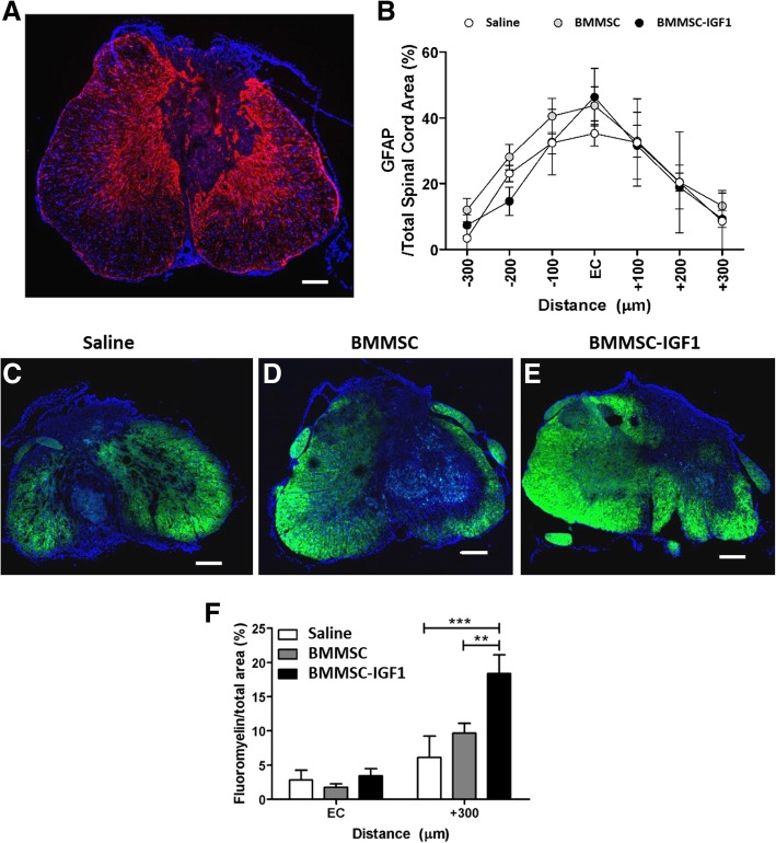
Results: BMMSC-IGF1 transplantation was associated with increased cell survival and recruitment of endogenous neural progenitor cells compared to BMMSC- or saline-treated controls. Modulation of gene expression of pro- and anti-inflammatory mediators was observed after BMMSC-IGF1 and compared to saline- and BMMSC-treated mice. Treatment with BMMSC-IGF1 restored spinal cord redox homeostasis by upregulating antioxidant defense genes. BMMSC-IGF1 protected against SCI-induced myelin loss, showing more compact myelin 28 days after SCI. Functional analyses demonstrated significant gains in BMS score and gait analysis in BMMSC-IGF1, compared to BMMSC or saline treatment.

Conclusions: Overexpression of IGF-1 in BMMSC resulted in increased cell survival, immunomodulation, myelination, and functional improvements, suggesting that IGF-1 facilitates the regenerative actions of BMMSC in acute SCI.

Keywords: Bone marrow-derived mesenchymal stem cells; Gene and cell therapy; IGF-1; Spinal cord injury.


Conflict of interest statement

The authors declare that they have no competing interests.

Figures

Fig. 1
Fig. 1
Increased survival of BMMSC-IGF1 and induction of cell proliferation in the injured spinal cord. Presence of GFP+ BMMSCs was detected in spinal cord sections of mice transplanted with wild-type BMMSCs (a) or BMMSC-IGF1 (b), visualized in green by confocal microscopy, 5 days after spinal cord injury and cell transplantation. Nuclei were stained with DAPI (blue). Scale bars = 50 μm. c Quantification of GFP mRNA in the injured spinal cord segments, isolated at 5 days post-injury and transplant, measured by qRT-PCR. Confocal microscopy of injured mouse spinal cords, 5 days post-injury and transplanted with wild-type BMMSC (d) or BMMSC-IGF1 (e), immunostained for the proliferation marker Ki-67 (red) and nuclei stained with DAPI (blue). Scale bars = 200 μm. f Quantification of Casp3+-labeled cells in spinal cord sections. Values represent mean ± SEM. *P < 0.05; **P < 0.01; ***P < 0.001
Fig. 2
Fig. 2
Cellular proliferation in injured mouse spinal cords following BMMSC-IGF transplantation. Spinal cord segments from a uninjured mouse and SCI mice treated with b saline, c BMMSCs and d, e BMMSC-IGF1, observed by confocal microscopy, 2 days following spinal cord injury and cell transplantation, immunostained for Ki-67. Scale bars = 200 μm (ad); scale bar = 50 μm (e). Central canal (highlighted/insert, ad). f Quantitative percentage of Ki-67+ in the spinal cord 5 days post-injury. Values represent mean ± SEM. *P < 0.05; **P < 0.01; ***P < 0.001
Fig. 3
Fig. 3
SCI mice treated with BMMSC-IGF1 demonstrate elevated progenitor cell presence, 5 days following SCI and transplant. Injured spinal cord section from BMMSC-IGF1-treated mouse immunostained for a DCX (red; scale bar = 10 μm), b double immunostained for DCX and PCNA (green; scale bar = 10 μm), and c detailed region with DCX (red; scale bar = 100 μm). Quantitative analysis of d DCX-positive cells and e percentage of IBA1 cells from injured spinal cords. RT-qPCR analysis of expression of f Mbp, g Olig1, h Olig2, and i NKx2.2, genes associated with oligodendrocyte progenitor cells differentiation. Values represent mean ± SEM. *P < 0.05; **P < 0.01; ***P < 0.001
Fig. 4
Fig. 4
Modulation of inflammatory mediators after SCI and treatment. Transcripts for a iNOS, b Arg1, c Chi3I3, d Mrc1, e Nfe2I2, f Cat, and g Gpx3 were determined in SCI segment homogenates from saline (n = 5), BMMSC (n = 5), or BMMSC-IGF1-treated mice, by RT-qPCR. Values represent means ± SEM. Concentrations of h malondialdehyde (MDA), measured by MDA Oxidative Stress Assay, and i nitrite, determined by the Griess method, in SCI segment homogenates from naive, (n = 5), saline (n = 5), BMMSC (n = 5), or BMMSC-IGF1-treated mice. Values represent mean ± SEM. *P < 0.05; **P < 0.01; ***P < 0.001
Fig. 5
Fig. 5
Functional analysis following SCI and treatment. Functional analysis (a) of saline-, BMMSC-, and BMMSC-IGF1-treated mice, evaluated weekly from day 1 to 28 days using the Basso Mouse Score (BMS). Body weight (b) was assessed with BMS and is represented as percent change in body weight, based on weight prior to injury (baseline). DigiGait-derived functional measurements of c gait symmetry, d stride length, e stride duration, and f stride frequency were evaluated at weeks 2, 3, and 4. Values represent mean ± SEM. *P < 0.05; **P < 0.01; and ***P < 0.001
Fig. 6
Fig. 6
Lesion volume and myelination 4 weeks following SCI and treatment. Lesion volume (a) as measured by GFAP staining, which is concentrated around the lesion site, measured against total spinal cord area bilaterally from the epicenter (EC) of the injury. b Quantification of GFAP derived lesion volume. Myelination immunostaining with fluoromyelin was performed in saline (c), BMMSC (d), and BMMSC-IGF1 (e) treated SCI sections. Quantitative analysis (f) of fluoromyelin-labeled oligodendrocytes at the EC and 300 μm caudally. Bars represent means ± SEM of five mice/group. Values represent mean ± SEM. *P < 0.05 (BMMSC-IGF1: EC vs + 300), #P < 0.05 (+ 300: BMMSC-IGF1 vs BMMSC)
Fig. 7
Fig. 7
Ultrastructural changes at the site of lesion in animals with SCI visualized by transmission electron microscopy. Representative images of spinal cord sections obtained from uninjured or SCI-mice admistered with saline, BMMCs or BMMC-IGF1 are shown. Morphological patterns of collagen fibers (a-d), degeneration vacuoles (e-h) and myelin sheath (i-l) were evaluated, as indicated by black arrows. Scale bar = 5 μm
Fig. 8
Fig. 8
Quantitative ultrastructural analyses of spinal cord sections in uninjured and SCI mice. a-d Linear regression analysis between myelin thickness and axon diameter from control, saline, BMMSC, and BMMSC-IGF1 groups. Three animals were used in each group. e Axon distribution by g-ratio range in control (N = 1193), saline (N = 218), BMMSC (940), and BMMSC-IGF1 (942) groups. Analysis was done as described in the “Materials and methods” section. Values represent the mean ± SEM in each range. *P < 0.05; ***P < 0.001

References

    1. McDonald JW, Sadowsky C. Spinal-cord injury. Lancet. 2002;359:417–425. doi: 10.1016/S0140-6736(02)07603-1. - DOI - PubMed
    1. Ribeiro TB, Duarte AS, Longhini AL, et al. Neuroprotection and immunomodulation by xenografted human mesenchymal stem cells following spinal cord ventral root avulsion. Sci Rep. 2015;5:16167. doi: 10.1038/srep16167. - DOI - PMC - PubMed
    1. Shende P, Subedi M. Pathophysiology, mechanisms and applications of mesenchymal stem cells for the treatment of spinal cord injury. Biomed Pharmacother. 2017;91:693–706. doi: 10.1016/j.biopha.2017.04.126. - DOI - PubMed
    1. Qu J, Zhang H. Roles of mesenchymal stem cells in spinal cord injury. Stem Cells Int. 2017;10:1155. - PMC - PubMed
    1. Moya A, Paquet J, Deschepper M, Larochette N, Oudina K, Denoeud C, Bensidhoum M, et al. Mesenchymal stem cell failure to adapt to glucose shortage and rapidly use intracellular energy reserves through glycolysis explains poor cell survival after implantation. Stem Cells. 2018;36(3):363–376. doi: 10.1002/stem.2763. - DOI - PubMed

Publication types

Substances