. 2019 May 21;10(1):142.
doi: 10.1186/s13287-019-1253-6.

Human embryonic stem cell-derived exosomes promote pressure ulcer healing in aged mice by rejuvenating senescent endothelial cells

Affiliations

Human embryonic stem cell-derived exosomes promote pressure ulcer healing in aged mice by rejuvenating senescent endothelial cells

Bi Chen et al. Stem Cell Res Ther. .

Abstract

Background: Angiogenesis, as an endogenous repair mechanism, plays crucial roles in wound healing and tissue regeneration. However, this process is impaired in the elderly due to aging-related vascular endothelial dysfunction. This study was aimed to explore the pro-angiogenic effects of exosomes from human embryonic stem cells (ESC-Exos) in aged mice of pressure-induced ulcer model and the underlying mechanism.

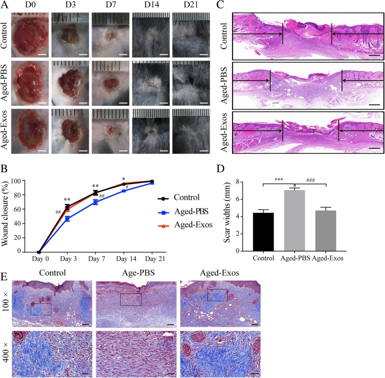
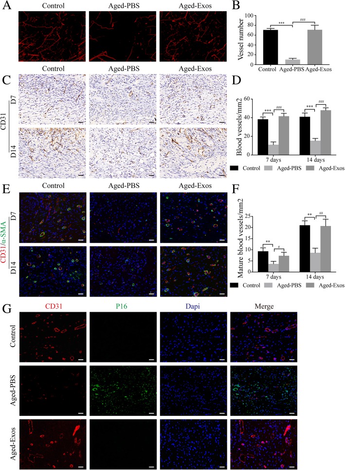
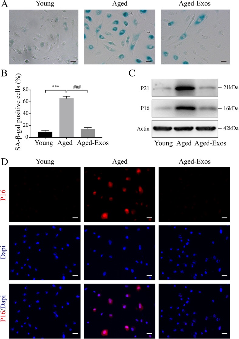
Methods: Pressure ulcer wounds were created on the back of D-galactose-induced aging mice. ESC-Exos were locally applied onto the wound beds, with PBS as control. The effects of ESC-Exos on wound healing were analyzed by measuring wound closure rates, histological and immunofluorescence analyses. Then, the anti-aging effect of ESC-Exos on vascular endothelial cells was tested in an in vitro D-galactose-induced HUVEC senescence model.

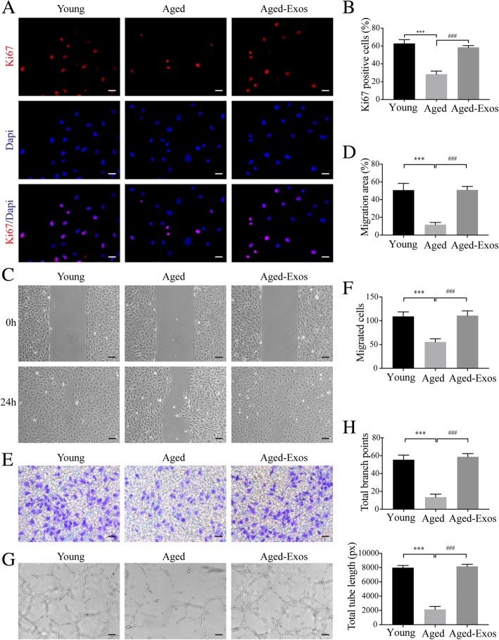
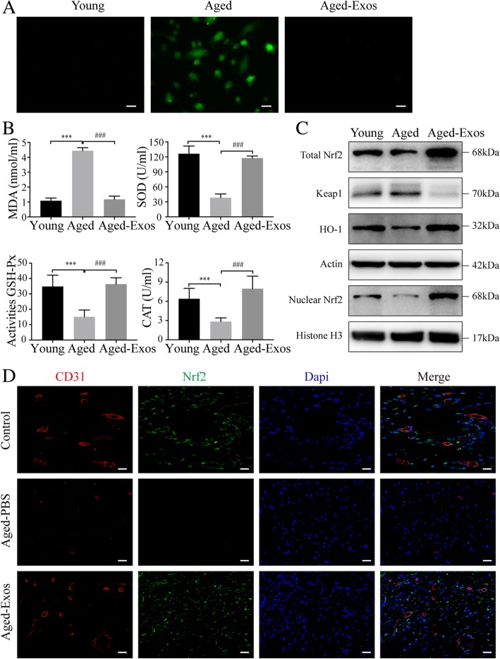
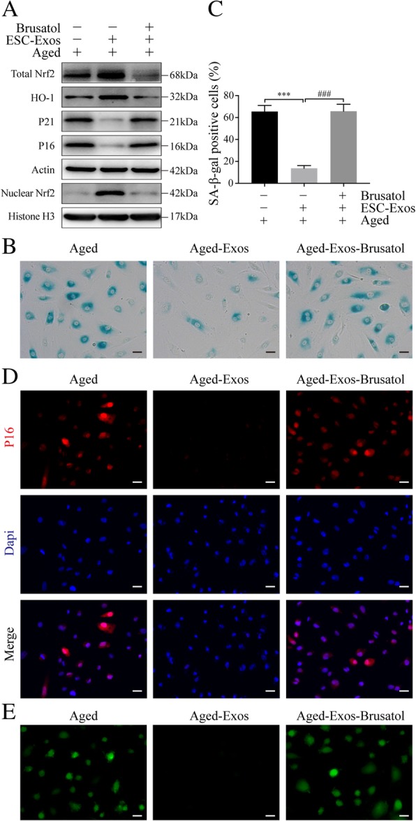
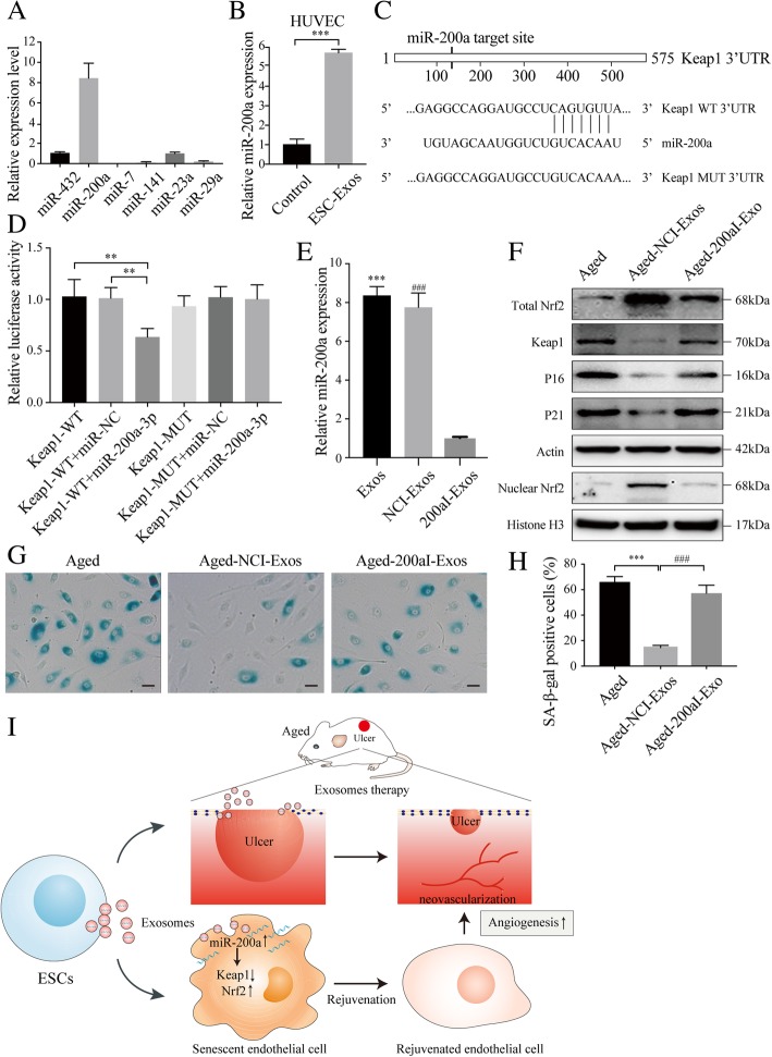
Results: ESC-Exos could accelerate wound closure and enhance angiogenesis, and the senescence of vascular endothelial cells was significantly ameliorated after ESC-Exos treatment. In vitro, ESC-Exos could rejuvenate the senescence of endothelial cells and recover compromised proliferation, migratory capacity, and tube formation. This recovery was Nrf2-activation-dependent, since cotreatment with Nrf2 inhibitor Brusatol could abolish the rejuvenative effects of ESC-Exos. Further study revealed that miR-200a was highly enriched in ESC-Exos and played a crucial role in ESC-Exos-mediated rejuvenation through downregulating Keap1, which negatively regulates Nrf2 expression.

Conclusions: ESC-Exos ameliorate endothelial senescence by activating Nrf2 and recover aging-related angiogenic dysfunction, thereby accelerating wound healing in aged mice. ESC-Exos might be a natural nano-biomaterial for aging-related diseases therapy.

Keywords: Angiogenesis; Embryonic stem cells; Exosomes; Nrf2; Senescence.


Conflict of interest statement

The authors declare that they have no competing interests.

Figures

Fig. 1
Fig. 1
Characterization of ESCs and ESC-derived exosomes. a ESC colonies morphology. Scale bar, 100 μm. b ALP staining of ESCs. Scale bar, 100 μm. c IF staining analysis of pluripotency-related markers of ESCs. Scale bar, 50 μm. d Particle size distribution of ESC-Exos measured by qNano analysis. e Morphology of ESC-Exos observed by TEM. Scale bar, 100 nm. f Western blotting showed the presence of exosomal markers including CD9, CD63, and TSG101, but exosomes were negative for GM130, Actin, and Lamin A/C
Fig. 2
Fig. 2
ESC-Exos promoted pressure ulcer wound healing in aged mice. a Gross view of wounds treated with ESC-Exos or PBS in aging mice and wounds with PBS in the young control group, at days 3, 7, 14, and 21 post-wounding. Scale bar, 2 mm. b The rate of wound closure of three groups. n = 6 per group. **P < 0.01; *P < 0.05 Aged-PBS versus control group; ##P < 0.01; #P < 0.05 Aged-Exos versus Aged-PBS group. c H&E staining of wound sections from three groups at 7 days after initial treatment. The black arrows indicate the edges of the scar. n = 3 per group. Scale bar, 500 μm. d Quantification of the scar widths. n = 3 per group. ***P < 0.001 Aged-PBS versus control group; ###P < 0.001 Aged-Exos versus Aged-PBS group. e Masson’s trichrome staining of wound sections from three groups. n = 3 per group. Scale bar, 100 μm (top) or 25 μm (bottom)
Fig. 3
Fig. 3
ESC-Exos enhanced angiogenesis and ameliorated vascular endothelial cell senescence in the wound sites of aged mice. a Micro-CT images of blood vessel formation in pressure ulcer wounds from the indicated groups at 14 days after initial treatment. n = 3 per group. b Quantification analysis of the number of blood vessels. n = 3 per group. ***P < 0.001 Aged-PBS versus control group; ###P < 0.001 Aged-Exos versus Aged-PBS group. c IHC staining of CD31 of wound sections at 7 and 14 days after initial treatment. n = 3 per group. Scale bar, 50 μm. d Statistical results from c. n = 3 per group. ***P < 0.001 Aged-PBS versus control group; ###P < 0.001 Aged-Exos versus Aged-PBS group. e IF staining against CD31 and α-SMA. Endothelial cells (CD31), smooth muscle cells (α-SMA), and cell nuclei were stained red, green, and blue, respectively, at 7 and 14 days after initial treatment. n = 3 per group. Scale bar, 50 μm. f Statistical results from e. n = 3 per group. **P < 0.01 Aged-PBS versus control group; ##P < 0.01; #P < 0.05 Aged-Exos versus Aged-PBS group. g IF staining against CD31 and P16. Endothelial cells (CD31), senescent cells (P16), and cell nuclei were stained red, green, and blue, respectively, at 14 days after initial treatment. n = 3 per group. Scale bar, 50 μm
Fig. 4
Fig. 4
ESC-Exos can ameliorate endothelial senescence induced by D-gal in vitro. HUVECs were treated with 10 g/L D-gal to induce senescence, and aged HUVECs were then treated with 1 × 1010 particles/mL ESC-Exos or PBS, while young HUVECs (without D-gal treatment) were set as the control. a SA-β-gal staining. SA-β-gal-positive cells are shown in blue when observed under an optical microscope. n = 3 per group. Scale bar, 50 μm. b Statistical results from (a). n = 3 per group. ***P < 0.001 Aged versus Young group; ###P < 0.001 Aged-Exos versus Aged group. c Western blotting analysis of P16 and P21 expression. n = 3 per group. d IF staining was performed to assess the expression level of P16 (red). P16-positive cells were significantly reduced in number after ESC-Exos treatment. n = 3 per group. Scale bar, 50 μm
Fig. 5
Fig. 5
ESC-Exos can ameliorate aging-related angiogenic dysfunction induced by D-gal. HUVECs were treated with D-gal to induce senescence, followed by treatment with ESC-Exos or PBS, and young HUVECs were set as the control. a IF staining against Ki67 was performed to assess the proliferative capacity of HUVECs. n = 3 per group. Scale bar, 50 μm. b Quantification of the number of ki67-positive cells in (a). n = 3 per group. ***P < 0.001 Aged versus Young group; ###P < 0.001 Aged-Exos versus Aged group. Wound healing assay (c, d) (scale bar, 100 μm) and transwell assay (e, f) (scale bar, 100 μm) revealed that ESC-Exos treatment could recover the compromised migratory ability of aged HUVECs. n = 3 per group. ***P < 0.001 Aged versus Young group; ###P < 0.001 Aged-Exos versus Aged group. g Representative images of the tube formation assay in young HUVECs or aged HUVECs after treatment with ESC-Exos or PBS. n = 3 per group. Scale bar, 200 μm. h Quantitative analyses of the total tube length and branch points. n = 3 per group. ***P < 0.001 Aged versus Young group; ###P < 0.001 Aged-Exos versus Aged group
Fig. 6
Fig. 6
ESC-Exos could reduce oxidative stress and enhance the activity of the endogenous Nrf2 anti-oxidative system. a Intracellular ROS levels were determined by green fluorescent intensity after cells were labeled with DCFH-DA. n = 3 per group. Scale bar, 50 μm. b Oxidative stress levels were evaluated by the activity measurement of MDA, SOD, GSH-Px and CAT. n = 3 per group. ***P < 0.001 Aged versus Young group; ###P < 0.001 Aged-Exos versus Aged group. c Protein expression levels of Nrf2 and HO1 in indicated groups. n = 3 per group. d IF staining against CD31 and Nrf2. Endothelial cells (CD31), Nrf2-positive cells, and cell nuclei were stained red, green, and blue, respectively, at 14 days after initial treatment. The white arrows indicate the Nrf2+ vascular endothelial cells. n = 3 per group. Scale bar, 50 μm
Fig. 7
Fig. 7
ESC-Exos rejuvenate endothelial senescent cells through activating Nrf2. Aged HUVECs were treated with ESC-Exos or co-treated with ESC-Exos and Nrf2 inhibitor (Brusatol), while aged HUVECs without treatment were set as control. a Western blot analysis of Total Nrf2, Nuclear Nrf2, HO1, P21, and P16 protein levels. b, c SA-β-gal kit was used to evaluate the SA-β-gal activity and percentages of SA-β-gal-positive cells were quantified. n = 3 per group. ***P < 0.001 Aged-Exos versus Aged group; ###P < 0.001 Aged-Exos-Brusatol versus Aged-Exos group. Scale bar, 50 μm. d IF staining against P16 was performed to assess the expression level of P16. n = 3 per group. Scale bar, 50 μm. e ROS levels were determined by green fluorescent intensity after cells were labeled with DCFH-DA. n = 3 per group. Scale bar, 50 μm
Fig. 8
Fig. 8
ESC-Exos activated Nrf2 signaling by downregulating the expression of Keap1 via transferring miR-200a. a Detection of the expression of the indicated miRNAs in ESC-Exos by qRT-PCR analysis. b Aged HUVECs incubated with ESC-Exos for 6 h and the expression level of miR-200a were determined by qPCR analysis. n = 3 per group. ***P < 0.001. c The Keap1 3′-UTR contains one putative miR-200a binding site. The first eight nucleotides of miR-200a are complementary to the binding site in the 3′-UTR. All of these eight nucleotides were mutated to abrogate miR-200a binding. d Dual-luciferase reporter assay of miR-200a with 3′-UTR vectors (WT or MUT) of human Keap1 in HUVECs was performed. We found that miR-200a directly targets the 3′-UTR of Keap1. e Levels of miR-200a in exosomes from treated ESCs were analyzed by qRT-PCR. ***P < 0.001 Exos versus NCI-Exo; ###P < 0.001 NCI-Exo versus 200aI-Exos. f Western blot analysis of Nrf2, Keap1, P21, and P16 protein levels. Aged HUVECs were treated with NCI-ESC-Exos or 200aI-ESC-Exos, while aged HUVECs without treatment were set as the control. n = 3 per group. g, h SA-β-gal staining. The SA-β-gal activity and percentages of SA-β-gal-positive cells were quantified. n = 3 per group. ***P < 0.001 Aged-NCI-Exos versus Aged group; ###P < 0.001 Aged-NCI-Exos versus Aged-200aI-Exos. Scale bar, 50 μm. i Schematic diagram depicts rejuvenative effects of exosomes derived from embryonic stem cells. Exosomes secreted by ESCs induce enhanced angiogenesis and promoted pressure ulcer repair in aged mice. ESC-Exos-delivered miR-200a rejuvenates senescent endothelial cells by downregulating Keap1 and recovering Nrf2 activation

References

    1. López-Otín C, Blasco MA, Partridge L, Serrano M, Kroemer G. The hallmarks of aging. Cell. 2013;153(6):1194–1217. doi: 10.1016/j.cell.2013.05.039. - DOI - PMC - PubMed
    1. van Deursen JM. The role of senescent cells in ageing. Nature. 2014;509(7501):439–446. doi: 10.1038/nature13193. - DOI - PMC - PubMed
    1. Eming SA, Martin P, Tomic-Canic M. Wound repair and regeneration: mechanisms, signaling, and translation. Sci Transl Med. 2014;6(265):265sr6. doi: 10.1126/scitranslmed.3009337. - DOI - PMC - PubMed
    1. Zhao P, Sui BD, Liu N, Lv YJ, Zheng CX, Lu YB, et al. Anti-aging pharmacology in cutaneous wound healing: effects of metformin, resveratrol, and rapamycin by local application. Aging Cell. 2017;16(5):1083–1093. doi: 10.1111/acel.12635. - DOI - PMC - PubMed
    1. Valcarcel-Ares MN, Gautam T, Warrington JP, Bailey-Downs L, Sosnowska D, de Cabo R, et al. Disruption of Nrf2 signaling impairs angiogenic capacity of endothelial cells: implications for microvascular aging. J Gerontol A Biol Sci Med Sci. 2012;67(8):821–829. doi: 10.1093/gerona/glr229. - DOI - PMC - PubMed

Publication types

MeSH terms