

Quantitative computed tomography texture analysis: can it improve diagnostic accuracy to differentiate malignant lymph nodes?
- PMID: 31113494
- PMCID: PMC6530003
- DOI: 10.1186/s40644-019-0214-8
Quantitative computed tomography texture analysis: can it improve diagnostic accuracy to differentiate malignant lymph nodes?
Abstract
Background and objective: Mediastinal lymph node (LN) staging in individuals with non-small-cell lung cancer plays an important role in staging and treatment planning. This study aimed to assess the accuracy of computed tomography (CT) texture analysis (CTTA) in differentiating benign and malignant mediastinal LNs.
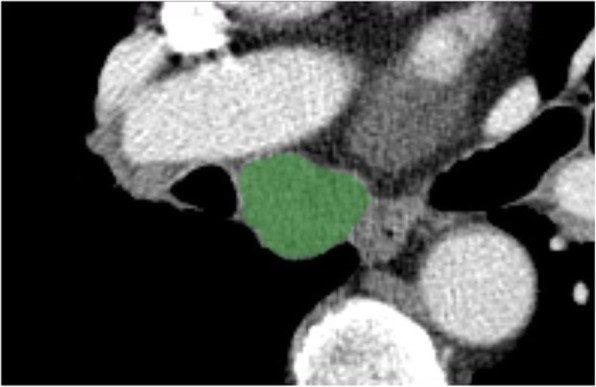
Methods: Pathologically confirmed malignant and benign mediastinal LN samples, obtained using endobronchial ultrasound-guided transbronchial needle aspiration (EBUS-TBNA), were retrospectively reviewed, in addition to chest CT and 18-fluorodeoxyglucose (FDG) uptake positron emission tomography (PET) data. For each LN, CTTA was performed using "AVIEW" software (Coreline Soft, Republic of Korea) by drawing a region of interest.
Results: A total of 132 LNs from 80 patients were included and classified into two groups according to pathology results: malignant (n = 61) and benign (n = 71). In EBUS, size > 1 cm, round shape, heterogeneous echogenicity, and presence of coagulation necrosis sign were more prevalent in malignant than in benign LNs; length was the only feature that distinguished the two groups. Among CTTA features, compactness and normalized standard deviation (SD) showed differences between the two groups. The ability to distinguish malignant LNs was higher using high standard uptake value (SUV) on FDG PET/CT (SUVmax ≥ 5) and normalized SD on CTTA (area under the receiver operating characteristic curve 0.739 versus 0.742, respectively); however, normalized SD demonstrated very low sensitivity despite high specificity.
Conclusions: CTTA may be helpful in distinguishing between benign and malignant LNs; however, the diagnostic value was not high. Therefore, integrated evaluation with other imaging modalities is needed.
Keywords: Malignancy; Mediastinal lymph node; Texture and shape analysis.
Conflict of interest statement
The authors declare that they have no competing interests.
Figures

References
-
- Silvestri GA, Gonzalez AV, Jantz MA, Margolis ML, Gould MK, Tanoue LT, Harris LJ, Detterbeck FC. Methods for staging non-small cell lung cancer: diagnosis and management of lung cancer, 3rd ed: American College of Chest Physicians evidence-based clinical practice guidelines. Chest. 2013;143:e211S–e250S. doi: 10.1378/chest.12-2355. - DOI - PubMed
Publication types
MeSH terms
LinkOut - more resources
Full Text Sources
Medical
