. 2019 May 21;10(3):e00273-19.
doi: 10.1128/mBio.00273-19.

Mycobacterial SigA and SigB Cotranscribe Essential Housekeeping Genes during Exponential Growth

Affiliations

Mycobacterial SigA and SigB Cotranscribe Essential Housekeeping Genes during Exponential Growth

Kelley Hurst-Hess et al. mBio. .

Abstract

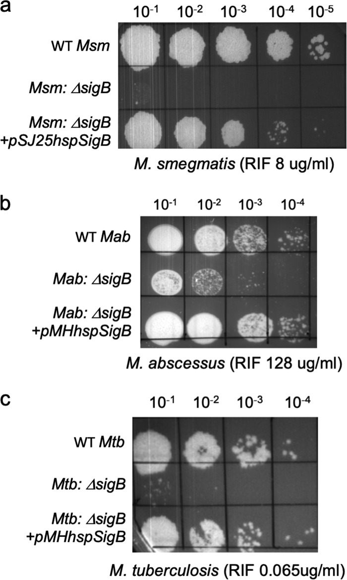
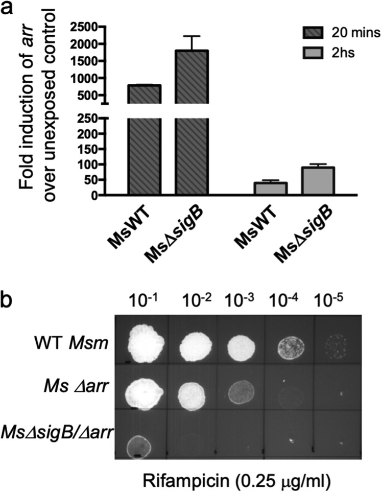
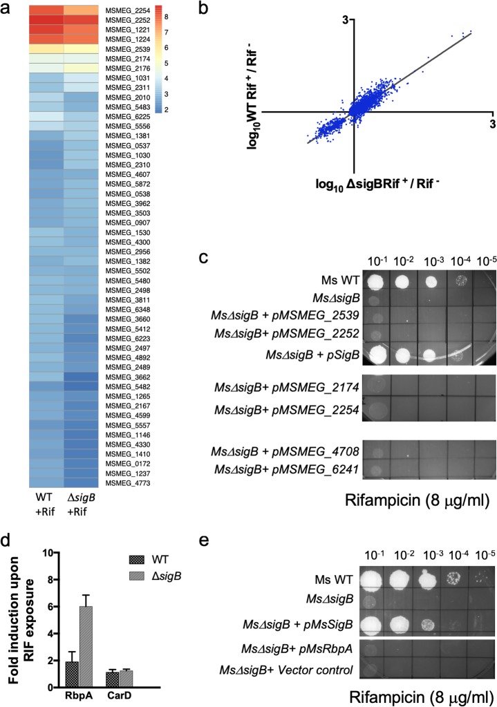
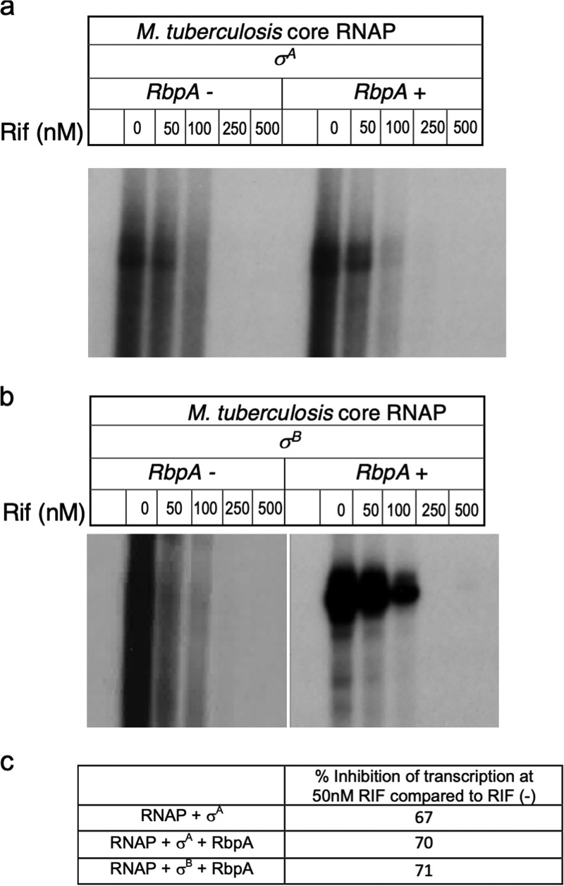
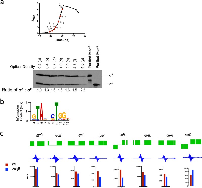
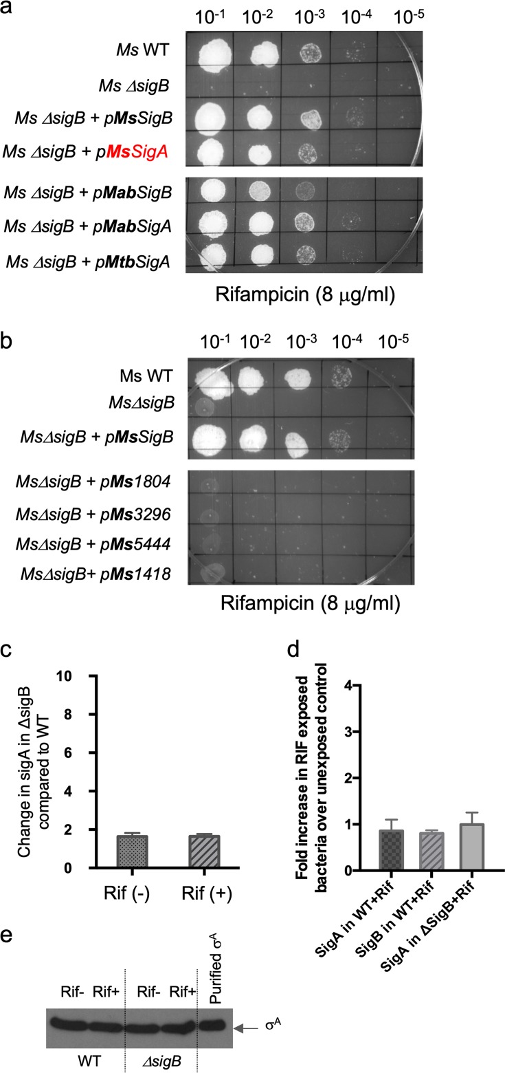
Mycobacterial σB belongs to the group II family of sigma factors, which are widely considered to transcribe genes required for stationary-phase survival and the response to stress. Here we explored the mechanism underlying the observed hypersensitivity of ΔsigB deletion mutants of Mycobacteriumsmegmatis, M. abscessus, and M. tuberculosis to rifampin (RIF) and uncovered an additional constitutive role of σB during exponential growth of mycobacteria that complements the function of the primary sigma factor, σA Using chromatin immunoprecipitation sequencing (ChIP-Seq), we show that during exponential phase, σB binds to over 200 promoter regions, including those driving expression of essential housekeeping genes, like the rRNA gene. ChIP-Seq of ectopically expressed σA-FLAG demonstrated that at least 61 promoter sites are recognized by both σA and σB These results together suggest that RNA polymerase holoenzymes containing either σA or σB transcribe housekeeping genes in exponentially growing mycobacteria. The RIF sensitivity of the ΔsigB mutant possibly reflects a decrease in the effective housekeeping holoenzyme pool, which results in susceptibility of the mutant to lower doses of RIF. Consistent with this model, overexpression of σA restores the RIF tolerance of the ΔsigB mutant to that of the wild type, concomitantly ruling out a specialized role of σB in RIF tolerance. Although the properties of mycobacterial σB parallel those of Escherichiacoli σ38 in its ability to transcribe a subset of housekeeping genes, σB presents a clear departure from the E. coli paradigm, wherein the cellular levels of σ38 are tightly controlled during exponential growth, such that the transcription of housekeeping genes is initiated exclusively by a holoenzyme containing σ70 (E.σ70).IMPORTANCE All mycobacteria encode a group II sigma factor, σB, closely related to the group I principal housekeeping sigma factor, σA Group II sigma factors are widely believed to play specialized roles in the general stress response and stationary-phase transition in the bacteria that encode them. Contrary to this widely accepted view, we show an additional housekeeping function of σB that complements the function of σA in logarithmically growing cells. These findings implicate a novel and dynamic partnership between σA and σB in maintaining the expression of housekeeping genes in mycobacteria and can perhaps be extended to other bacterial species that possess multiple group II sigma factors.

Keywords: ChIP-Seq; Mycobacterium; rifampin; sigB; sigma factor.


Figures

FIG 1
FIG 1
Deletion of σB confers RIF sensitivity in M. smegmatis (Msm), M. abscessus (Mab), and M. tuberculosis (Mtb). (a to c) Tenfold serial dilutions of M. smegmatis mc2155, M. abscessus ATCC 19977, M. tuberculosis mc27000, and their respective ΔsigB and complemented strains were grown to an A600 of 0.7 and spotted on Middlebrook 7H10 ADC or OADC containing the indicated concentrations of RIF. Deletion of sigB results in RIF sensitivity in all three strains. The mutant phenotype can be complemented by constitutive expression of the respective sigB gene.
FIG 2
FIG 2
σB-mediated resistance to RIF is independent of ADP-ribosyltransferase (Arr) and putative effector genes. (a) Wild-type M. smegmatis (MsWT) and the MsΔsigB strain were grown to an A600 of 0.7 and exposed to 4 μg/ml RIF for either 20 min or 2 h, and the amount of M. smegmatis arr transcripts was determined by qPCR and plotted as the fold induction over the level of expression for an unexposed control. Data represent the mean ± SD (n = 3). sigA was used as an endogenous control. (b) Tenfold serial dilutions of WT strain M. smegmatis mc2155 and the MsΔarr and MsΔsigB MsΔarr strains were grown to an A600 of 0.7 and spotted on Middlebrook 7H10 ADC containing the indicated concentration of RIF.
FIG 3
FIG 3
Transcriptomic changes accompanying RIF exposure in the wild-type and ΔsigB mutant M. smegmatis strains. (a) Wild-type M. smegmatis and the ΔsigB mutant were exposed to 4 μg/ml of RIF for 20 min and analyzed using RNA-seq. Unexposed samples of both strains were used as controls. Two biological replicates of each sample were used. Genes induced >4-fold with a q value of <0.001 were analyzed further, and the 50 most induced genes are represented as a heat map. (Left) WT; (right) ΔsigB mutant. (b) RIF-induced changes in gene expression in the WT versus ΔsigB mutant are shown using genes with a q value of <0.1. (c) Complemented strains were created by integrating MSMEG_2539, MSMEG_2252, MSMEG_2254, and MSMEG_2174 (genes highly upregulated in the presence of RIF) and MSMEG_4708 and MSMEG_6241(two SigB-dependent genes identified by RNA-seq) at the Bxb1 attB site of mc2155 ΔsigB. Tenfold serial dilutions of M. smegmatis mc2155, the MsΔsigB mutant, and the complemented strains were grown to an A600 of 0.7 and spotted on Middlebrook 7H10 ADC plates containing the indicated concentration of RIF. Overexpression of the genes listed above did not restore the RIF-sensitive phenotype of mc2155 ΔsigB. (d) Wild-type M. smegmatis and the MsΔsigB strain were grown to an A600 of 0.7 and exposed to 4 μg/ml RIF for 30 min, and the amounts of the rbpA and carD transcripts were determined by qPCR and plotted as the fold induction over the level of expression for an unexposed control. Data represent the mean ± SD (n = 3). sigA was used as an endogenous control. (e) A strain complemented with rbpA was created by integrating MSMEG_3858 at the Bxb1 attB site of mc2155 ΔsigB. Tenfold serial dilutions of M. smegmatis mc2155, the MsΔsigB mutant, and the rbpA complemented strain were grown to an A600 of 0.7 and spotted on Middlebrook 7H10 ADC plates containing the indicated concentration of RIF.
FIG 4
FIG 4
σA and σB containing holoenzymes are equally RIF susceptible. (a and b) Multiple-round in vitro transcription assays were performed on the sigA promoter using 200 nM σA-RNAP/σB-RNAP. RbpA (600 nM) was added where indicated. RIF was added to the indicated concentrations for 30 min at 37°C. Transcription was initiated by addition of 2 μl of an NTP mix (1.5 mM ATP, GTP, and CTP and 0.5 mM UTP) plus 2 μCi of [α-32P]UTP. The reaction mixtures were incubated at 37°C for 30 min, and the reactions were terminated by the addition of 5 mM EDTA and 100 μg/ml tRNA. Samples were ethanol precipitated and separated using denaturing PAGE (6% urea polyacrylamide gel). (c) The products were visualized using a Typhoon imager (GE Healthcare) and quantitated using ImageQuant software. Inhibition of RNAP activity at 50 nM RIF is expressed as a ratio of the activity in the presence and absence of RIF.
FIG 5
FIG 5
σB is transcriptionally active in exponentially growing M. smegmatis. (a) (Top) Growth kinetics of wild-type M. smegmatis indicating the growth phase and samples used for Western blotting. (Bottom) Relative levels of the σA and σB proteins at the indicated optical densities determined by Western blotting using an anti-σ70 monoclonal antibody. Samples were normalized by wet weight and protein concentration to ensure equivalent loading at each OD. Purified σA and σB proteins were used as controls. The ratio of σAB was quantitated using ImageJ software and is shown below. Equivalent amounts of protein were loaded in each lane of the Coomassie-stained gel (see Fig. S4b in the supplemental material). (b) Sequence logo of enriched motif in σB-FLAG-bound sites identified using the MEME Suite of tools (MEME E value = 7.0e−003). (c) The ChIP-Seq peaks of σB bound to promoters of key housekeeping genes visualized with SignalMap software are shown. The transcript levels of the corresponding genes (RPKM values) in the wild type (red) and the ΔsigB strain (blue) are plotted.
FIG 6
FIG 6
Overexpression of σA restores the RIF sensitivity of the MsΔsigB mutant. (a to c) Complemented strains were created by integrating Ms_SigA, Mtb_SigA, Mab_SigA, Ms_SigB, Mtb_SigB, Mab_SigB, Ms_Ms1804, Ms_Ms3296, Ms_Ms5444, and MSMEG_1418 at the Bxb1 attB site of mc2155 ΔsigB. Tenfold serial dilutions of M. smegmatis mc2155, the mc2155 ΔsigB mutant, and the complemented strains were grown to an A600 of 0.7 and spotted on Middlebrook 7H10 ADC plates containing the indicated concentrations of RIF. The RIF sensitivity of mc2155 ΔsigB could be complemented by the constitutive expression of sigA and sigB from all mycobacterial strains but not by ECF sigma factors. (b) Wild-type M. smegmatis and the MsΔsigB strain were grown to an A600 of 0.7 and exposed to 4 μg/ml RIF for 30 min, and the amount of the M. smegmatis sigA transcript was determined by qPCR and plotted as the fold induction of sigA levels in the MsΔsigB strain over the level of expression in the wild-type strain. The data represent the mean ± SD (n = 3). MSMEG_4936 was used as an endogenous control, as its levels were unchanged under various conditions in RNA-seq experiments. (d) Wild-type M. smegmatis and the MsΔsigB strain were grown to an A600 of 0.7 and exposed to 4 μg/ml RIF for 30 min, and the amounts of the M. smegmatis sigA and sigB transcripts were determined by qPCR and plotted as the fold induction upon RIF exposure over the level of expression for an unexposed control. The data represent the mean ± SD (n = 3). MSMEG_4936 was used as an endogenous control. (e) Wild-type M. smegmatis and the MsΔsigB strain were grown to an A600 of 0.7 and exposed to 4 μg/ml RIF for 30 min. The levels of σA protein were determined by Western blotting using an anti-σ70 monoclonal antibody. Samples were normalized by wet weight and protein concentration to ensure equivalent loading of each sample. Purified σA was used as a control.

Similar articles

Cited by

References

    1. Gruber TM, Gross CA. 2003. Multiple sigma subunits and the partitioning of bacterial transcription space. Annu Rev Microbiol 57:441–466. doi:10.1146/annurev.micro.57.030502.090913. - DOI - PubMed
    1. Saecker RM, Record MT Jr, Dehaseth PL. 2011. Mechanism of bacterial transcription initiation: RNA polymerase-promoter binding, isomerization to initiation-competent open complexes, and initiation of RNA synthesis. J Mol Biol 412:754–771. doi:10.1016/j.jmb.2011.01.018. - DOI - PMC - PubMed
    1. Paget MS, Helmann JD. 2003. The sigma70 family of sigma factors. Genome Biol 4:203. doi:10.1186/gb-2003-4-1-203. - DOI - PMC - PubMed
    1. Murakami KS, Masuda S, Campbell EA, Muzzin O, Darst SA. 2002. Structural basis of transcription initiation: an RNA polymerase holoenzyme-DNA complex. Science 296:1285–1290. doi:10.1126/science.1069595. - DOI - PubMed
    1. Manganelli R, Provvedi R, Rodrigue S, Beaucher J, Gaudreau L, Smith I, Proveddi R. 2004. Sigma factors and global gene regulation in Mycobacterium tuberculosis. J Bacteriol 186:895–902. doi:10.1128/JB.186.4.895-902.2004. - DOI - PMC - PubMed

Publication types

MeSH terms

LinkOut - more resources