. 2019 Jun;18(6):627-637.
doi: 10.1038/s41563-019-0368-6. Epub 2019 May 21.

Parenchymal and stromal tissue regeneration of tooth organ by pivotal signals reinstated in decellularized matrix

Affiliations

Parenchymal and stromal tissue regeneration of tooth organ by pivotal signals reinstated in decellularized matrix

Ling He et al. Nat Mater. 2019 Jun.

Abstract

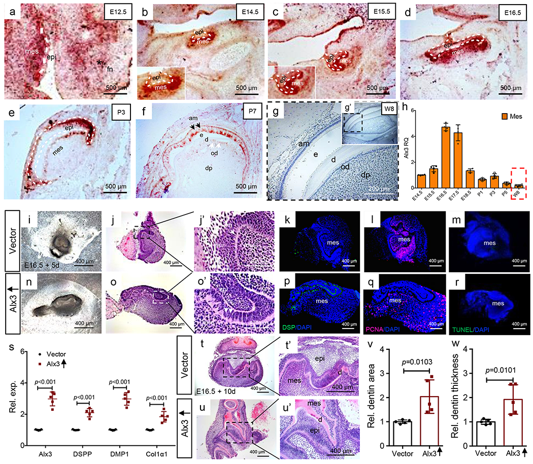
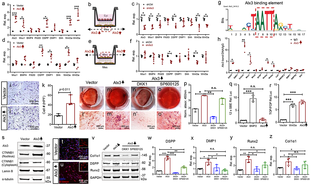
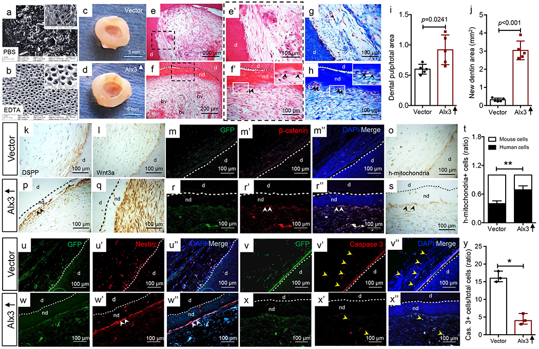
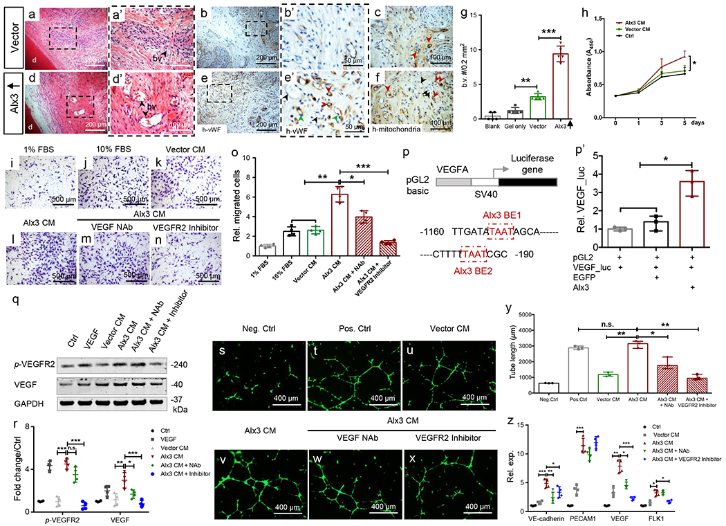
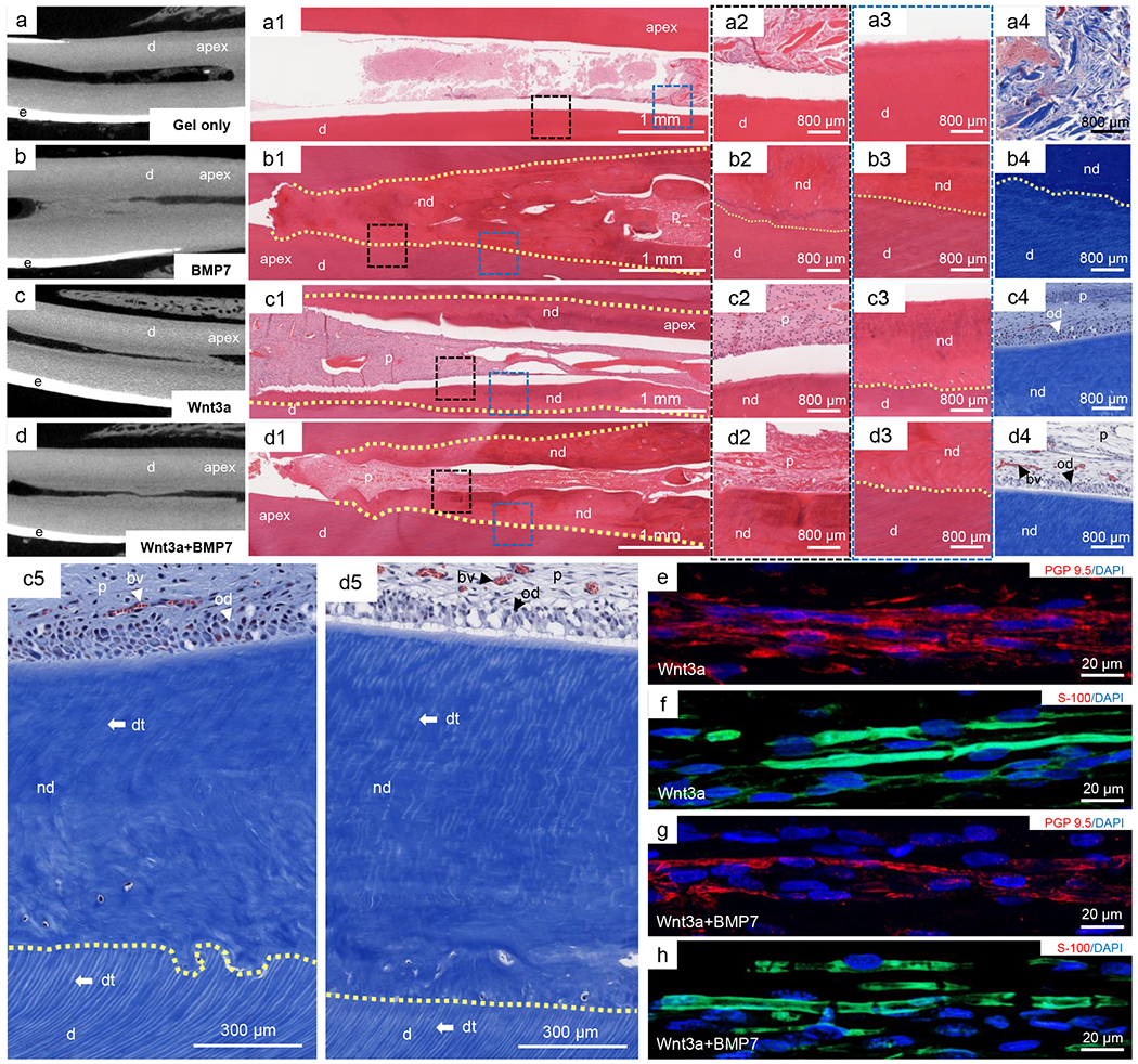
Cells are transplanted to regenerate an organs' parenchyma, but how transplanted parenchymal cells induce stromal regeneration is elusive. Despite the common use of a decellularized matrix, little is known as to the pivotal signals that must be restored for tissue or organ regeneration. We report that Alx3, a developmentally important gene, orchestrated adult parenchymal and stromal regeneration by directly transactivating Wnt3a and vascular endothelial growth factor. In contrast to the modest parenchyma formed by native adult progenitors, Alx3-restored cells in decellularized scaffolds not only produced vascularized stroma that involved vascular endothelial growth factor signalling, but also parenchymal dentin via the Wnt/β-catenin pathway. In an orthotopic large-animal model following parenchyma and stroma ablation, Wnt3a-recruited endogenous cells regenerated neurovascular stroma and differentiated into parenchymal odontoblast-like cells that extended the processes into newly formed dentin with a structure-mechanical equivalency to native dentin. Thus, the Alx3-Wnt3a axis enables postnatal progenitors with a modest innate regenerative capacity to regenerate adult tissues. Depleted signals in the decellularized matrix may be reinstated by a developmentally pivotal gene or corresponding protein.


Conflict of interest statement

Competing Financial Interests

The authors declare no competing financial interests in this article. J.J.M. has co-founded Innovative Elements and Xinkewo with goals to develop regenerative products.

Figures

Fig. 1
Fig. 1
Alx3 immunomapping and tissue reconstitution, a-f: Alx3 in situ hybridization from E12.5 to postnatal day 7 tooth organs. White dash lines: boundary of epithelium and mesenchyme. Scale bar, 500 μm. 3 independent biological samples. g,g’: Alx3 immunohistochemistry of molar tooth organ of an 8-wk-old CD1 mouse and higher magnification (insert). Scale bar, 200 μm. 2 independent biological samples, h: Real-time PCR of mesenchyme Alx3 expression (n=4 independent biological samples). Presented as mean±SD, bars represent standard deviation (SD). Red dash box: Mesenchyme Alx3’s expression of 8-wk-old adult molar (w8). i-o’: Photomicrograph and HE images of reconstituted E16.5 tooth organs following 5-day culture. A total of 5 independent biological samples. k,r: Dentin sialoprotein (DSP) immunofluorescence; Proliferating cell nuclear antigen (PCNA) immunofluorescence; TUNEL staining. Scale bar, 400 μm. 3 independent biological samples, s: Real-time PCR of Alx3, DSPP (dentin sialophosphoprotein), DMP1 (dentin matrix protein-1) and Col1a1 (collagen 1 alpha 1) in reconstituted tooth organs following 5-day culture (n=5 independent biological samples; presented as mean±SD and p value; p values calculated by two-sided Student’s t-tests). t-u’: Photomicrographs and HE images of E16.5 reconstituted tooth organs following 10-day culture. 5 independent biological samples. Scale bar, 400 μm. v,w: Quantified dentin area and thickness (n=5 independent biological samples; presented as mean±SD and p value; p values calculated by two-sided Student’s t-tests). mes: mesenchyme; epi: epithelium; fn: frontonasal process; e: enamel; d: dentin; od: odontoblasts; dp: dental pulp.
Fig. 2
Fig. 2
Alx3 signaling pathways, a-c: Epithelium and mesenchyme cells isolated from E16.5 tooth organs followed by mesenchymal Alx3 overexpression (a) or knockdown (c) and Transwell co-culture with isolated epithelium cells (b). d-f: E16.5 mesenchyme cells isolated, followed by mesenchymal Alx3 overexpression (d) or knockdown (f) and Transwell co-culture with un-manipulated mesenchyme cells (e) (a-f: n=5 independent biological samples; presented as mean±SD; p values calculated by multiple two-sided Student’s t-tests with Holm-Bonferroni correction; *: p<0.05; **: p<0.01; ***: p<0.001). g,h: ChIP assay of adult human dental-pulp mesenchyme stem/progenitor cells (MSCs) (n=4 independent biological samples; presented as median with range; p values calculated by Mann-Whitney tests with Bonferroni correction; *: p<0.05; **: p<0.01). Red dash box: Alx3 binding element from JASPAR CORE database, i-k: Transwell cell migration and quantification (n=3 independent biological samples; presented as median with range; p values calculated by Mann-Whitney tests with Bonferroni correction). Scale bar, 400 μm. l-p: Alizarin Red-S staining and quantification (n=3 independent biological samples; presented as median with range; p values calculated by Kruskal-Wallis tests; *: p<0.05; **: p<0.01; n.s.: not significant). Scale bar, 800 μm. q: BMP/Smad luciferase activity upon BMP2 treatment (100 ng/ml) or Alx3 overexpression (n=4 independent biological samples; presented as mean±SD; p values calculated by one-way ANOVA with Bonferroni; ***: p<0.001; n.s.: not significant), r: Wnt luciferase activity upon Wnt3a treatment (100 ng/ml) or Alx3 overexpression (n=4 independent biological samples; presented as mean±SD; p values calculated by one-way ANOVA with Bonferroni; ***: p<0.001). s: Western blot of nuclear and cytoplasmic β-catenin (CTNNB1) upon Alx3 overexpression. t,u: β-catenin transnucleation by immunofluorescence. Scale bar, 400 μm. v: Western blot of Col1α1, DSPP and Runx2; DKK1 (200 ng/ml); SP600125 (10 μW). w-z: Real-time PCR of DSPP, Runx2 and collagen1α1 (n=4 independent biological samples; presented as mean±SD; p values calculated by one-way ANOVA with Bonferroni; *: p<0.05; **: p<0.01; ***: p<0.001; n.s.: not significant).
Fig. 3
Fig. 3
Transplanted, Alx3-restored adult human dental-pulp mesenchyme stem/progenitor cells (MSCs) regenerated both parenchyma and stroma with enhanced Wnt signaling and improved cell survival. a,b: PBS and EDTA processed, decellularized scaffolds (inserts with higher magnification); n=3 independent biological samples. c,d: Decellularized scaffolds retrieved following 8-wk in vivo implantation: vector control sample (c); Alx3 restored MSC sample (d). Scale bar, 5 mm. HE images of both vector control (e,e’) and Alx3-restored MSC samples (f,f’) samples. Black arrowheads: odontoblast-like cells that aligned newly-formed dentin surface (f’ insert); e,f: Scale bar, 200 μm; e’,f’: Scale bar, 100 μm. g,h: Masson’s Trichrome staining. Black arrowheads: odontoblast-like cells that aligned newly-formed dentin surface (h insert). Scale bar, 100 μm. c-h: n=5 independent biological samples, i: Dental pulp-like tissue area over total root-canal area (n=5 independent biological samples; presented as mean±SD and p value; p value calculated by two-side Student t-tests). j: Newly-formed dentin area (n=5 independent biological samples; presented as mean±SD and p value; p value calculated by two-side Student t-tests). k,p: DSPP immunohistochemistry (black arrowheads: DSPP). Scale bar, 100 μm. l,q: Wnt3a immunohistochemistry (black arrowheads: Wnt3a). Scale bar, 100 μm. m-r”: β-catenin-positive cells (white arrowheads), k-r”: n=3 independent biological samples. o,s: Human mitochondria staining of transplanted human cells and quantification (t) (n=5 independent biological samples; presented as mean±SD; p value calculated by two-sided Student’s t-tests; **: p<0.01). u-w”: Nestin-positive cells (white arrowheads), v-x”: Caspase-3 positive cells and quantification, u-x”: n=3 independent biological samples, (y) apoptotic cells exemplified by yellow arrowheads (n=3 independent biological samples; presented as median with range; p value calculated by Mann-Whitney tests with Bonferroni correction; *: p<0.05). Scale bar, 100 μm. d: dentin; nd: newly-formed dentin; bv: blood vessels; black dash lines: boundary between native dentin and newly-formed dentin (p,q,s); blue dash lines: boundary between newly-formed dentin and dental-pulp (f,f’,h); white dash lines: boundary between native dentin and newly-formed dentin (m-r”,u-x”).
Fig. 4
Fig. 4
Alx3-restored adult mesenchyme stem/progenitor cells (MSCs) induced angiogenesis and VEGF signaling. a,a’,d,d’: Angiogenesis in regenerated tissues. a,d: scale bar, 200 μm. a’,d’: scale bar, 100 μm. d: native dentin. b,b’,e,e’: Human von Willebrand factor immunohistochemistry. Green arrowheads: endothelial cells of human origin (e’); black arrowheads: endothelial cells of host (mouse) origin (e’); red arrowheads: chimeric blood vessels (e’). b,e: scale bar, 200 μm. b’,e’: scale bar, 50 μm. c,f: Human mitochondria staining; red arrowheads: human cells; black arrowheads: host (mouse) cells. Scale bar, 100 μm. a-h: n=5 independent biological samples, g: Blood vessel (b.v.) quantification (n=5 independent biological samples; presented as mean±SD; p values calculated by one-way ANOVA with Bonferroni; **: p<0.01; ***: p<0.001). h: CCK8 of HUVECs treated with conditioned medium by vector control or Alx3-restored MSCs (n=4 independent biological samples; Presented as mean±SD; p values calculated by one-way ANOVA with Bonferroni; *: p<0.05). i-o: Transwell migration assay by Alx3 conditioned medium or with neutralizing antibody or VEGFR2 inhibitor (n=4 independent biological samples; presented as mean±SD; p values calculated by one-way ANOVA with Bonferroni; *: p<0.05; **; p<0.01; ***: p<0.001). Scale bar, 500 μm. p: Mouse VEGFA promoter luciferase reporter with two Alx3-binding elements in red boxes. BE: binding element, p’: VEGF luciferase assay upon Alx3 transfection (n=3 independent biological samples; presented as median with range; p values calculated by Kruskal-Wallis test; *: p<0.05). q,r: VEGF production and p-VEGFR2 activation by western blot and quantification (n=4 independent biological samples; presented as mean±SD; p values calculated by one-way ANOVA with Bonferroni; *: p<0.05; **; p<0.01; ***: p<0.001; n.s.: not significant), s-x: Tube formation of HUVECs treated by conditioned medium, Alx3 CM with or without NAb or VEGFR2 inhibitor. Scale bar, 400 μm. 3 independent biological samples, y: Tube length quantification (n=3 independent biological samples; presented as median with range; p values calculated by Kruskal-Wallis test; *: p<0.05; **; p<0.01; n.s.: not significant), z: Real-time PCR of VE-cadherin, PECAM1, VEGF and FLK1 (n=4 independent biological samples; presented as mean±SD; p values calculated by one-way ANOVA with Bonferroni; *: p<0.05; **: p<0.01; ***: p<0.001).
Fig. 5
Fig. 5
Parenchymal and stromal regeneration orthotopically in a preclinical, large animal model (total of 58 teeth in 10 minipigs), a-d: Micro-computed tomography (μ-CT) scans of porcine central incisors showing dentin (d), enamel (e) and the enclosed root canal, apex: root tip of teeth. a1-a4: HE and Masson’s Trichrome staining of collagen gel infusion alone, d: native dentin. b1-b4: HE and Masson’s Trichrome staining of BMP7 delivery in collagen gel. p: dental pulp; nd: newly-formed dentin. c1-c4: HE and Masson’s Trichrome staining of Wnt3a delivery in collagen gel. a1,b1,c1,d1: scale bar, 1 mm. a2-a4,b2-b4,c2-c4: scale bar, 800 μm. c5: High magnification of pulp-dentin boundary in Wnt3a alone group, p: dental pulp; dt: dentinal tubules; od: odontoblasts; nd: newly-formed dentin; d: native dentin. d5: High magnification of pulp-dentin boundary in Wnt3a and BMP7 combined delivery group, bv: blood vessel. Scale bar, 300 μm. e-h: Sprouts of nerve-like structures in regenerated dental pulp. e,f: PGP9.5 and S-100 immunolocalized fiber sprouts upon Wnt3a delivery. g,h: PGP9.5 and S-100 immunolocalized fiber sprouts upon combined Wnt3a and BMP7 delivery, e-h: scale bar, 20 μm.
Fig. 6
Fig. 6
Structural and mechanical properties of regenerated dentin and overall schematic of Alx3/Wnt/VEGF cascades. a: Scanning electron microscopy of dental pulp (p), newly-formed dentin (nd) and native dentin (d) in cross-section. Scale bar: 200 μm. b,c: Dentinal tubules and resin casts in the newly-formed dentin (blue dashed box in a) and native dentin (yellow dashed box in a) by SEM. Scale bar: 50 μm. d,e: Odontoblast processes casts in the native dentin (d,e) and newly-formed dentin (f,g) of a native porcine incisor by SEM. d: dentin; dt: dentinal tubules; OdP: odontoblast process. d,f: scale bar: 50 μm. e,g: scale bar: 10 μm. h: Newly-formed dentin volume; i: Dentin mineral density, j: Ratio of newly-formed dentin (N.D.) and native dentin; k: Harness (MPa); I: Young’s modules (GPa). (h-j: n=5 independent biological tooth samples; k,l: n=4 independent biological tooth samples; Presented as mean±SD; p values calculated by one-way ANOVA with Bonferroni; *p<0.05, n.s.: not significant). m: Schematic diagram showing Alx3 regulation of Wnt3a and VEGF as its direct targets, leading to parenchymal and stromal regeneration, with multiple cellular processes including improved cell survival, parenchymal progenitor migration and differentiation, endothelial cells migration, angiogenesis and neural sprouting.

Comment in

  • A pulpy story.
    Millar SE. Millar SE. Nat Mater. 2019 Jun;18(6):530-531. doi: 10.1038/s41563-019-0372-x. Nat Mater. 2019. PMID: 31114070 No abstract available.

References

    1. Sacco A, Doyonnas R, Kraft R, Vitorovic S & Blau HM Self-renewal and expansion of single transplanted muscle stem cells. Nature 456, 502–506, doi:10.1038/nature07384 (2008). - DOI - PMC - PubMed
    1. Shiba Y et al. Allogeneic transplantation of iPS cell-derived cardiomyocytes regenerates primate hearts. Nature 538, 388–391, doi:10.1038/nature19815 (2016). - DOI - PubMed
    1. Fox IJ et al. Stem cell therapy. Use of differentiated pluripotent stem cells as replacement therapy fortreating disease. Science 345, 1247391, doi:10.1126/science.1247391 (2014). - DOI - PMC - PubMed
    1. Simmons PJ, Przepiorka D, Thomas ED & Torok-Storb B Host origin of marrow stromal cells following allogeneic bone marrow transplantation. Nature 328, 429–432, doi:10.1038/328429a0 (1987). - DOI - PubMed
    1. Mao JJ & Prockop DJ Stem cells in the face: tooth regeneration and beyond. Cell Stem Cell 11,291–301, doi:10.1016/j.stem.2012.08.010 (2012). - DOI - PMC - PubMed

Publication types

MeSH terms