. 2019 Apr 10:12:2729-2735.
doi: 10.2147/OTT.S190409. eCollection 2019.

Successful treatment of brain radiation necrosis resulting from triple-negative breast cancer with Endostar and short-term hyperbaric oxygen therapy: a case report

Affiliations

Successful treatment of brain radiation necrosis resulting from triple-negative breast cancer with Endostar and short-term hyperbaric oxygen therapy: a case report

Shiyun Xing et al. Onco Targets Ther. .

Abstract

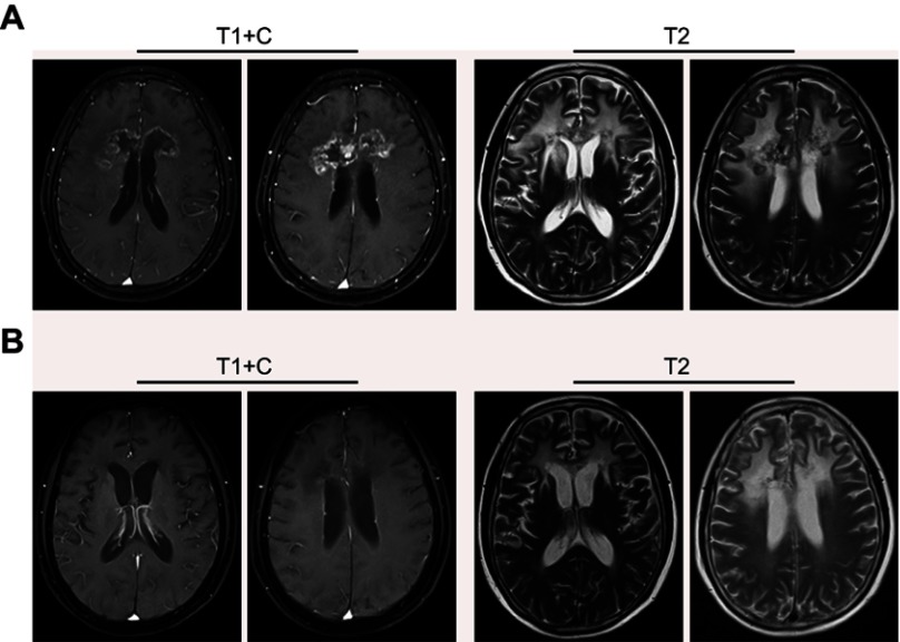
Radiation necrosis (RN) is one of the complications of radiotherapy. Angiogenesis is a key factor underlying the development of RN, and Endostar, a safe and well-tolerated recombinant human endostatin, has been used to treat a variety of tumors. Thus far, however, no definitive reports on the use of Endostar for RN treatment have been reported. Here, we report the successful treatment of one patient with symptomatic brain radiation necrosis (BRN) using Endostar in combination with short-term hyperbaric oxygen therapy (HBO). One triple-negative breast cancer patient with recurrent brain metastatic lesions after standard chemoradiotherapy was referred to a specialty center outside our hospital for stereotaxic radiotherapy. Two months later, the patient showed deteriorating clinical symptoms, and magnetic resonance imaging (MRI) showed radiation necrosis with significant surrounding edema. The patient had a poor response to mannitol and steroids. After diagnosing this patient with BRN, we began short-term HBO therapy and intravenously administered Endostar for 4 cycles. The patient responded well to this strategy, showing rapidly and dramatically improved MRI findings and clinical symptoms. No tumor progression was observed at 10 months after treatment. Endostar in combination with short-term HBO therapy had marked effects on symptomatic BRN. However, additional large-scale, double-blinded, controlled trials are necessary to confirm the clinical effect of Endostar in combination with a short-term HBO therapy regimen on BRN.

Keywords: Endostar; brain radiation necrosis; hyperbaric oxygen therapy; triple-negative breast cancer.


Conflict of interest statement

The authors report no conflicts of interest in this work.

Figures

Figure 1
Figure 1
MRI detection of brain metastasis and an MRI scan after curative radiotherapy. (A) Contrast-enhancement T1-weighted images revealed an enhanced solitary mass 19×22×25 mm in size with a surrounding area of low intensity in the right basal ganglia, associated with left frontal lobe metastases. (B) One month after curative radiotherapy, the MRI scan showed that the brain metastatic tumoral volume was significantly reduced and had disappeared. Abbreviations: MRI, magnetic resonance imaging; T1+C, contrast-enhancement T1-weighted images; T2, T2-weighted images.
Figure 2
Figure 2
MRI detection of brain metastasis recurrence and MRI scanning after administration of the temozolomide regimen. (A) An MRI scan revealed an irregularly strengthened signal in the anterior corpus callosum and peripheral edema of the lesions. (B) One year after administration of the temozolomide regimen for four cycles, an MRI scan revealed that the lesions in the anterior corpus callosum had decreased, but the size of the lesions in the right lateral ventricle had significantly increased. Abbreviations: MRI, magnetic resonance imaging; T1+C, contrast-enhancement T1-weighted images; T2, T2-weighted images.
Figure 3
Figure 3
MRI scan after stereotactic radiotherapy. (A and B) After treating with mannitol and steroids to reduce intracranial pressure and to limit further damage caused by cerebral edema, an MRI scan revealed an enhanced solitary mass (53×28×20 mm) with a surrounding area of low intensity in the bilateral frontal lobe and genu of the corpus callosum. Abbreviations: MRI, magnetic resonance imaging; T1, T1-weighted images; T2, T2-weighted images.
Figure 4
Figure 4
MRI scan after treating with a vascular endostatin and short-term HBO therapy. (A and B) After treating with a vascular endostatin and short-term HBO therapy, an MRI scan revealed that the clumps of matter in the bilateral frontal lobes and the genu of the corpus callosum were smaller than those on the MRI scan from 2017–07 and especially smaller than those shown on the MRI scan from 2018–06; however, the edema in the surrounding tissue was still present. Abbreviations: MRI, magnetic resonance imaging; HBO therapy, hyperbaric oxygen therapy; T1+C, contrast-enhancement T1-weighted images; T2, T2-weighted images.

References

    1. Buonomo OC, Caredda E, Portarena I, et al. New insights into the metastatic behavior after breast cancer surgery, according to well-established clinicopathological variables and molecular subtypes. PLoS One. 2017;12(9):e0184680. doi:10.1371/journal.pone.0184680 - DOI - PMC - PubMed
    1. Fulford LG, Easton DF, Reis-Filho JS, et al. Specific morphological features predictive for the basal phenotype in grade 3 invasive ductal carcinoma of breast. Histopathology. 2006;49(1):22–34. doi:10.1111/j.1365-2559.2006.02453.x - DOI - PubMed
    1. Rostami R, Mittal S, Rostami P, Tavassoli F, Jabbari B. Brain metastasis in breast cancer: a comprehensive literature review. J Neurooncol. 2016;127(3):407–414. doi:10.1007/s11060-016-2075-3 - DOI - PubMed
    1. Soliman H, Das S, Larson DA, Sahgal A. Stereotactic radiosurgery (SRS) in the modern management of patients with brain metastases. Oncotarget. 2016;7(11):12318–12330. doi:10.18632/oncotarget.7131 - DOI - PMC - PubMed
    1. Kondziolka D, Shin SM, Brunswick A, Kim I, Silverman JS. The biology of radiosurgery and its clinical applications for brain tumors. Neuro Oncol. 2015;17(1):29–44. doi:10.1093/neuonc/nou284 - DOI - PMC - PubMed