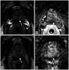
Diagnostic Accuracy of a MR Protocol Acquired with and without Endorectal Coil for Detection of Prostate Cancer: A Multicenter Study
- PMID: 31114466
- PMCID: PMC6504800
- DOI: 10.1159/000489425
Diagnostic Accuracy of a MR Protocol Acquired with and without Endorectal Coil for Detection of Prostate Cancer: A Multicenter Study
Abstract
Introduction: The purpose of this study was to compare diagnostic accuracy of a prostate multiparametric magnetic resonance imaging (mpMRI) protocol for detection of prostate cancer between images acquired with and without en-dorectal coil (ERC).
Materials: This study was approved by the regional ethics committee. Between 2014 and 2015, 33 patients (median age 51.3 years; range 42.1-77.3 years) who underwent prostate-MRI at 3T scanners at 2 different institutions, acquired with (mpMRIERC) and without (mpMRIPPA) ERC and who received radical prostatectomy, were included in this retrospective study. Two expert readers (R1, R2) attributed a PI-RADS version 2 score for the most suspect (i. e. index) lesion for mpMRIPPA and mpMRIERC. Sensitivity and positive predictive value for detection of index lesions were assessed using 2 × 2 contingency tables. Differences between groups were tested using the McNemar test. Whole-mount histopathology served as reference standard.
Results: On a quadrant-basis cumulative sensitivity ranged between 0.61-0.67 and 0.76-0.88 for mpMRIPPA and mpMRIERC protocols, respectively (p > 0.05). Cumulative positive predictive value ranged between 0.80-0.81 and 0.89-0.91 for mpMRIPPA and mpMRIERC protocols, respectively. The differences were not statistically significant for R1 (p = 0.267) or R2 (p = 0.508).
Conclusion: Our results suggest that there may be no significant differences for detection of prostate cancer between images acquired with and without an ERC.
Keywords: Diagnostic accuracy; Endorectal coil; Index lesion; Prostate MRI; Prostate cancer.
Figures


References
-
- Scheenen TW, Rosenkrantz AB, Haider MA, Futterer JJ. Multiparametric magnetic resonance imaging in prostate cancer management: current status and future perspectives. Invest Radiol. 2015;50:594–600. - PubMed
-
- Rosenkrantz AB, Shanbhogue AK, Wang A, Kong MX, Babb JS, Taneja SS. Length of capsular contact for diagnosing extraprostatic extension on prostate MRI: assessment at an optimal threshold. J Magn Reson Imaging. 2016;43:990–997. - PubMed
-
- Donati OF, Afaq A, Vargas HA, Mazaheri Y, Zheng J, Moskowitz CS, Hricak H, Akin O. Prostate MRI: evaluating tumor volume and apparent diffusion coefficient as surrogate biomarkers for predicting tumor Gleason score. Clin Cancer Res. 2014;20:3705–3711. - PubMed
-
- Donati OF, Mazaheri Y, Afaq A, Vargas HA, Zheng J, Moskowitz CS, Hricak H, Akin O. Prostate cancer aggressiveness: assessment with whole-lesion histogram analysis of the apparent diffusion coefficient. Radiology. 2014;271:143–152. - PubMed
LinkOut - more resources
Full Text Sources
Research Materials
Miscellaneous
