. 2019 Apr 30:10:951.
doi: 10.3389/fimmu.2019.00951. eCollection 2019.

TRAIL-R1 and TRAIL-R2 Mediate TRAIL-Dependent Apoptosis in Activated Primary Human B Lymphocytes

Affiliations

TRAIL-R1 and TRAIL-R2 Mediate TRAIL-Dependent Apoptosis in Activated Primary Human B Lymphocytes

Julian Staniek et al. Front Immunol. .

Abstract

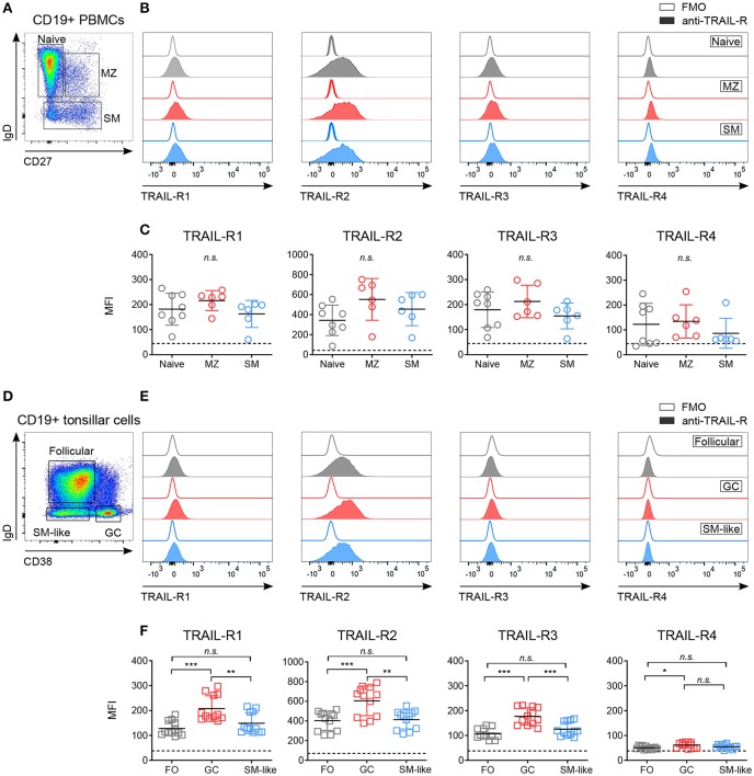
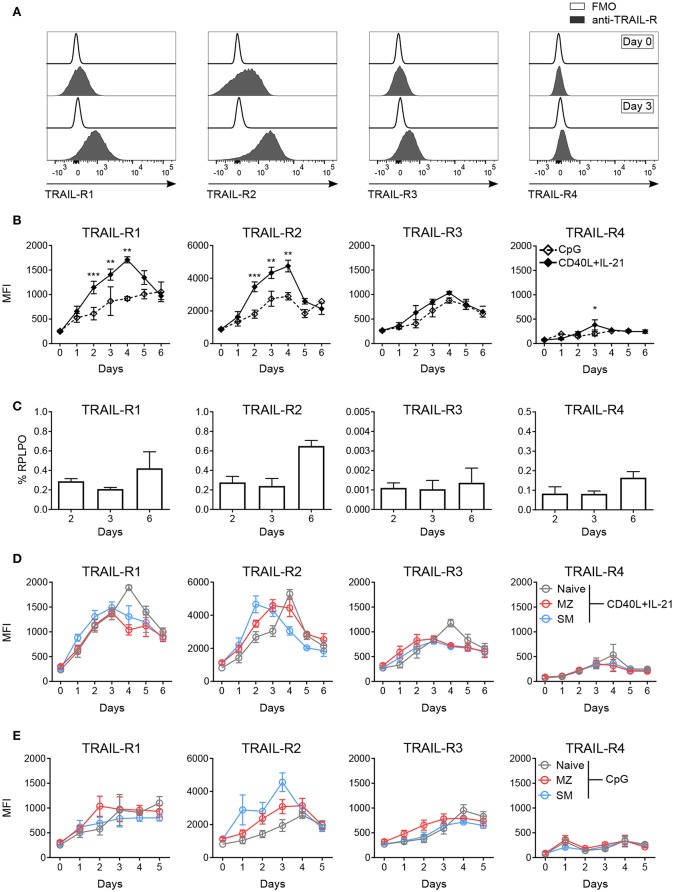
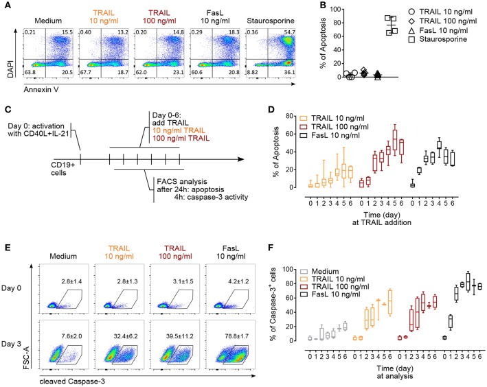
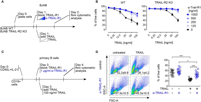
The maintenance of B cell homeostasis requires a tight control of B cell generation, survival, activation, and maturation. In lymphocytes upon activation, increased sensitivity to apoptotic signals helps controlling differentiation and proliferation. The death receptor Fas is important in this context because genetic Fas mutations in humans lead to an autoimmune lymphoproliferative syndrome that is similar to lymphoproliferation observed in Fas-deficient mice. In contrast, the physiological role of TNF-related apoptosis-inducing ligand receptors (TRAIL-Rs) in humans has been poorly studied so far. Indeed, most studies have focused on tumor cell lines and on mouse models whose results are difficult to transpose to primary human B cells. In the present work, the expression of apoptosis-inducing TRAIL-R1 and TRAIL-R2 and of the decoy receptors TRAIL-R3 and TRAIL-R4 was systematically studied in all developmental stages of peripheral B cells isolated from the blood and secondary lymphoid organs. Expression of TRAIL-Rs is modulated along development, with highest levels observed in germinal center B cells. In addition, T-dependent and T-independent signals elicited induction of TRAIL-Rs with distinct kinetics, which differed among B cell subpopulations: switched memory cells rapidly upregulated TRAIL-R1 and -2 upon activation while naïve B cells only reached similar expression levels at later time points in culture. Increased expression of TRAIL-R1 and -2 coincided with a caspase-3-dependent sensitivity to TRAIL-induced apoptosis in activated B cells but not in freshly isolated resting B cells. Finally, both TRAIL-R1 and TRAIL-R2 could signal actively and both contributed to TRAIL-induced apoptosis. In conclusion, this study provides a systematic analysis of the expression of TRAIL-Rs in human primary B cells and of their capacity to signal and induce apoptosis. This dataset forms a basis to further study and understand the dysregulation of TRAIL-Rs and TRAIL expression observed in autoimmune diseases. Additionally, it will be important to foresee potential bystander immunomodulation when TRAIL-R agonists are used in cancer treatment.

Keywords: B lymphocytes; TRAIL; TRAIL-R; apoptosis; human.


Figures

Figure 1
Figure 1
Differential expression of TRAIL-Rs in human B cell subpopulations. Expression of TRAIL-R1, TRAIL-R2, TRAIL-R3 and TRAIL-R4 was characterized by flow cytometry in ex vivo CD19+ human B cells isolated from PBMCs (A–C) and tonsillar CD19+ cells (D–F). (A) Gating strategy to identify naïve (IgD+ CD27), marginal zone (MZ; IgD+ CD27+) and switched memory (SM; IgD CD27) cells from CD19+ live B cells. (B) Histograms show expression levels of indicated TRAIL-Rs in naïve, MZ and SM B cells compared to respective fluorescent minus one (FMO) controls. (C) Mean fluorescence intensity (MFI) of TRAIL-Rs among indicated B cell subsets. Each dot represents a biological replicate. (D) Gating strategy to identify follicular (FO; IgD+ CD38), germinal center (GC; IgD CD38+) and switched memory-like (SM-like; IgD CD38) cells among CD19+ live B cells. (E) TRAIL-R expression in FO, GC and SM-like B cells compared to respective FMO controls. (F) Quantifications of TRAIL-R expression among tonsillar B cell subpopulations depicted as MFI. Dots represent duplicates of six biological replicates. (C,F) Dashed lines indicate averaged FMO controls. Mean values ± SD are shown. n.s., not significant; *adj.p < 0.05; **adj.p < 0.01; ***adj.p < 0.001. Statistical analysis was performed using one-way ANOVA, followed by Tukey's multiple comparison test where appropriate.
Figure 2
Figure 2
In vitro activation induces TRAIL-R expression in human B cells. Expression of TRAIL-Rs was analyzed by flow cytometry and RT-qPCR in in vitro activated human B cells isolated from PBMCs. (A) Representative histograms depict expression of TRAIL-Rs in CD19+ live B cells at day 0 and at day 3 after stimulation with CD40L+IL-21 compared to respective fluorescent minus one (FMO) controls. (B) Quantification of TRAIL-R expression in CD19+ live B cells upon stimulation with CpG or CD40L+IL-21 up to day 6 of culture depicted as MFI (n ≥ 3). (C) RT-qPCR analysis of all TRAIL-R transcripts in isolated CD19+ B cells upon stimulation with CD40L+IL-21 at day 2, 3 and 6 (n = 3). (D) TRAIL-R expression in naïve, MZ and SM B cells upon T cell-dependent (CD40L+IL-21) activation up to day 6 of culture shown as MFI (n ≥ 3). (E) TRAIL-R expression in naïve, MZ and SM B cells upon T cell-independent (CpG) stimulation up to day 5 of culture depicted as MFI (n ≥ 3). Mean values ± SEM (B,D,E) or ± SD (C) are shown. n, biological replicates. (B) *p < 0.05; **p < 0.01; ***p < 0.001. Statistical analysis was done with two-tailed unpaired Student's t-test.
Figure 3
Figure 3
TRAIL induces caspase-3 activation and promotes apoptosis in activated human B cells. (A) Apoptosis of resting CD19+ B cells in response to TRAIL, FasL or staurosporine (1 μM) for 24 h was determined by Annexin V and DAPI staining and flow cytometric analysis. (B) Percentage of apoptotic resting CD19+ B cells after incubation for 24 h with TRAIL, FasL or staurosporine was quantified by flow cytometry. The percentage of apoptotic B cells is shown, see Material and Methods for formula. Each dot represents a biological replicate. (C) Experimental setup to analyze TRAIL-induced apoptosis and caspase-3 induction in T cell-dependent activated B cells by flow cytometry. (D) Apoptosis was quantified after 24 h of TRAIL or FasL stimulation of activated B cells until day 6 of culture, as described in (C). The percentage of apoptotic B cells is shown (n ≥ 6). (E) Representative flow cytometric analysis of FSC-A and cleaved caspase-3 in resting and 3-days in vitro activated CD19+ B cells after 4 h of TRAIL or FasL treatment. Numbers reflect mean percentage of cleaved caspase-3+ cells ± SEM (F) percentages of caspase-3+ B cells after 4 h of TRAIL or FasL stimulation in activated B cells until day 6 of culture (n ≥ 3). (B,E) Mean values ± SD or SEM are shown, respectively. (D,F) Data are expressed as 10–90 percentile.
Figure 4
Figure 4
Both TRAIL-R1 and TRAIL-R2 mediate apoptosis in primary human B cells and tumor B cell lines. (A) Experimental approach, describing the addition of reagents to the culture to study TRAIL-induced apoptosis in BJAB cells by flow cytometry. (B) Percentages of live BJAB WT (left) and TRAIL-R2 KO (right) cells after 24h of scalar concentrations of TRAIL ± scalar concentrations of TRAIL-R1 blocking antibody (n = 3). (C) Experimental setup to analyze contribution of TRAIL-R1 and TRAIL-R2 to TRAIL-induced apoptosis in activated B cells by flow cytometry. (D) Representative identification of live primary B cells by FSC-A and SSC-A (left) and quantification (right) of live B cells after 24 h of TRAIL stimulation ± TRAIL-R1 blocking antibody (1μg/ml) at day 4 of culture by flow cytometry. Dots represent triplicates of four biological replicates. (B, D) Mean values ± SD are shown. (B) n, independent experiments. (D) *adj.p < 0.05; ***adj.p < 0.001. Statistical analysis was performed using two-way ANOVA, followed by Tukey's multiple comparison test where appropriate.

References

    1. Mesin L, Ersching J, Victora GD. Germinal center B cell dynamics. Immunity. (2016) 45:471–82. 10.1016/j.immuni.2016.09.001 - DOI - PMC - PubMed
    1. Shlomchik MJ, Marshak-Rothstein A, Wolfowicz CB, Rothstein TL, Weigert MG. The role of clonal selection and somatic mutation in autoimmunity. Nature. (1987) 328:805–11. 10.1038/328805a0 - DOI - PubMed
    1. Shokat KM, Goodnow CC. Antigen-induced B-cell death and elimination during germinal-centre immune responses. Nature. (1995) 375:334–8. 10.1038/375334a0 - DOI - PubMed
    1. Hao Z, Duncan GS, Seagal J, Su YW, Hong C, Haight J, et al. . Fas receptor expression in germinal-center B cells is essential for T and B lymphocyte homeostasis. Immunity. (2008) 29:615–27. 10.1016/j.immuni.2008.07.016 - DOI - PMC - PubMed
    1. Fisher GH, Rosenberg FJ, Straus SE, Dale JK, Middleton LA, Lin AY, et al. . Dominant interfering Fas gene mutations impair apoptosis in a human autoimmune lymphoproliferative syndrome. Cell. (1995) 81:935–46. 10.1016/0092-8674(95)90013-6 - DOI - PubMed

Publication types

MeSH terms

Substances