Review
. 2019 Apr 30:9:127.
doi: 10.3389/fcimb.2019.00127. eCollection 2019.

Herpes Simplex Virus Evasion of Early Host Antiviral Responses

Affiliations
Review

Herpes Simplex Virus Evasion of Early Host Antiviral Responses

Eduardo I Tognarelli et al. Front Cell Infect Microbiol. .

Abstract

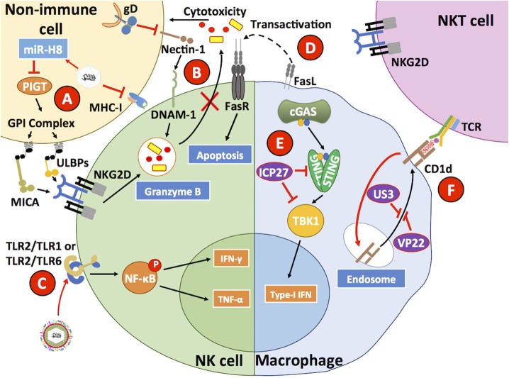
Herpes simplex viruses type 1 (HSV-1) and type 2 (HSV-2) have co-evolved with humans for thousands of years and are present at a high prevalence in the population worldwide. HSV infections are responsible for several illnesses including skin and mucosal lesions, blindness and even life-threatening encephalitis in both, immunocompetent and immunocompromised individuals of all ages. Therefore, diseases caused by HSVs represent significant public health burdens. Similar to other herpesviruses, HSV-1 and HSV-2 produce lifelong infections in the host by establishing latency in neurons and sporadically reactivating from these cells, eliciting recurrences that are accompanied by viral shedding in both, symptomatic and asymptomatic individuals. The ability of HSVs to persist and recur in otherwise healthy individuals is likely given by the numerous virulence factors that these viruses have evolved to evade host antiviral responses. Here, we review and discuss molecular mechanisms used by HSVs to evade early innate antiviral responses, which are the first lines of defense against these viruses. A comprehensive understanding of how HSVs evade host early antiviral responses could contribute to the development of novel therapies and vaccines to counteract these viruses.

Keywords: apoptosis; cytosolic nucleic acid receptors; dendritic cells (DCs); inflammasome; innate immunity; interferon (IFN); natural killer cells (NK cells); toll-like receptors (TLRs).


Figures

Figure 1
Figure 1
HSV virion structure. HSVs possess linear, double-stranded DNA genomes (152–154 kbp) encoding more than 70 ORFs. The viral genomes are contained within icosahedral capsids of ~125 nm, which in turn are surrounded by complex meshes of viral proteins known as the tegument. The tegument is enveloped by lipid membranes, which harbor numerous transmembrane glycoproteins. A table with tegument proteins involved in immune evasion is shown on the right, ordered from highest to lowest molecular weight (MW).
Figure 2
Figure 2
HSVs modulate antiviral mechanisms related to cell death in non-immune cells. HSVs utilize numerous mechanisms to hamper the capacity of host cells to restrict viral infection. (A) The engagement of the TNFR receptor leads to the activation of caspase-8 eliciting apoptosis, or eventually RIP1/3 to induce necroptosis. However, HSV proteins ICP6 and ICP10 hamper signaling events related to these pathways, thus prolonging cell survival during infection. (B) Engagement of the Fas receptor with Fas ligand (FasL) generally leads to extrinsic apoptosis events mediated by the activation of caspase-8. However, HSV glycoproteins J (gJ) and gD block signaling events by this receptor. Additionally, the LAT transcript also interferes with caspase-8 mediated signaling that usually leads to apoptosis. (C) However, HSV infection has been described to upregulate the expression of PUMA in the mitochondria of HSV-infected cells, which leads to BAX/BAK-dependent apoptosis mediated by caspase-3. Thus, HSV may induce the intrinsic apoptotic pathway at later time points of infection after inhibiting apoptosis. (D) Another antiviral mechanism hampered by HSV infection is inhibition of cell-induced apoptosis due to translation arrest. Upon detection of viral components, host PKR triggers eIF2α phosphorylation, which inhibits its function and consequently mRNA translation, leading to global protein synthesis arrest and caspase-3 activation. However, the viral proteins US11 and γ34.5 impair eIF2α phosphorylation, allowing viral gene translation to ensue during infection and limiting apoptosis through this pathway. (E) The host protein ZAP can act as an antiviral factor that promotes degradation of viral mRNAs. However, its function is inhibited by the HSV protein UL41 (VHS), which promotes ZAP mRNA degradation. (F) Finally, infected cells may attempt to prevent the release of mature virion through a membrane protein called tetherin that is capable of binding to enveloped virions. As a countermeasure, the viral glycoprotein gD interacts with tetherin which ultimately provokes degradation of the latter. Black lines show cellular processes. Red lines show processes modulated by HSVs.
Figure 3
Figure 3
HSVs interfere with viral sensing. (A) cGAS is a cytosolic DNA sensor that triggers the activation of STING, which can lead to the phosphorylation of the transcription factor NF-κB and the transcription factor IRF3 through the activity of TBK1. HSV proteins, such as UL37 and UL41 interfere with cGAS activity. VP11/12 and US3 modulate Akt signaling to promote cGAS phosphorylation and suppress its activity, further impairing the capacity of cGAS to mediate STING activation. (B) Toll-like receptors (TLRs) are involved in recognizing pathogen and danger signals. Engagement of TLRs with agonists leads to improved antiviral responses due to increased type-I IFN secretion, which is dependent on IRF3/7 and leads to the production of cytokines dependent on NF-κB activation. Importantly, VP24 can target TBK1 to block IRF3 phosphorylation. Downstream of TBK1, ICP0 binds IRF3, and IRF7 to inhibit their activity. (C) US3 also blocks IRF3 activation and its translocation to the nucleus reducing type-I IFN production by HSV-infected cells. (D) MDA5 and RIG-1 can recognize dsRNA products elicited during viral infection and replication. HSV proteins UL37 and UL41 can impair the function of these cellular sensors, which signal through MAVS to activate NF-κB and promote cytokine production. (E) DNA-dependent activator of interferon (DAI) can sense HSV likely through the recognition of HSV dsDNA and inhibit the activity of ICP0, leading to a decrease in viral genome replication. However, after DAI recognition downstream signaling events from STING, through NF-κB are blocked by the viral protein UL24. (F) The inflammasome is a multiprotein complex that assembles upon host sensor (e.g., AIM2, IFI16, NLRP3) encounter with viral determinants. The HSV protein VP22 has been reported to block AIM2 sensing of HSV and hence, block pro-caspase-1 activation by adaptor protein apoptosis-associated speck-like protein containing CARD (ASC). By blocking pro-caspase-1 activation, HSV inhibits the production of the pro-inflammatory cytokine IL-1β. Although the host sensor IFI16 has been reported to signal mainly through STING, it can also participate in inflammasome activation. (G) The HSV-2 protein ICP0 can direct IFI16 to degradation compartments, thus blocking downstream signaling events by this sensor. Black lines show cellular processes. Red lines show processes modulated by HSVs.
Figure 4
Figure 4
HSV proteins modulate key steps in interferon-related pathways. HSV proteins inhibit interferon-related pathways. Engagement of Toll-like receptors (TLRs) by viral determinants leads to the activation of transcription factors that induce the expression of type-I IFNs. (A) The HSV ICP0 protein can block IRF7 activation by hampering its phosphorylation and consequently inhibit its translocation to the nucleus. (B) Additionally, UL36 inhibits the ubiquitination of TRAF3 which is required for positive downstream signaling and activation of the transcription factors NF-κB and IRF3. (C) HSV proteins US3 and ICP0 can interfere with IRF3 activation at this stage, thus blocking this signaling pathway that otherwise would lead to type-I IFN expression. (D) Furthermore, VP16 inhibits the formation of the IRF3-CREBBP/p300 complex hampering signaling events that would lead to IFN-I expression. (E) Upon IFN-I engagement, IFNR on the cell surface elicits intracellular signaling cascades mediated by STAT1, STAT2, and JAK1. However, the viral protein ICP27 interferes with STAT1 activation and the viral protein VHS hampers STAT2- and JAK1-related signaling pathways that otherwise would induce the expression of ISGs, which elicit antiviral effects. Black lines show cellular processes. Red lines show processes modulated by HSVs.
Figure 5
Figure 5
HSVs interfere with antiviral processes in innate immune cells. (A) HSV has been described to reduce MHC-I expression on the surface of infected cells. In addition, HSV also reduces MICA and ULBP1-3 expression through the inhibition of PIGT, a member of the GPI anchoring complex by the HSV-1-encoded microRNA H8 (miR-H8). (B) The HSV glycoprotein gD reduces nectin-1 expression on the surface of infected cells, hampering DNAM-1 binding to this host factor and diminishing the capacity of NK cells to mediate the lysis of HSV-infected cells, which is normally mediated by granzymes. (C) HSV has been reported to directly engage TLR2 on the surface of NK cells, which leads to IFN-γ and TNF-α secretion. (D) FasL expressed on the surface of HSV-infected macrophages has been reported to induce Fas-mediated apoptosis in NK cells. (E) Within HSV-infected macrophages, the HSV protein ICP27 has been reported to inhibit STING and TBK1 activation, thus interfering with this signaling pathway that generally leads to IRF3-dependent type-I IFN secretion by virus-infected cells. (F) HSV infection of macrophages reduces the surface expression of CD1d, which in combination with a glycolipid acts as a receptor for NKT cell TCRs. CD1d is sequestered by the HSV proteins US3 and VP22. Thus, HSV reduces NKT expansion and function by hiding its activating ligand. Black lines show cellular processes. Red lines show processes modulated by HSVs.
Figure 6
Figure 6
HSVs interfere with dendritic cell function. Dendritic cells (DCs) are susceptible to HSV-1 and HSV-2 infection. (A) Upon infection with HSV, the host protein CYTIP is degraded, which causes the upregulation of LFA-1 and reduces the capacity of DCs to migrate to draining lymph nodes and activate T cells. (B) HSV infection hampers the capacity of DCs to present virus-derived antigens to T cells on MHC-I molecules by interfering with the activity of transporters associated with antigen presentation (TAP proteins). Inhibition of TAPs is mediated by the viral protein ICP47. (C) HSVs elicit apoptosis in DCs through the downregulation of c-FLIP, a potent anti-apoptotic protein, which is directed to the proteasome during infection of these cells. (D) HSV infection hampers the activity of the autophagosome, which has been reported to reduce antigen presentation to CD8+ T cells. (E) CD80 and CD86 are co-stimulatory molecules that are commonly upregulated during infection, and along with MHC-peptide complexes enable DCs to activate T cells. HSV inhibits the expression of CD80 and CD86 on the DC surface thanks to the viral proteins γ34.5. The viral protein ICP22 also inhibits the expression of CD80 on the cell surface. (F) HSV infection inhibits inducible nitric oxide synthase (iNOS) in DCs through the downregulation of caveolin-1, which will reduce the antiviral capacity of these cells. Black lines show cellular processes. Red lines show processes modulated by HSVs.

References

    1. Abaitua F., Hollinshead M., Bolstad M., Crump C. M., O'hare P. (2012). A nuclear localization signal in herpesvirus protein VP1-2 is essential for infection via capsid routing to the nuclear pore. J. Virol. 86, 8998–9014. 10.1128/JVI.01209-12 - DOI - PMC - PubMed
    1. Alexopoulou L., Holt A. C., Medzhitov R., Flavell R. A. (2001). Recognition of double-stranded RNA and activation of NF-kappaB by Toll-like receptor 3. Nature 413, 732–738. 10.1038/35099560 - DOI - PubMed
    1. Al-Khatib K., Williams B. R., Silverman R. H., Halford W., Carr D. J. (2004). Distinctive roles for 2',5'-oligoadenylate synthetases and double-stranded RNA-dependent protein kinase R in the in vivo antiviral effect of an adenoviral vector expressing murine IFN-beta. J. Immunol. 172, 5638–5647. 10.4049/jimmunol.172.9.5638 - DOI - PMC - PubMed
    1. Anipindi V. C., Bagri P., Roth K., Dizzell S. E., Nguyen P. V., Shaler C. R., et al. (2016). Estradiol enhances CD4+ T-cell anti-viral immunity by priming vaginal DCs to induce Th17 responses via an IL-1-dependent pathway. PLoS Pathog. 12:e1005589 10.1371/journal.ppat.1005589 - DOI - PMC - PubMed
    1. Ansari M. A., Dutta S., Veettil M. V., Dutta D., Iqbal J., Kumar B., et al. (2015). Herpesvirus genome recognition induced acetylation of nuclear IFI16 is essential for its cytoplasmic translocation, inflammasome and IFN-beta responses. PLoS Pathog. 11:e1005019. 10.1371/journal.ppat.1005019 - DOI - PMC - PubMed

Publication types

MeSH terms

LinkOut - more resources