Review
. 2019 Jul;20(1):5-15.
doi: 10.3892/mmr.2019.10218. Epub 2019 May 9.

Advances in nanomaterials for use in photothermal and photodynamic therapeutics (Review)

Affiliations
Review

Advances in nanomaterials for use in photothermal and photodynamic therapeutics (Review)

Zhizhou Yang et al. Mol Med Rep. 2019 Jul.

Abstract

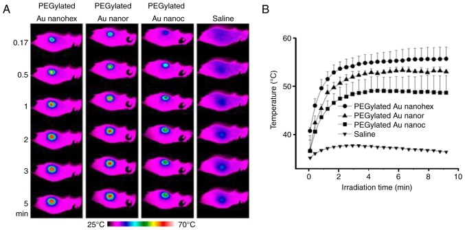
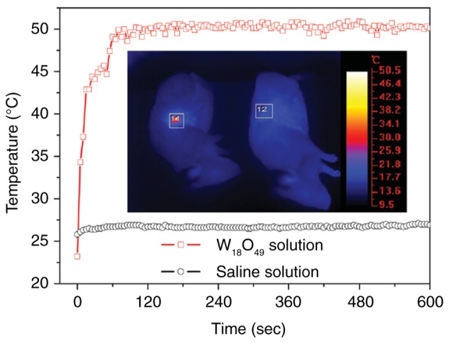
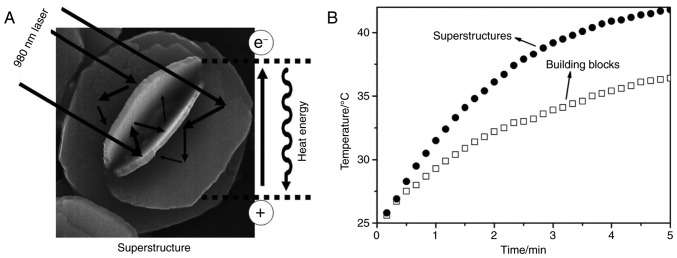
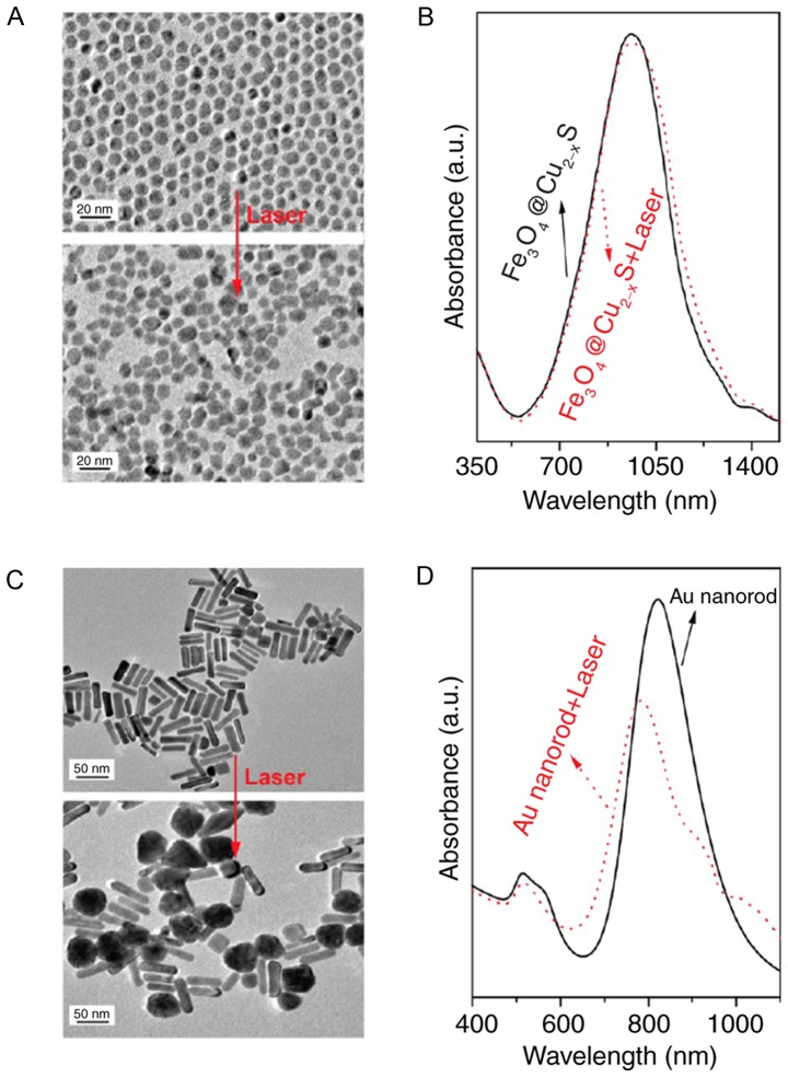
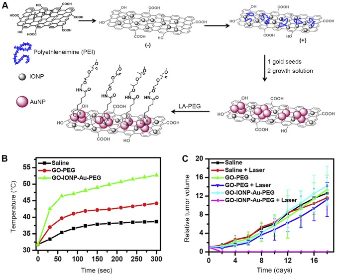
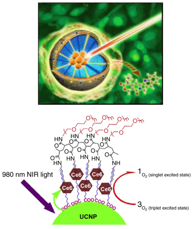
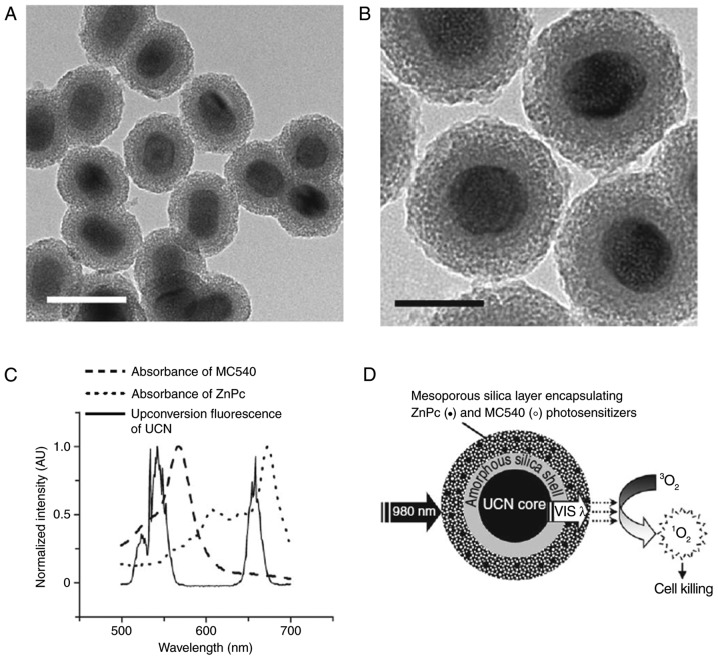
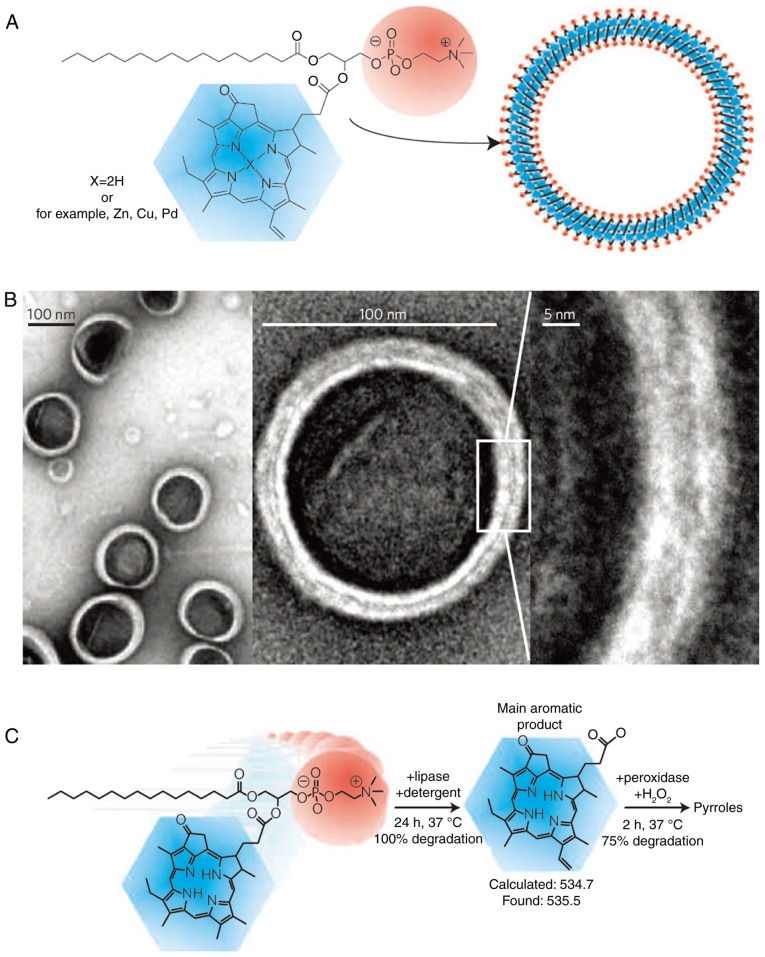
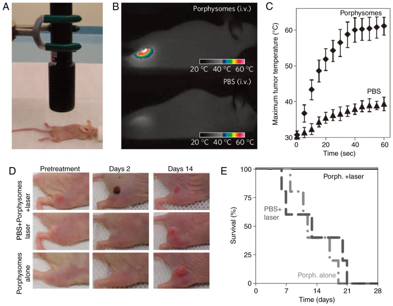
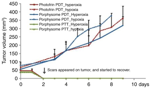
Nanomaterials play crucial roles in the diagnosis and treatment of diseases. Photothermal and photodynamic therapy, as two minimally invasive therapeutic methods, have promising potential in the diagnosis and prevention of cancer. Recently, many photothermal materials (such as noble metal material, transition metal sulfur oxides, carbon material and upconversion nanomaterial) and photodynamic materials (such as phthalein cyanogen, porphyrins and other dye molecules) have been applied in photothermal therapy (PTT) and photodynamic therapy (PDT). Moreover, as nanomaterials have suitable biocompatibility, these materials have been applied in cancer therapy. In the present review, we summarized the effects of different material types, synthesis methods, material morphologies and surface modifications on the outcomes of cancer therapy. The application of nanomaterials in PTT and PDT was introduced and the advantages and disadvantages of PTT and PDT in the prevention of cancer were discussed. Finally, we discussed the application of nanomaterials in the combination of PTT and PDT in cancer treatment.


Figures

Figure 1.
Figure 1.
(A) Thermographs indicating the temperature of tumor tissues in mice for different periods of time in PTT. The mice were intravenously injected with aqueous suspensions of PEGylated nanohexapods, nanorods, nanocages and saline. (B) The relation of average temperature within the tumor tissues and the irradiation time. Laser power density, 1.2 W·cm2 (Reprinted from ref 18 with permission. Copyright 2013, American Chemical Society). PTT, photothermal therapy; nanohex, nanohexapods; nanor, nanorods; nanoc, nanocages.
Figure 2.
Figure 2.
Temperature of PEGylated W18O49 nanowire in vivo and in vitro. Laser, 980 nm; power density, 0.72W·cm2. The concentration of W18O49 nanowire PBS solution was 2 g/l (Reprinted from ref 28 with permission. Copyright 2013, John Wiley and Sons).
Figure 3.
Figure 3.
(A) Schematic diagram for mirror and photothermal transformation of superstructure nanoCuS. (B) The relation between temperature and superstructure nanoCuS for different periods of time. Laser, 980 nm; power density, 0.51 W·cm2 (Reprinted from ref 30 with permission. Copyright 2011, John Wiley and Sons).
Figure 4.
Figure 4.
Photothermal stability comparison for (A and B) Fe3O4@Cu2-xS core-shell nanomaterials and (C and D) Au nanorods (50×15 nm). Laser, 980 nm; power density, 2 W·cm2; irradiation time, 30 min (Reprinted from ref 36 with permission. Copyright 2013, American Chemical Society).
Figure 5.
Figure 5.
(A) The formulation of PEGylated GO-IONP-Au. (B) The temperature for different periods of time in GO-PEG, GO-IONP-Au-PEG and saline. (C) The tumor growth curves of tumor-bearing mice in different periods of time (Reprinted from ref. 74 with permission. Copyright 2013, Elsevier Ltd. All rights reserved).
Figure 6.
Figure 6.
Schematic diagram for the application of upconversion nanoparticles in PDT (Reprinted from ref 7 with permission. Copyright 2011, Elsevier Ltd. All rights reserved). PDT, photodynamic therapy; NIR, near-infrared light; UCNP, up-converting nanophosphors.
Figure 7.
Figure 7.
(A and B) Scanning electron microscopy (SEM) images of NaYF4:Yb/Er upconversion nanoparticles coated with mesoporous-silica. (C) Photoluminescence spectroscopy of MC540, ZnPc and upconversion fluorescence of UCN (dashed lines indicate the curve of photosensitizer light absorption). (D) The treatment mechanism of MC540 and ZnPc (Reprinted from ref 53 with permission. Copyright 2012, Springer Nature). UCN, upconversion nanomaterials.
Figure 8.
Figure 8.
(A) The self-assembling mechanism of porphysomes. (B) Scanning electron microscopy (SEM) images of porphysomes. (C) The intracellular degradation mechanisms of the porphysome (Reprinted from ref 75 with permission. Copyright 2011, Springer Nature).
Figure 9.
Figure 9.
The application of porphysomes in the treatment of tumor-bearing mice. (A and B) Thermal images of mice-bearing tumor xenografts after being intravenously administered porphysomes or PBS and irradiated with a laser for photothermal therapy (PTT). (C) Quantification of increase in temperature in mice from A. (D) Resulting tumor response after PTT treatment at day 2 and 14. (E) Survival curve of mice receiving PTT (Reprinted from ref 75 with permission. Copyright 2011, Springer Nature).
Figure 10.
Figure 10.
The tumor volume at different periods of time in hypoxia/hyperoxia tumor tissues treated with porphyrins and porphysomes (Reprinted from ref 75 with permission. Copyright 2013, American Chemical Society). PTT, photothermal therapy; PDT, photodynamic therapy (PDT).

References

    1. Oh J, Yoon H, Park JH. Nanoparticle platforms for combined photothermal and photodynamic therapy. Biomed Eng Lett. 2013;3:67–73. doi: 10.1007/s13534-013-0097-8. - DOI
    1. Cao J, An H, Huang X, Fu G, Zhuang R, Zhu L, Xie J, Zhang F. Monitoring of the tumor response to nano-graphene oxide-mediated photothermal/photodynamic therapy by diffusion-weighted and BOLD MRI. Nanoscale. 2016;8:10152–10159. doi: 10.1039/C6NR02012G. - DOI - PubMed
    1. Lin J, Wang S, Huang P, Wang Z, Chen S, Niu G, Li W, He J, Cui D, Lu G, et al. Photosensitizer-loaded gold vesicles with strong plasmonic coupling effect for imaging-guided photothermal/photodynamic therapy. ACS Nano. 2013;7:5320–5329. doi: 10.1021/nn4011686. - DOI - PMC - PubMed
    1. Xiong LQ, Chen ZG, Yu MX, Li FY, Liu C, Huang CH. Synthesis, characterization, and in vivo targeted imaging of amine-functionalized rare-earth up-converting nanophosphors. Biomaterials. 2009;30:5592–5600. doi: 10.1016/j.biomaterials.2009.06.015. - DOI - PubMed
    1. Shibu ES, Hamada M, Murase N, Biju V. Nanomaterials formulations for photothermal and photodynamic therapy of cancer. J Photochem Photobiol C: Photochem Rev. 2013;15:53–72. doi: 10.1016/j.jphotochemrev.2012.09.004. - DOI

MeSH terms