Review
. 2019 May 21;10(1):56.
doi: 10.1186/s13244-019-0733-7.

An update on Burkitt lymphoma: a review of pathogenesis and multimodality imaging assessment of disease presentation, treatment response, and recurrence

Affiliations
Review

An update on Burkitt lymphoma: a review of pathogenesis and multimodality imaging assessment of disease presentation, treatment response, and recurrence

Kevin Kalisz et al. Insights Imaging. .

Abstract

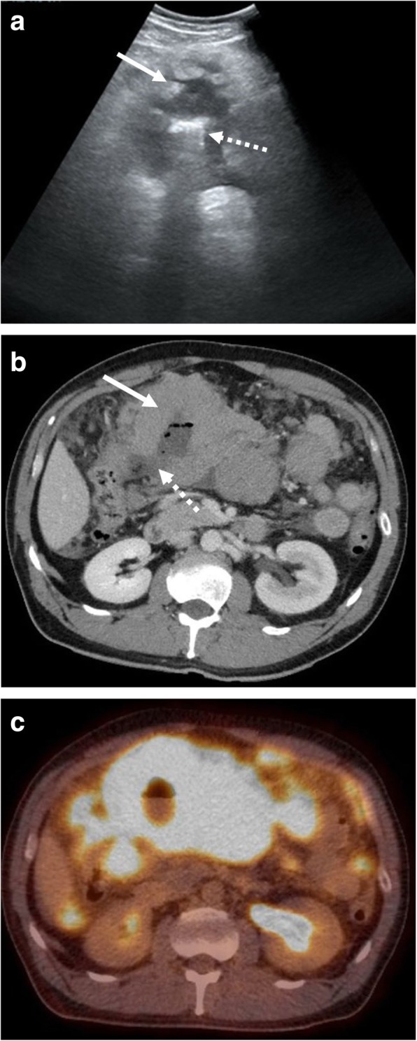
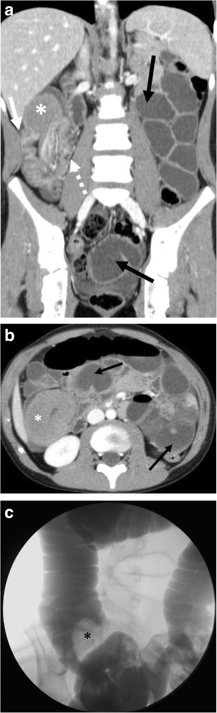
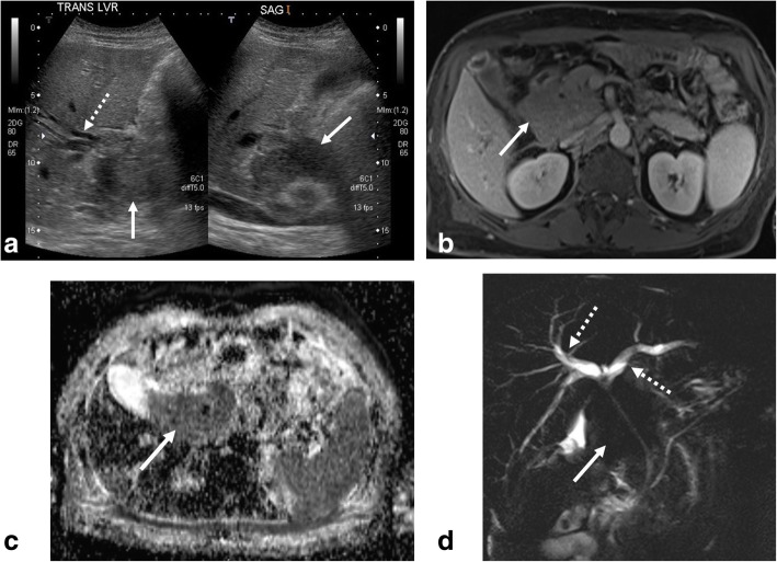
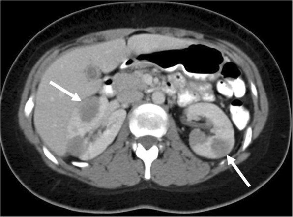
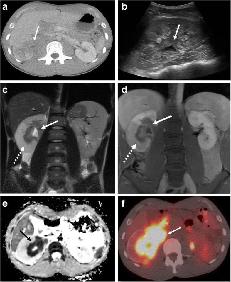
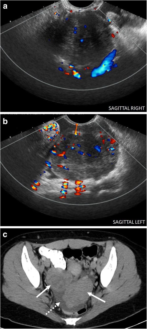
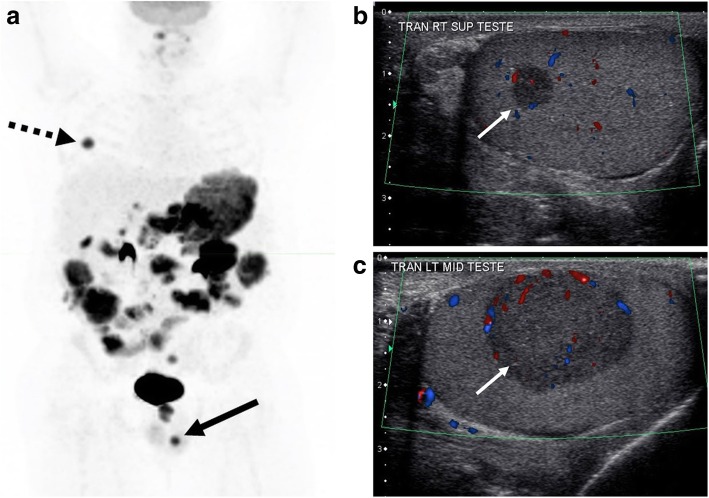
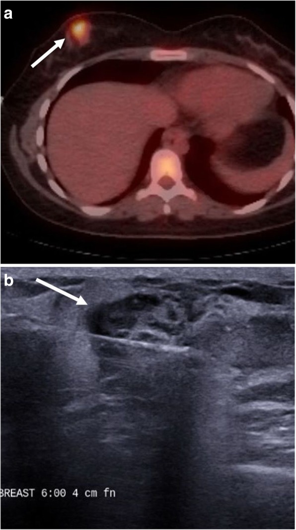
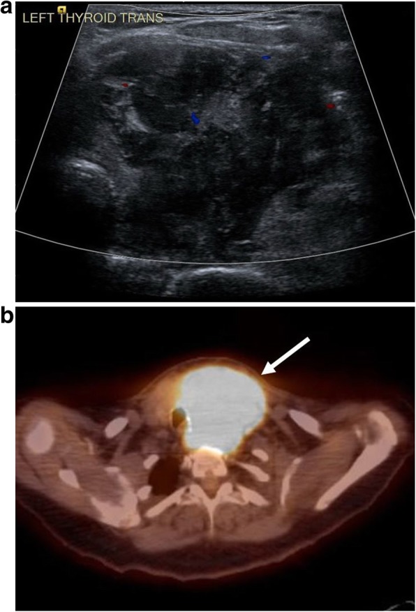
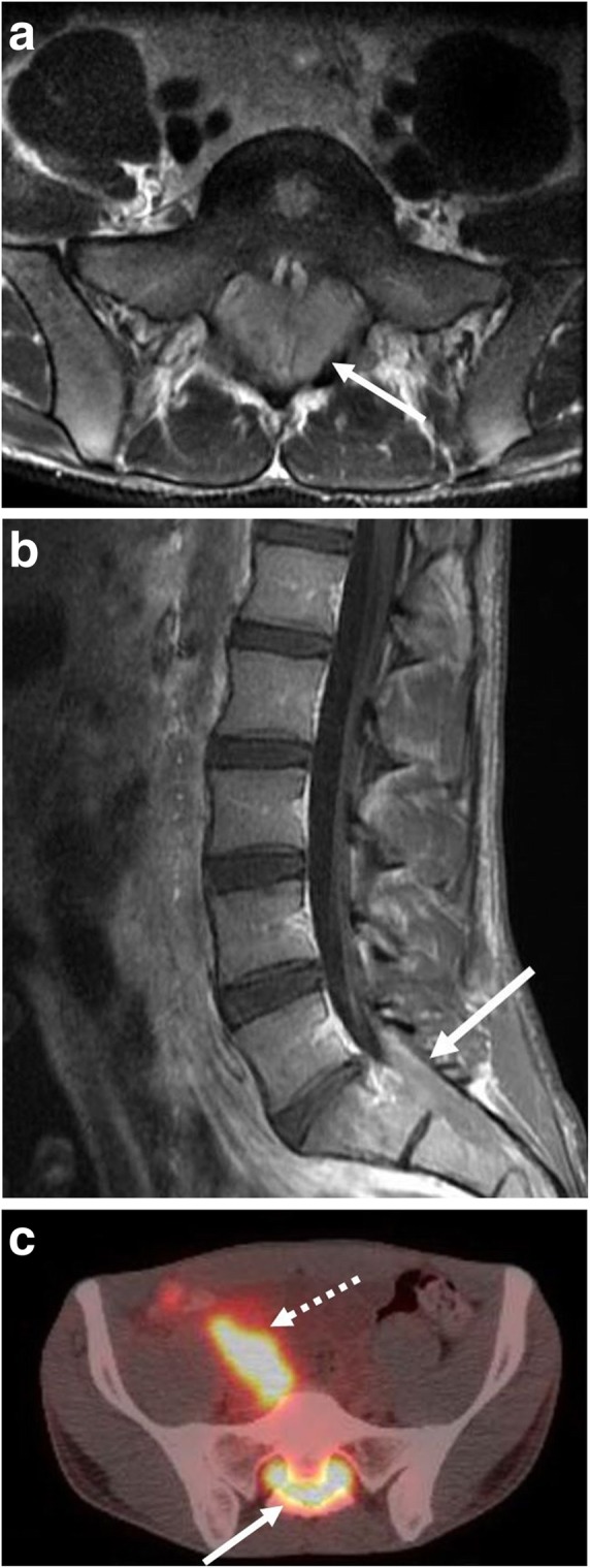
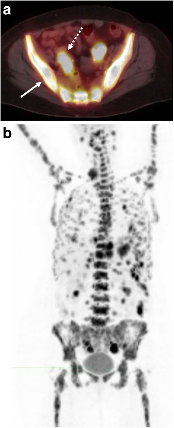
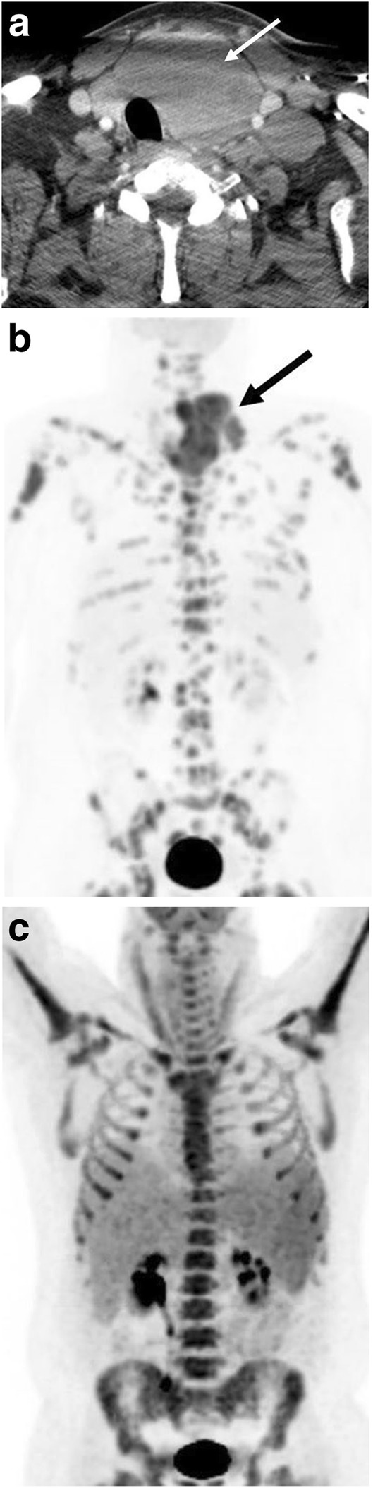
Burkitt lymphoma (BL) is a highly aggressive, rapidly growing B cell non-Hodgkin lymphoma, which manifests in several subtypes including sporadic, endemic, and immunodeficiency-associated forms. Pathologically, BL is classically characterized by translocations of chromosomes 8 and 14 resulting in upregulation of the c-myc protein transcription factor with upregulation of cell proliferation. BL affects nearly every organ system, most commonly the abdomen and pelvis in the sporadic form. Imaging using a multimodality approach plays a crucial role in the management of BL from diagnosis, staging, and evaluation of treatment response to therapy-related complications with ultrasound, computed tomography, magnetic resonance imaging, and positron emission tomography playing roles. In this article, we review the pathobiology and classification of BL, illustrate a multimodality imaging approach in evaluating common and uncommon sites of involvement within the trunk and head and neck, and review common therapies and treatment-related complications.

Keywords: B cell; Burkitt lymphoma; Computed tomography; Diagnostic imaging; Drug therapy; Lymphoma.


Conflict of interest statement

The authors declare that they have no competing interests.

Figures

Fig. 1
Fig. 1
Bowel involvement with perforation. A 49-year-old male presenting to the emergency department with abdominal pain. a Transverse grayscale ultrasound image demonstrates mass-like thickening of a bowel loop within the mid-abdomen (solid arrow) with central air and associated dirty shadowing (dashed arrow). b Subsequently performed axial CT shows a large central mass (solid arrow) with associated perforation (dashed arrow). c Follow-up fused axial PET-CT demonstrates associated hypermetabolic activity and extensive background abdominal disease. Burkitt lymphoma was confirmed at surgery
Fig. 2
Fig. 2
Bowel involvement with intussusception. A 10-year-old male presenting to the emergency department with abdominal pain. a Coronal and (b) axial contrast enhanced CT images demonstrate ileo-colic intussusception with intussusceptum (dashed arrow) and intussuscipiens (solid arrow) and dilated, fluid-filled small bowel loops (black arrows) from an associated small bowel obstruction. Lead point mass (asterisks) is also seen. c Subsequent fluoroscopic-guided contrast reduction enema demonstrating filling defect (asterisks) corresponding to the lead point mass was ultimately unsuccessful. The patient underwent surgical resection with pathology demonstrating Burkitt lymphoma
Fig. 3
Fig. 3
Pathology findings of Burkitt lymphoma. A 10-year-old male (presented in Fig. 2). a Gross exam shows a 6 cm firm mass at the ileo-cecal junction. Microscopically, (b) (× 40) the lymphoma is composed of sheets of monotonous lymphocytes invading the bowel mucosa and cells (c) (× 200) has a characteristic “starry sky” appearance due to the presence of scattered histiocytes engulfing apoptotic lymphoma
Fig. 4
Fig. 4
Nodal mass with biliary obstruction. A 55-year-old male presenting with jaundice. a Initial grayscale transverse and sagittal ultrasound images demonstrate an ill-defined hypoechoic mass near the porta hepatis (solid arrow) with associated biliary dilatation. Follow-up MRI with (b) axial post contrast, (c) apparent diffusion coefficient map, and (d) coronal MRCP images demonstrate an enhancing mass with diffusion restriction (solid arrows) encasing and narrowing the extrahepatic bile ducts causing intrahepatic biliary dilatation (dashed arrow)
Fig. 5
Fig. 5
Renal involvement with renal masses. Contrast-enhanced axial CT image of a 23-year-old female with known Burkitt lymphoma demonstrates multiple circumscribed hypoattenuating renal masses (arrows)
Fig. 6
Fig. 6
Renal collecting system involvement. A 24-year-old male presenting with abdominal and lower back pain. a Contrast-enhanced axial CT image demonstrates an infiltrative soft tissue mass (arrow) encasing the right renal collecting system with resultant dilated renal pelvis and urothelial thickening. b Corresponding sagittal grayscale ultrasound image (b) demonstrates obliteration of the normal renal sinus fat by hypoechoic tissue encasing the renal pelvis (arrow). Follow-up MRI with (c) coronal T2-weighted, (d) coronal T1-weighted post contrast, and (e) axial apparent diffusion coefficient images shows a right renal pelvis mass with associated diffusion restriction (solid arrows) with edema and delayed enhancement of portions of the right renal parenchyma (dashed arrows). f Follow-up fused axial PET-CT demonstrates associated hypermetabolic activity (arrow)
Fig. 7
Fig. 7
Bilateral ovarian masses. A 23-year-old female presenting with pelvic pain. Sagittal color Doppler images through the (a) right and (b) left ovaries demonstrate heterogeneous enlargement of the bilateral ovaries containing internal flow, greater on the left. c Subsequent contrast-enhanced CT shows markedly enlarged bilateral ovaries (solid arrows) with adjacent pelvic free fluid (dashed arrow). Subsequent exploratory laparotomy with bilateral salpingo-oophorectomy revealed Burkitt lymphoma
Fig. 8
Fig. 8
Bilateral testicular masses. A 39-year-old HIV-positive male with known Burkitt lymphoma. a Whole body planar PET image demonstrates widespread abdominal disease with a small focus of hypermetabolic activity within the region of the left testicle (solid arrow). Less intense, linear activity within the abdomen corresponds to physiologic bowel activity. Small focus of uptake in the right hemithorax (dashed arrow) corresponds to pleural disease. Sagittal color Doppler images through the (b) right and (c) left testicles demonstrate bilateral hypoechoic testicular masses, left larger than right (arrows)
Fig. 9
Fig. 9
Pleural and pericardial involvement. A 62-year-old female presenting with chest pain and shortness of breath. a Contrast-enhanced axial CT image demonstrates a partially loculated right pleural effusion (solid arrow) and mass-like thickening of the pericardium extending into the epicardial fat encasing the right coronary artery (dashed arrow). b Fused axial PET-CT demonstrates associated hypermetabolic uptake within the pericardial mass (white dashed arrows) and better depicts multifocal right-sided pleural involvement (white solid arrow). There is also sternal involvement (black solid arrow)
Fig. 10
Fig. 10
Breast involvement. A 22-year-old female with known Burkitt lymphoma. a Staging fused axial PET-CT demonstrates hypermetabolic activity within the inferior and medial right breast (arrow). b Grayscale ultrasound during percutaneous biopsy demonstrates an irregular, heterogeneous mass with indistinct margins (arrow). Biopsy results revealed Burkitt lymphoma
Fig. 11
Fig. 11
Thyroid involvement. A 58-year-old female presenting with dysphagia. a Transverse color Doppler image through the left thyroid demonstrates a large, heterogeneous hypovascular mass. b Fused axial PET-CT demonstrates the thyroid mass with rightward tracheal displacement and associated hypermetabolic activity. Biopsy of the thyroid mass revealed Burkitt lymphoma
Fig. 12
Fig. 12
Central nervous system involvement. A 24-year-old male presenting with abdominal and lower back pain. a Axial T2-weighted and (b) sagittal post contrast images of the lumbar spine and sacrum demonstrate an enhancing soft tissue mass within the lower spinal canal (arrows) with extension into bilateral sacral neural foramina. c Fused axial PET-CT demonstrates associated hypermetabolic activity within the sacral mass (solid arrow). Involvement of the right retroperitoneum is also seen (dashed arrow)
Fig. 13
Fig. 13
Diffuse marrow involvement. A 74-year-old female with known Burkitt lymphoma. a Fused axial PET-CT demonstrates diffuse hypermetabolic activity within the visualized marrow of the pelvis (solid arrow). Soft tissue activity is also seen within the pelvis (dashed arrow). b Whole body planar PET image demonstrates both diffuse marrow and soft tissue disease within the lower neck, thorax, abdomen, and pelvis
Fig. 14
Fig. 14
Treatment response. A 42-year-old man initially presenting with neck mass and constitutional symptoms. Contrast-enhanced axial CT at presentation (a) demonstrates a large left neck mass (arrow) replacing the left thyroid with tracheal deviation. b Staging whole body planar PET image demonstrates a hypermetabolic left lower neck mass (arrow) and heterogeneous marrow involvement. The patient underwent R-CHOP with intrathecal methotrexate, 4 cycles of hyper-CVAD and intrathecal methotrexate. Follow-up (c) whole-body planar PET image imaging show resolution of the cervical mass without evidence of distant disease. Diffuse homogeneous marrow uptake on the PET scan is likely related to post-therapy marrow activation
Fig. 15
Fig. 15
Pulmonary recurrence. An 83-year-old man initially presenting with GI bleeding and was found to have conglomerate gastric and intra-abdominal masses. a Initial staging whole body planar PET image demonstrates extensive abdominal disease involvement (arrow). He went on to complete 4 cycles of bendamustine and rituximab. b Follow-up whole body planar PET image demonstrates complete disease response. c A subsequent restaging whole body planar PET image demonstrates a new hypermetabolic focus within the left upper thorax (arrow). d Corresponding fused axial PET-CT demonstrates a pleural-based lesion (arrow). Percutaneous biopsy revealed Burkitt lymphoma
Fig. 16
Fig. 16
Treatment-related neutropenic colitis. A 58-year-old male initially presenting with a left neck mass. a Coronal fused PET-CT demonstrates a large, hypermetabolic left neck mass (arrow) causing tracheal deviation. The mass was subsequently biopsied as Burkitt lymphoma, and the patient was subsequently started on hyper-CVAD therapy. After cycle #3, the patient was admitted for abdominal pain and neutropenic fever. b Axial and (c) coronal non-contrast CT images obtained in the emergency department show diffuse colonic wall thickening and inflammatory stranding (arrows). Findings are consistent with treatment-related colitis

References

    1. Mbulaiteye SM, Anderson WF, Ferlay J, et al. Pediatric, elderly, and emerging adult-onset peaks in Burkitt's lymphoma incidence diagnosed in four continents, excluding Africa. Am J Hematol. 2012;87(6):573–578. doi: 10.1002/ajh.23187. - DOI - PMC - PubMed
    1. Morton LM, Wang SS, Devesa SS, Hartge P, Weisenburger DD, Linet MS. Lymphoma incidence patterns by WHO subtype in the United States, 1992-2001. Blood. 2006;107(1):265–276. doi: 10.1182/blood-2005-06-2508. - DOI - PMC - PubMed
    1. Ferry JA. Burkitt’s lymphoma: clinicopathologic features and differential diagnosis. Oncologist. 2006;11(4):375–383. doi: 10.1634/theoncologist.11-4-375. - DOI - PubMed
    1. Ogwang MD, Bhatia K, Biggar RJ, Mbulaiteye SM. Incidence and geographic distribution of endemic Burkitt lymphoma in northern Uganda revisited. Int J Cancer. 2008;123(11):2658–2663. doi: 10.1002/ijc.23800. - DOI - PMC - PubMed
    1. Magrath I. Epidemiology: clues to the pathogenesis of Burkitt lymphoma. Br J Haematol. 2012;156(6):744–756. doi: 10.1111/j.1365-2141.2011.09013.x. - DOI - PubMed

LinkOut - more resources