Review
. 2019 Jul;286(14):2645-2663.
doi: 10.1111/febs.14932. Epub 2019 Jun 10.

ER-phagy: shaping up and destressing the endoplasmic reticulum

Affiliations
Review

ER-phagy: shaping up and destressing the endoplasmic reticulum

Simon Wilkinson. FEBS J. 2019 Jul.

Abstract

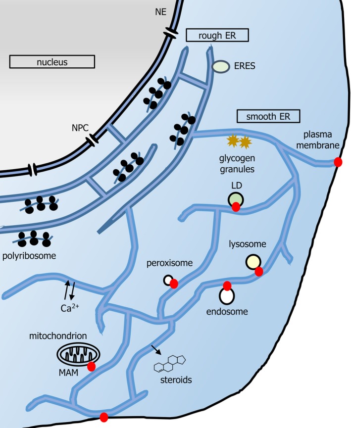
The endoplasmic reticulum (ER) network has central roles in metabolism and cellular organization. The ER undergoes dynamic alterations in morphology, molecular composition and functional specification. Remodelling of the network under fluctuating conditions enables the continual performance of ER functions and minimizes stress. Recent data have revealed that selective autophagy-mediated degradation of ER fragments, or ER-phagy, fundamentally contributes to this remodelling. This review provides a perspective on established views of selective autophagy, comparing these with emerging mechanisms of ER-phagy and related processes. The text discusses the impact of ER-phagy on the function of the ER- and the cell, both in normal physiology and when dysregulated within disease settings. Finally, unanswered questions regarding the mechanisms and significance of ER-phagy are highlighted.

Keywords: ERLAD; ER-phagy; FAM134B; microautophagy; recovER-phagy.


Conflict of interest statement

The authors declare no conflict of interest.

Figures

Figure 1
Figure 1
A schematic of mammalian ER. Nuclear pore complexes (NPCs) gate nucleocytoplasmic transport at the NE. The pER is composed of the rER and sER. rER (darker blue) is composed of flattened, stacked, frequently fenestrated, sheets, connected by helicoidal junctions (shown in cross section here). rER is studded with polyribosomes synthesizing secretory protein and functions in import, folding, glycosylation and onward secretion of such protein. Onward transport originates from ribosome‐free subdomains of rER (ER exit sites, ERESs). Smooth ER (sER, lighter blue) extends in a reticular network throughout the cell, characterized by three‐way junctions. ER tubules also exist in dense arrays in the perinuclear region (not shown for simplicity). The smooth ER functions in detoxification reactions, and lipid and steroid synthesis. Lipid synthesis contributes to organellar membrane generation, for example, during formation of LDs. Organelle contact sites (red dots) may also regulate signalling, for example, immune signalling and transfer of calcium to mitochondria both occur at MAMs. Contact sites may also regulate membrane dynamics, for example, endosome budding and mitochondrial fission. Although contact sites may be at the rER or sER, depending upon the organelle (e.g. both in the case of MAMs), for clarity they are depicted herein at the cell periphery. Note that the yeast ER has a different morphology; extensive cortical ER runs parallel to the plasma membrane, and is connected by tubules to the perinuclear ER, which delimits the nucleoplasm.
Figure 2
Figure 2
Pathways by which ER material transits to the lysosome. This review will reference five distinct routes via which ER fragments or lumenal material may be delivered to the lysosome. These include processes that are either bona fide ER‐phagy pathways, or related processes. Firstly, in macroautophagy (macroER‐phagy (1)), fragments of ER are sequestered by the growth of an encircling double‐membraned phagophore, which then forms an enclosed autophagosome and fuses with lysosomes. MacroER‐phagy can participate in ERLAD (ER‐to‐lysosome‐associated degradation) if particular proteasome‐resistant ER proteins are concentrated within the cargo fragment of ER. MicroER‐phagy is said to occur when lysosomal invagination or protrusion engulfs portions of ER. In yeast microER‐phagy (2), the ER expels whorls of membrane prior to vacuolar invagination. In mammals (3), procollagen‐enriched buds of ER forming from ER exit sites (ERESs) may be targeted in a microautophagy‐mediated ERLAD pathway. In contrast, non‐ER‐phagy processes that involve some or all of the core autophagy machinery are (4) an ER‐phagy–related ERLAD pathway in which single membrane ER‐derived vesicles packed with misfolded lumenal protein species, such as mutant α‐1‐antitryspin, fuse with lysosomes and (5) hypothetic autophagy‐dependent but non‐ER‐phagy ERLAD pathways, wherein aggregated or mutant protein would be expelled from the ER prior to cytosolic sequestration by autophagy or be incorporated directly from the ER membrane into the delimiting membrane of the autophagosome.
Figure 3
Figure 3
Essential mechanism of autophagosome generation in mammals. A phagophore is shown here (double black lines represent the dual lipid bilayer), notionally extending from an ER cradle (blue tubules). The hierarchy of ATG protein action that initiates and matures the phagophore is depicted as described in the text. Briefly, the ULK1/2 complex activity drives VPS34 complex‐mediated phosphorylation of phosphatidylinositol to phosphatidyl‐3′‐inositolphosphate (PI3P), which in turn recruits WIPI2. WIPI2 and FIP200 recruit the ATG5 complex. The ATG5 complex acts with ATG3 and ATG7 to attach phosphatidylethanolamine in the phagophore to the exposed C‐terminal glycine of proteolytically processed LC3/GABARAP. Further lipid is delivered from various sources, such as tubular endosomes; the transmembrane ATG proteins ATG9L1/2 co‐ordinate this. Note that while LC3/GABARAP plays a role in accelerating expansion and closure of phagophores, it is also required for selection of cargo via interaction with cargo receptors.
Figure 4
Figure 4
A model of FAM134B and Atlastin function in delivery of ER content to lysosomes. In the key at the top of the diagram, the core sequence of each LIR motif is shown for each receptor (RHD, reticulon homology domain; GTPase, dynamin‐like GTPase domain). The box provides a schematic overview of FAM134B‐ and Atlastin‐dependent macroER‐phagy and of ER‐phagy‐related ERLAD of mutant α‐1‐antitrypsin (ATZ). Note that the cartoon of macroER‐phagy is shown particularly in the context of clearance of specific ER lumenal moieties (procollagen, PC), to illustrate the full breadth of our knowledge of this process, but macroER‐phagy likely operates in other contexts to functionally remodel the ER in different ways. In macroER‐phagy, the LIR motif of FAM134B drives clustering at sites of autophagosome genesis, probably aided by initial phagophore generation and recruitment of lipidated LC3/GABARAP. RHD‐mediated curvature in conjunction with the GTPase activity of the Atlastins (ATL2 and ATL3 depicted here), results in ER fragmentation. For tubular ER degradation (which is largely FAM134B‐independent, but RTN3L dependent) fragmentation may also be promoted by LC3/GABARAP‐mediated recruitment of ATL3 via LIR motifs (also known GIM motifs due to selectivity for GABARAP subfamily proteins). LC3/GABARAP‐mediated recognition of FAM134B (and ATL3 for tubular ER) also ensures that the ER fragment is incorporated into the mature autophagosome. In contrast, in ER‐phagy–related ERLAD, single‐membraned vesicles derive from the ER, incorporating FAM134B. However, interaction of FAM134B with LC3/GABARAP is only required for lysosomal fusion, along with the SNARE pairing of STX17 and VAMP8. This delivers the ER lumenal contents into the lysosome, although the membrane is donated to the lysosome, whereas in macroautophagy the entire fragment of ER, membrane and lumen, is internalized and degraded. A minimal LC3/GABARAP lipidation machinery, excluding ATG proteins such as the ULK complex, is required for ER‐phagy–related ERLAD. Selectivity for lumenal content in macroER‐phagy or ER‐phagy–related ERLAD is at least partly mediated via binding of FAM134B to the chaperone calnexin, which can in turn bind to misfolded or polymerized PC or ATZ.
Figure 5
Figure 5
Models of action of RTN3L, SEC62 and CCPG1 in macroER‐phagy. Schematics of receptors RTN3L, SEC62 and CCPG1 are depicted on the left‐hand side of the diagram. Core LIR motif sequences are shown. Additionally, in CCPG1, minor (light blue) and major (bold blue) sequences supporting FIP200‐binding activity are shown (FIR = FIP200 interacting region). LC3/GABARAP molecules (green circles) bind LIR motifs in RTN3L to mediate focal recruitment at the nascent phagophore and oligomer formation, promoting ER curvature and incorporation of the eventual ER fragment into the mature autophagosome. The LIR motif of SEC62 mediates binding of ER fragments containing UPR‐upregulated proteins to the nascent phagophore. How these subregions of ER are generated or how SEC62 recognizes specific lumenal cargoes is unknown. When the UPR is at basal levels, SEC63 may bind SEC62 and compete for LC3/GABARAP interaction. Finally, CCPG1 uses a LIR to bind LC3/GABARAP on the phagophore and FIR regions to bind FIP200 on either the ER or the phagophore. Both interactions are required for sequestration of CCPG1‐enriched ER into autophagosomes. CCPG1 has a substantial (> 450 amino acid) lumenal domain that could hypothetically participate in recognition of specific lumenal cargoes.

References

    1. English AR, Zurek N & Voeltz GK (2009) Peripheral ER structure and function. Curr Opin Cell Biol 21, 596–602. - PMC - PubMed
    1. Phillips MJ & Voeltz GK (2016) Structure and function of ER membrane contact sites with other organelles. Nat Rev Mol Cell Biol 17, 69–82. - PMC - PubMed
    1. Walter P & Ron D (2011) The unfolded protein response: from stress pathway to homeostatic regulation. Science 334, 1081–1086. - PubMed
    1. Ruggiano A, Foresti O & Carvalho P (2014) Quality control: ER‐associated degradation: protein quality control and beyond. J Cell Biol 204, 869–879. - PMC - PubMed
    1. Caldieri G, Barbieri E, Nappo G, Raimondi A, Bonora M, Conte A, Verhoef L, Confalonieri S, Malabarba MG, Bianchi F, et al. (2017) Reticulon 3-dependent ER-PM contact sites control EGFR nonclathrin endocytosis. Science 356, 617–624. - PMC - PubMed

Publication types

MeSH terms