. 2019 May 22;14(5):e0217030.
doi: 10.1371/journal.pone.0217030. eCollection 2019.

Losartan and isoproterenol promote alterations in the local renin-angiotensin system of rat salivary glands

Affiliations

Losartan and isoproterenol promote alterations in the local renin-angiotensin system of rat salivary glands

Isadora Prado Cano et al. PLoS One. .

Abstract

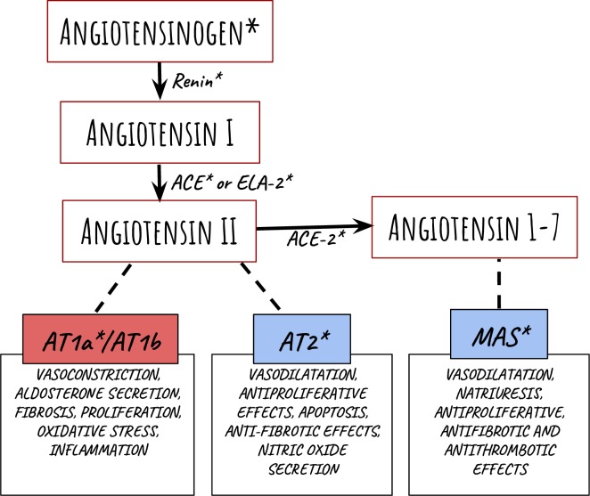
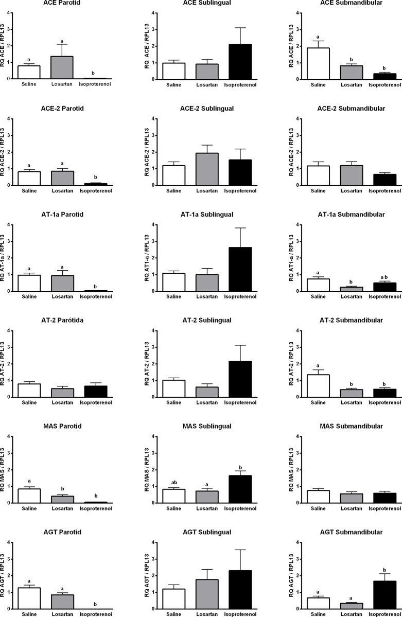
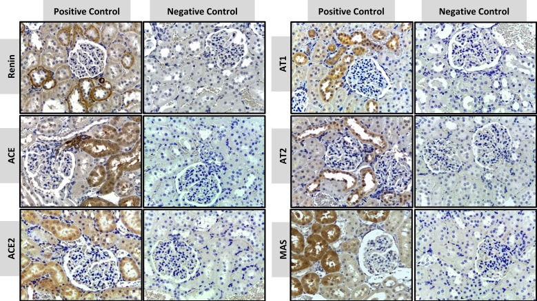
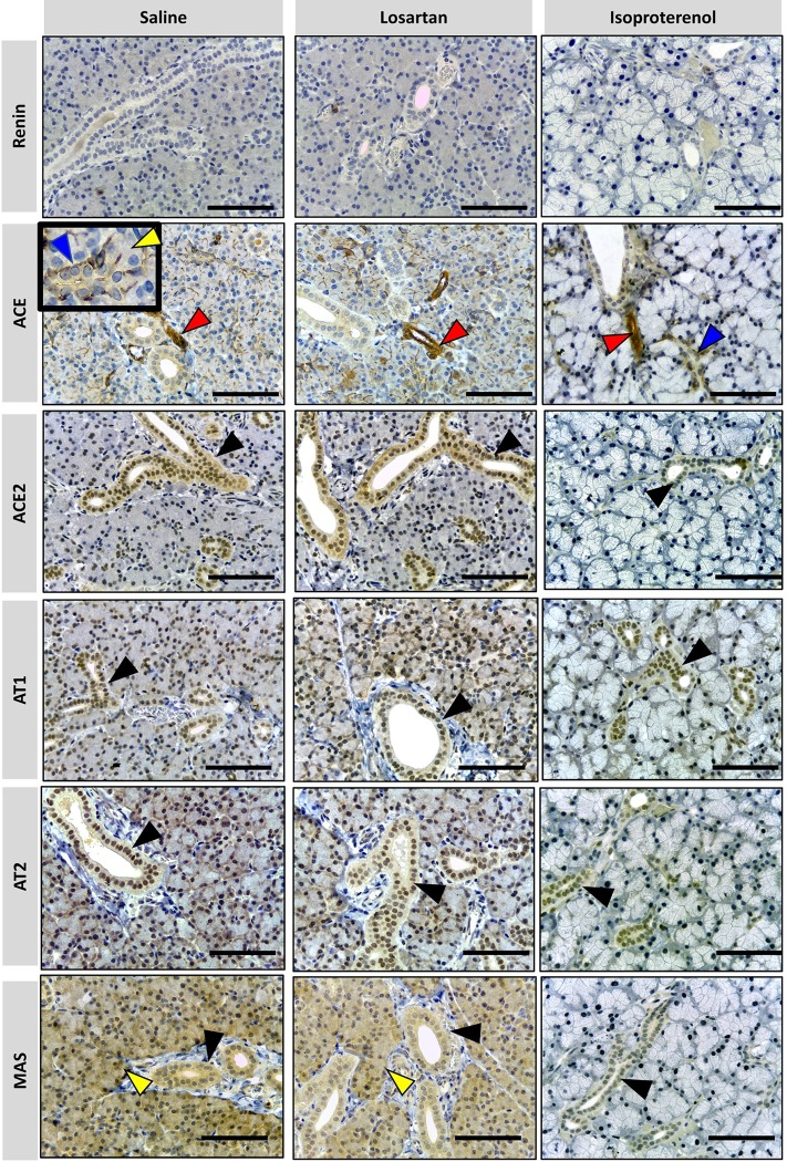
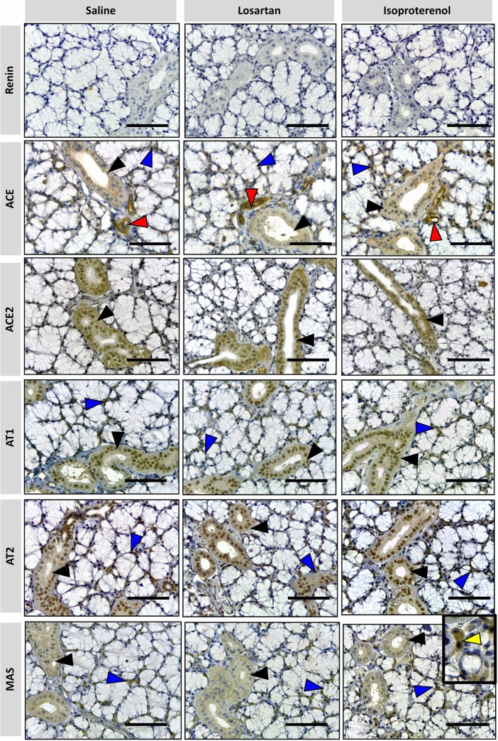
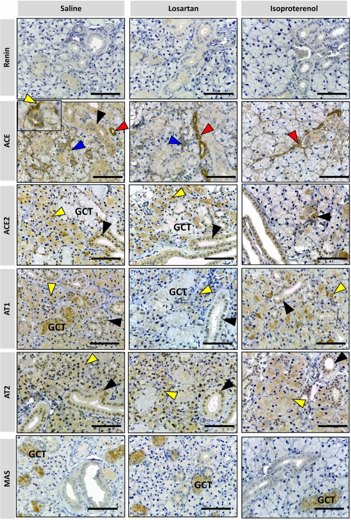
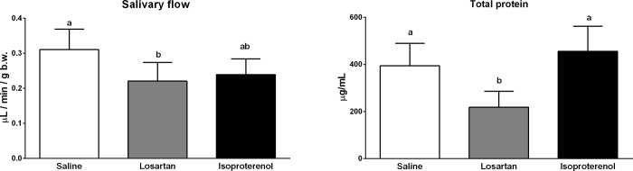
Renin-angiotensin system (RAS) systemically or locally collaborates with tissue homeostasis, growth and development, which has been extensively studied for its pharmacological implications. This study was primarily aimed at finding and characterizing local RAS in rat parotid, sublingual and submandibular glands. It was also hypothesized that vasoactive drugs could affect the expression of RAS targets, as well as saliva flow and its composition. Therefore, another objective of this study was to compare the effects of losartan (angiotensin II receptor blocker) and isoproterenol (β-adrenergic receptor agonist). Forty-one Wistar rats were divided into three groups and administered a daily intraperitoneal dose of saline, losartan or isoproterenol solutions for one week. The following RAS targets were studied using qPCR: renin (REN), angiotensinogen (AGT), angiotensin converting enzyme (ACE), ACE-2, elastase-2 (ELA-2), AT1-a and MAS receptors, using RPL-13 as a reference gene. Morphology of glands was analyzed by immunohistochemistry using REN, ACE, ACE-2, AT1, AT2 and MAS antibodies. The volume and total protein content of saliva were measured. Our results revealed that ACE, ACE-2, AT1-a, AT2 and MAS receptors were expressed in all salivary gland samples, but REN and ELA-2 were absent. Losartan decreased mRNA expression of RAS targets in parotid (MAS) and submandibular glands (ACE and both AT receptors), without affecting morphological alterations, and significantly decreased saliva and total protein secretions. Isoproterenol treatment affected gene expression profiles in parotid (ACE, ACE-2, AT1-a, MAS, AGT), and submandibular (ACE, AT2, AGT) glands, thus promoting acinar hypertrophy in serous acini, without significant changes in salivary flow or total protein content. These drugs affected mainly acini, followed by duct systems and myoepithelial cells, whereas blood vessels were not affected. In conclusion, there is a local RAS in major rat salivary glands and losartan, an angiotensin II receptor blocker, affected not only the RAS-target gene expression but also decreased salivary flow and total protein content.


Conflict of interest statement

The authors have declared that no competing interests exist.

Figures

Fig 1
Fig 1. Representation of the metabolic RAS cascade.
Angiotensinogen and the peptide sequences are shown as a result of specific enzyme cleavages, which are included next to their represented arrows. The receptors are represented as boxes on the bottom and the function described is a result of the linked angiotensin binding. The RAS targets used in this study are indicated with an asterisk. Graphic illustration is adapted from Ferrario and Strawn [1].
Fig 2
Fig 2. Relative quantification of each target in rat parotid, sublingual and submandibular glands.
Data are means ± standard deviation. One-way ANOVA/Tukey’s multiple comparison tests were performed for parametric results whereas Kruskal-Wallis/ Dunn’s post-test was applied for non-parametric results. Differences were considered statistically significant when p<0.05. Different letters indicate a statistically significant difference between groups and when such differences were calculated between two groups but not in a third one as compared to both, the latter is represented as ab. Letters were not used in the graph when differences were not significant between saline, losartan and isoproterenol.
Fig 3
Fig 3. Positive and negative controls in immunohistochemical staining of RAS targets in the granular cortex of the kidney.
Immunostains are shown as a brown color in positive controls of REN, ACE, ACE-2, AT1, AT2 and MAS. (x40 objective and scale bar = 50 μm).
Fig 4
Fig 4. Photomicrographs of immunohistochemical staining (brown) of RAS targets in rat parotid gland.
Targets: REN, ACE, ACE-2, AT1, AT2 and MAS were analyzed. Arrows indicated by colors: blue = ducts; yellow = acini; red = blood vessels; black = epithelial cells of the duct system. No REN expression in parotid glands was observed under all treatments. ACE is present in myoepithelial cells around intercalated ducts (indicated by blue arrowhead in detail), acini (indicated by yellow arrowhead in detail) and in blood vessels (indicated by red arrowhead). ACE-2, AT1 and AT2 are present in epithelial cells of the duct system (indicated by black arrowhead), while MAS is present in duct cells (black arrowhead) and acinar cells (blue arrowhead) in all treatments except for isoproterenol. Comparatively, the parotid glands of the animals which received saline and losartan show a similar pattern of immunolabeling for RAS targets but have more expression than in animals which received isoproterenol. (x40 objective and scale bar = 50 μm).
Fig 5
Fig 5. Photomicrographs of immunohistochemical staining (brown) of RAS targets in rat sublingual gland.
Targets: REN, ACE, ACE-2, AT1, AT2 and MAS were analyzed. Arrows indicated by colors: blue = myoepithelial cell; black = duct cells; red: blood vessels; yellow: acinar cells. No REN expression was observed in sublingual glands in all treatments. ACE is present in myoepithelial (blue arrow), duct cells (black arrowhead) and blood vessels (red arrowhead). ACE-2, AT1, AT2, and MAS are present in epithelial cells of the duct system (black arrowhead), myoephitelial cells (blue arrowhead) and more rarely in serous acinar cells (yellow arrowhead) under isoproterenol treatment. Comparatively, the immunostaining patterns of the RAS targets are similar in the sublingual gland in all treatments. (x40 objective and scale bar = 50 μm).
Fig 6
Fig 6. Photomicrographs of immunohistochemical staining (brown) of RAS in rat submandibular gland.
Targets: REN, ACE, ACE-2, AT1, AT2 and MAS were analyzed. Arrow colors indicate as follows: blue = myoepithelial cell; black = duct cells; red = blood vessels; yellow: acinar cells. GCT = granular convoluted tubule. No REN expression was observed in submandibular glands under all treatments. ACE shows higher expression in blood vessels (red arrowhead) under all treatments. ACE expression in duct cells (black arrowhead) and myoepithelial cells around intercalated ducts (blue arrowhead) and acini (yellow arrowhead) are higher in saline treatment than those in rat glands treated with losartan and isoproterenol. ACE-2 is present in acinar cells (yellow arrowhead) and the duct system (black arrowhead) except in the granular convoluted tubule cells (GCT). Mild ACE-2 expression in acinar cells can be observed in rat glands treated with isoproterenol. AT1 and AT2 are present in acinar cells (yellow arrowhead) and striated/excretory ducts (black arrow head), but only AT1 is expressed in GCT. AT1 is weakly expressed in rat glands treated with losartan and a strong immunostain for MAS is observed only in the GCT. (x40 objective and scale bar = 50 μm).
Fig 7
Fig 7. Salivary flow and total protein determination in control, losartan and isoproterenol groups.
Data are presented as means ± standard deviation. Different letters indicate a statistically significant difference (p<0.05) using One-way ANOVA/Tukey’s multiple comparison test for salivary flow analysis and Kruskal-Wallis/Dunn test for total protein analysis.

References

    1. Ferrario CM, Strawn WB. Role of the Renin-Angiotensin-Aldosterone System and Proinflammatory Mediators in Cardiovascular Disease. Am J Cardiol. 2006;98: 121–128. 10.1016/j.amjcard.2006.01.059 - DOI - PubMed
    1. Hall JE. O Papel dos Rins no Controle a Longo Prazo da Pressão Arterial e Na Hipertensão: O Sistema Integrado De Regulação da Pressão Arterial. Guyton e Hall: Tratado de Fisiologia Médica. 13th ed Rio de Janeiro: Elsevier Editora Ltda; 2017. pp. 227–41.
    1. Nehme A, Cerutti C, Dhaouadi N, Gustin MP, Courand P-Y, Zibara K, et al. Atlas of tissue renin-angiotensin-aldosterone system in human: A transcriptomic meta-analysis. Sci Rep. 2015;5: 10035 10.1038/srep10035 - DOI - PMC - PubMed
    1. Chappell MC. Biochemical evaluation of the renin-angiotensin system: the good, bad, and absolute? Am J Physiol Circ Physiol. 2016;310: H137–H152. 10.1152/ajpheart.00618.2015 - DOI - PMC - PubMed
    1. Mulinari dos Santos G, de Souza Batista FR, Kirchweger F, Tangl S, Gruber R, Okamoto R. Losartan reverses impaired osseointegration in spontaneously hypertensive rats. Clin Oral Implants Res. 2018;ahead of p. 10.1111/clr.13376 - DOI - PubMed

Publication types

MeSH terms