Review
. 2019 May 21;20(10):2505.
doi: 10.3390/ijms20102505.

ERK is a Pivotal Player of Chemo-Immune-Resistance in Cancer

Affiliations
Review

ERK is a Pivotal Player of Chemo-Immune-Resistance in Cancer

Iris C Salaroglio et al. Int J Mol Sci. .

Abstract

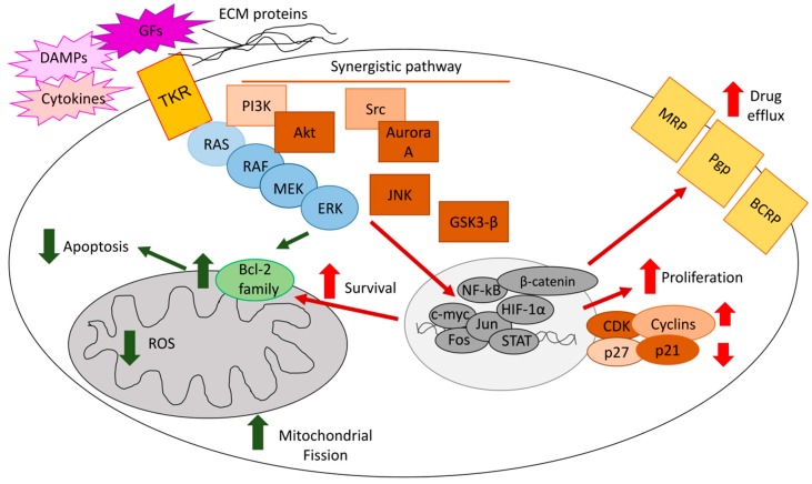
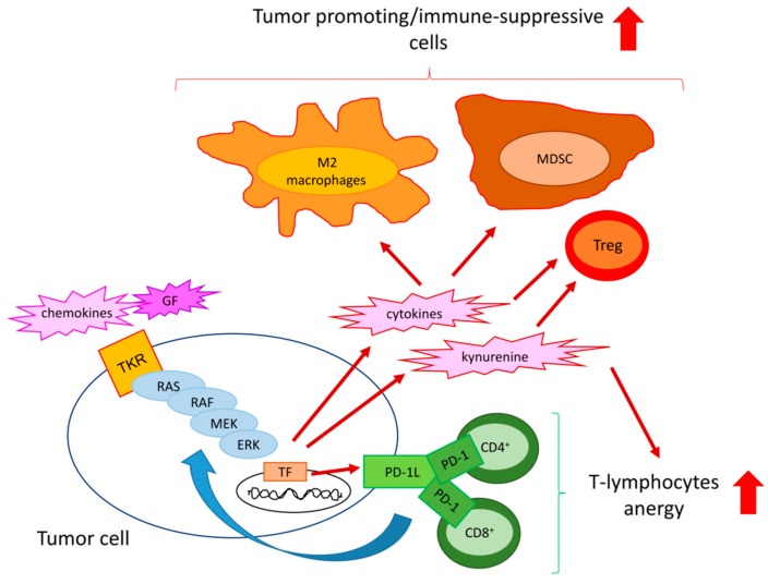
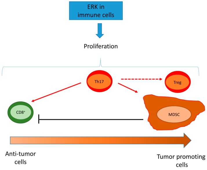
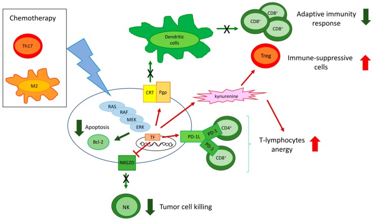
The extracellular signal-related kinases (ERKs) act as pleiotropic molecules in tumors, where they activate pro-survival pathways leading to cell proliferation and migration, as well as modulate apoptosis, differentiation, and senescence. Given its central role as sensor of extracellular signals, ERK transduction system is widely exploited by cancer cells subjected to environmental stresses, such as chemotherapy and anti-tumor activity of the host immune system. Aggressive tumors have a tremendous ability to adapt and survive in stressing and unfavorable conditions. The simultaneous resistance to chemotherapy and immune system responses is common, and ERK signaling plays a key role in both types of resistance. In this review, we dissect the main ERK-dependent mechanisms and feedback circuitries that simultaneously determine chemoresistance and immune-resistance/immune-escape in cancer cells. We discuss the pros and cons of targeting ERK signaling to induce chemo-immune-sensitization in refractory tumors.

Keywords: ERK; chemoresistance; immune-escape; immune-resistance.


Conflict of interest statement

The authors declare no conflict of interest. The founding sponsors had no role in the writing of the manuscript and in the decision to publish it.

Figures

Figure 1
Figure 1
Mechanisms of extracellular signal-regulated kinase (ERK)-induced chemoresistance. The RAS/RAF/MEK/ERK axis [rat sarcoma 2 viral oncogene homolog/rapidly accelerated fibrosarcoma/mitogen-activated protein kinases (MAPK)-extracelluar signal-regulated kinase], upon the activation of tyrosine kinase receptors (TKR) by growth factors (GFs), cytokines, or cell–extracellular matrix (ECM) interaction, cooperates with phosphoinositide 3-kinase (PI3K)/protein kinase B (AKT), c-Jun amino-terminal kinase (JNK), steroid receptor coactivator-1 (Src)/Aurora kinase, glycogen synthase kinase 3-β (GSK-3β)/β-catenin signals, activating a pool of transcription factors that determine: (i) increased cell proliferation by up-regulating cyclins and cyclin-dependent kinase (CDKs) and/or down-regulating the cell cycle check-points p21 and p27; (ii) prevention of apoptosis by decreasing Bcl-2 proteins, reducing mitochondrial reactive oxygen species (ROS) and mitochondrial fission; (iii) increased expression and activity of drug efflux transporters, such as P-glycoprotein (Pgp), multidrug resistance related proteins (MRPs), and breast cancer resistance proteins (BCRP). All these mechanisms make a tumor cell more resistant to chemotherapy. Green arrows: apotosis-related mechanisms; red arrow: pro-survival-related mechanisms.
Figure 2
Figure 2
Mechanisms of ERK-induced immune-resistance or immune-escape. In response to chemokines and growth factors (GFs), specific tyrosine kinase receptors (TKR) activate the RAS/RAF/MEK/ERK axis and the down-stream transcription factors (TF) in tumor cells. As a result, cancer cells produce immune-suppressive cytokines [e.g., interleukin (IL)-10 and transforming growth factor-β (TGF-β)] and metabolites (e.g., kynurenine) that reduce the proliferation of effector CD4+ and CD8+T-lymphocytes and expand tumor-promoting/immune-suppressive cells, such as M2-polarized macrophages, myeloid derived suppressor cells (MDSC), and T-regulatory (Treg) cells. The RAS/RAF/MEK/ERK axis also up-regulates the immune-checkpoint ligand programmed death-1 ligand (PD-1L), which expands PD-1 rich/anergic CD4+ and CD8+ T-cells and fuels ERK activity with a positive feedback mechanism. Exploiting these multiple strategies, ERK activation in tumor cells contributes to create a tumor-promoting/immune-suppressive environment.
Figure 3
Figure 3
Effects of ERK activation in tumor infiltrating cells. ERK induces the proliferation of anti-tumor immune cells, such as CD8+ T-lymphocytes, immune-suppressive cells, such as myeloid derived suppressor cells (MDSC) and T-regulatory (Treg) cells, and cells that have either pro-tumor or anti-tumor activities, such as T-helper 17 (Th17) cells. Th17 cells can promote the pro-inflammatory phenotype of CD8+T-lymphocytes, differentiate into cells with a Treg phenotype, and expand MDSCs that induce apoptosis of CD8+ T-cells. Depending on the type of immune-population characterized by high ERK activity, a wide range of effects (ranging from an anti-tumor to a tumor tolerant/immune-suppressive environment) is obtained. Dashed arrows indicate a direct activation by Th17 cells; the dotted arrow indicates the product of a differentiation of Th17 cells.
Figure 4
Figure 4
Cross-talks between tumor and immune cells determining chemo-immune-resistance. Chemotherapy and immune cells, e.g., T-helper 17 (Th17) cells producing IL-17 and M2-polarized macrophages producing colony stimulating factor-1, activate the RAS/RAF/MEK/ERK pathway in tumor cells, leading to the up-regulation of the Bcl-2 proteins that promote chemoresistance by reducing apoptosis. Moreover, in response to chemotherapy, resistant tumors activate the RAS/RAF/MEK/ERK axis that: (i) up-regulates P-glycoprotein (Pgp), a drug efflux transporter and an inhibitor of the calreticulin (CRT)-triggered phagocytosis by dendritic cells (DCs), avoiding the establishment of a durable adaptive immune response mediated by CD8+ T-cells; (ii) promotes the secretion of kynurenine that expands the immune-suppressive T-regulatory (Treg) cells; (iii) up-regulates the immune-checkpoint ligand programmed death-1 ligand (PD-1L) that expands PD-1 rich/anergic CD4+ and CD8+ T-cells; (iv) down-regulates the surface protein NKG2D, preventing the cytotoxic activity of natural killer (NK) cells. Chemo-immune-resistance is sustained by either chemotherapy or immune cell-induced mechanisms, all converging on an increased ERK activation. Green arrows: decreased pathways (thin arrows) and events (thick arrows); red arrow: increased pathways (thin arrows) and events (thick arrows).

References

    1. Roux P.P., Blenis J. ERK and p38 MAPK-activated protein kinases: A family of protein kinases with diverse biological functions. Microbiol. Mol. Biol. Rev. 2004;68:32–44. doi: 10.1128/MMBR.68.2.320-344.2004. - DOI - PMC - PubMed
    1. Buscà R., Pouysségur J., Lenormand P. ERK1 and ERK2 Map Kinases: Specific Roles or Functional Redundancy? Front. Cell. Dev. Biol. 2016;4:e53. doi: 10.3389/fcell.2016.00053. - DOI - PMC - PubMed
    1. Yoon S., Seger S. The extracellular signal-regulated kinase: Multiple substrates regulate diverse cellular functions. Growth Factors. 2006;24:21–44. doi: 10.1080/02699050500284218. - DOI - PubMed
    1. Roskoski R., Jr. ERK1/2 MAP kinases: Structure, function, and regulation. Pharmacol. Res. 2012;66:105–143. doi: 10.1016/j.phrs.2012.04.005. - DOI - PubMed
    1. Megiorni F., Camero S., Ceccarelli S., McDowell H.P., Mannarino O., Marampon F., Pizer B., Shukla R., Pizzuti A., Marchese C., et al. DNMT3B in vitro knocking-down is able to reverse embryonal rhabdomyosarcoma cell phenotype through inhibition of proliferation and induction of myogenic differentiation. Oncotarget. 2016;7:79342–79356. doi: 10.18632/oncotarget.12688. - DOI - PMC - PubMed

Substances