. 2019 May 28;73(20):2493-2502.
doi: 10.1016/j.jacc.2019.02.065.

Identification of Myocardial Disarray in Patients With Hypertrophic Cardiomyopathy and Ventricular Arrhythmias

Affiliations

Identification of Myocardial Disarray in Patients With Hypertrophic Cardiomyopathy and Ventricular Arrhythmias

Rina Ariga et al. J Am Coll Cardiol. .

Abstract

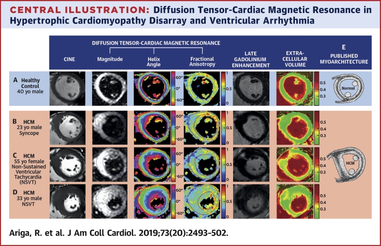
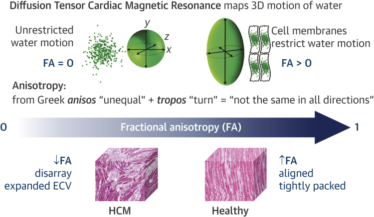
Background: Myocardial disarray is a likely focus for fatal arrhythmia in hypertrophic cardiomyopathy (HCM). This microstructural abnormality can be inferred by mapping the preferential diffusion of water along cardiac muscle fibers using diffusion tensor cardiac magnetic resonance (DT-CMR) imaging. Fractional anisotropy (FA) quantifies directionality of diffusion in 3 dimensions. The authors hypothesized that FA would be reduced in HCM due to disarray and fibrosis that may represent the anatomic substrate for ventricular arrhythmia.

Objectives: This study sought to assess FA as a noninvasive in vivo biomarker of HCM myoarchitecture and its association with ventricular arrhythmia.

Methods: A total of 50 HCM patients (47 ± 15 years of age, 77% male) and 30 healthy control subjects (46 ± 16 years of age, 70% male) underwent DT-CMR in diastole, cine, late gadolinium enhancement (LGE), and extracellular volume (ECV) imaging at 3-T.

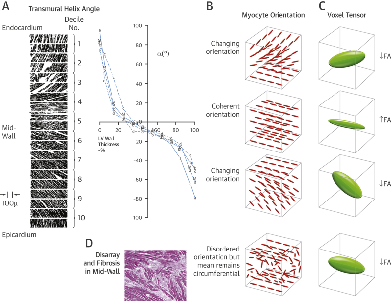
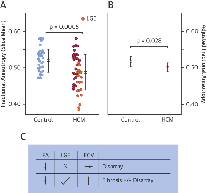
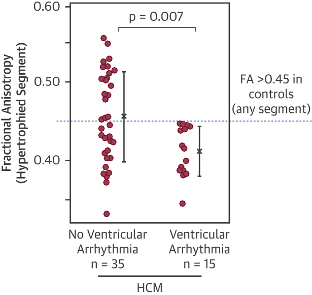
Results: Diastolic FA was reduced in HCM compared with control subjects (0.49 ± 0.05 vs. 0.52 ± 0.03; p = 0.0005). Control subjects had a mid-wall ring of high FA. In HCM, this ring was disrupted by reduced FA, consistent with published histology demonstrating that disarray and fibrosis invade circumferentially aligned mid-wall myocytes. LGE and ECV were significant predictors of FA, in line with fibrosis contributing to low FA. Yet FA adjusted for LGE and ECV remained reduced in HCM (p = 0.028). FA in the hypertrophied segment was reduced in HCM patients with ventricular arrhythmia compared to patients without (n = 15; 0.41 ± 0.03 vs. 0.46 ± 0.06; p = 0.007). A decrease in FA of 0.05 increased odds of ventricular arrhythmia by 2.5 (95% confidence interval: 1.2 to 5.3; p = 0.015) in HCM and remained significant even after correcting for LGE, ECV, and wall thickness (p = 0.036).

Conclusions: DT-CMR assessment of left ventricular myoarchitecture matched patterns reported previously on histology. Low diastolic FA in HCM was associated with ventricular arrhythmia and is likely to represent disarray after accounting for fibrosis. The authors propose that diastolic FA could be the first in vivo marker of disarray in HCM and a potential independent risk factor.

Keywords: diffusion tensor cardiac magnetic resonance imaging; disarray; fractional anisotropy; hypertrophic cardiomyopathy; risk stratification; sudden cardiac death; ventricular arrhythmia.


Figures

None
Graphical abstract
Figure 1
Figure 1
Diffusion Tensor Cardiac Magnetic Resonance Diffusion tensor cardiac magnetic resonance (DT-CMR) maps the diffusion of water molecules in 3 dimensions (3D). Fractional anisotropy (FA) calculated from the diffusion tensor quantifies the directionality of water diffusion within each imaging voxel (2.8 × 2.8 × 8 mm3) as its motion is impeded by several million myocytes and the surrounding interstitium. Without barriers, water motion is random and equal in all directions, which can be represented as a sphere using the diffusion tensor and has an FA of zero (perfect isotropy). Cell membranes act as barriers restricting water motion along the long axis of myocytes. Thus, FA is expected to be high in voxels with coherently aligned myocytes with a consistent orientation. Conversely, FA is expected to be low in voxels with differing myocyte orientations and in hypertrophic cardiomyopathy (HCM) due to disorganized cell orientations and expanded extracellular volume (ECV) .
Figure 2
Figure 2
The Proposed Effect of Transmural Helical Myocyte Orientation on FA Streeter’s classic micrograph of transmural variation of helix angles from endocardium (0% wall thickness) to epicardium in a canine left ventricle (A). Approximately 3 DT-CMR imaging voxels (2.8 × 2.8 × 8 mm3) span the myocardium transmurally in diastole (B). In the endo- and epicardial voxels, myocytes are progressively changing from a longitudinal to circumferential orientation and vice versa, respectively. There are no marked transmural differences in myocyte diameter or fibrous tissue in healthy myocardium . Therefore, the wide distribution of myocyte orientations in the endo- and epicardium will reduce FA. Conversely, the narrow distribution of orientations in the mid-wall where myocytes are consistently circumferentially orientated will elevate FA (C). Myocyte disarray and fibrosis in the mid-wall will reduce FA, due to the wide distribution of disorganized myocyte orientations and expanded extracellular space , but the overall mean voxel helix angle remains in the circumferential orientation (D). Thus, helix angle is expected to be normal despite abnormal FA. LV = left ventricular; other abbreviations as in Figure 1.
Central Illustration
Diffusion Tensor-Cardiac Magnetic Resonance in Hypertrophic Cardiomyopathy Disarray and Ventricular Arrhythmia Multimodal imaging of left ventricular myoarchitecture and disarray using diffusion tensor-cardiac magnetic resonance (DT-CMR), late gadolinium enhancement (LGE), and extracellular volume (ECV) mapping. DT-CMR can provide in vivo assessment of left ventricular myoarchitecture. The helix angle (HA) is the average myocyte orientation, and fractional anisotropy (FA) is a surrogate measure of underlying cell organization. The mid-ventricular slice at diastole in healthy control subjects (A) and patients with hypertrophic cardiomyopathy (HCM) (B–D) demonstrated similar HA distributions, but marked differences in FA. There was an almost complete mid-wall ring of high FA (yellow/orange) in control subjects (A), consistent with the classical description of circumferentially aligned mid-wall myocytes (E), which was also present in the HA map. By contrast, this ring was disrupted by reduced FA (B and C) or was absent (D) in HCM. These patterns were consistent with previously published HCM histology that shows disarray and fibrosis invading the mid-wall at the insertion point and hypertrophied segments (E). Low FA in the anteroseptum matched areas of focal LGE and elevated ECV (C) in keeping with fibrosis contributing to low FA. But low FA could not be explained by fibrosis in all cases. In some instances, low FA in the anteroseptum was present with no detectable LGE or elevated ECV (B). Low FA also extended beyond areas of patchy LGE and elevated ECV, with no remnants of a mid-wall ring (D). Thus, low diastolic FA is likely to represent disarray after accounting for fibrosis, which can, for the first time, be measured in vivo and noninvasively, providing a potentially independent marker for HCM risk stratification. NSVT = nonsustained ventricular tachycardia.
Figure 3
Figure 3
Reduced FA in HCM Even After Adjustment for Fibrosis Diastolic FA, averaged across the mid-ventricular slice, was reduced in HCM compared with healthy control subjects (A, slice mean ± SD). The wide standard deviation of FA in HCM demonstrates the heterogeneity within this disease population, and only a subset of patients had a lower FA than control subjects. HCM patients with late gadolinium enhancement (LGE) (n = 22; orange) had lower FA than those without LGE (p < 0.001). LGE and ECV, which are markers of fibrosis, were significant predictors of FA (p < 0.001). Yet FA adjusted for LGE and ECV remained reduced in HCM (B, linear mixed-effects model estimated marginal means with 95% confidence intervals), indicating that low FA may also demonstrate sensitivity to disarray. These results imply that fibrosis (presence of LGE and/or elevated ECV) contributes to low FA (C). However, in the absence of fibrosis, low FA may instead represent disarray. Abbreviations as in Figure 1.
Figure 4
Figure 4
Reduced FA in HCM With Ventricular Arrhythmias Diastolic FA, measured in the hypertrophied segment, was reduced in HCM with ventricular arrhythmia compared with those without (mean ± SD). Some patients with no apparent ventricular arrhythmia also had low FA, perhaps in part reflecting the low sensitivity of a 1-day Holter. Interestingly, the lowest FA in any segment in any control subject was 0.45. Thus, there was no overlap in segmental FA values between control subjects and HCM patients with ventricular arrhythmia. Abbreviations as in Figure 1.

Comment in

References

    1. Maron B.J., Olivotto I., Spirito P. Epidemiology of hypertrophic cardiomyopathy–related death. Circulation. 2000;102:858–864. - PubMed
    1. Maron B.J., Casey S.A., Chan R.H., Garberich R.F., Rowin E.J., Maron M.S. Independent assessment of the European Society of Cardiology sudden death risk model for hypertrophic cardiomyopathy. Am J Cardiol. 2015;116:757–764. - PubMed
    1. O'Mahony C., Tome-Esteban M., Lambiase P.D. A validation study of the 2003 American College of Cardiology/European Society of Cardiology and 2011 American College of Cardiology Foundation/American Heart Association risk stratification and treatment algorithms for sudden cardiac death in patients with hypertrophic cardiomyopathy. Heart. 2013;99:534–541. - PubMed
    1. Varnava A.M., Elliott P.M., Mahon N., Davies M.J., McKenna W.J. Relation between myocyte disarray and outcome in hypertrophic cardiomyopathy. Am J Cardiol. 2001;88:275–279. - PubMed
    1. Varnava A.M., Elliott P.M., Baboonian C., Davison F., Davies M.J., McKenna W.J. Hypertrophic cardiomyopathy: histopathological features of sudden death in cardiac troponin T disease. Circulation. 2001;104:1380–1384. - PubMed

Publication types

MeSH terms