. 2019 Jul 5;294(27):10471-10489.
doi: 10.1074/jbc.RA119.008684. Epub 2019 May 22.

The structural unit of melanin in the cell wall of the fungal pathogen Cryptococcus neoformans

Affiliations

The structural unit of melanin in the cell wall of the fungal pathogen Cryptococcus neoformans

Emma Camacho et al. J Biol Chem. .

Abstract

Melanins are synthesized macromolecules that are found in all biological kingdoms. These pigments have a myriad of roles that range from microbial virulence to key components of the innate immune response in invertebrates. Melanins also exhibit unique properties with potential applications in physics and material sciences, ranging from electrical batteries to novel therapeutics. In the fungi, melanins, such as eumelanins, are components of the cell wall that provide protection against biotic and abiotic elements. Elucidation of the smallest fungal cell wall-associated melanin unit that serves as a building block is critical to understand the architecture of these polymers, its interaction with surrounding components, and their functional versatility. In this study, we used isopycnic gradient sedimentation, NMR, EPR, high-resolution microscopy, and proteomics to analyze the melanin in the cell wall of the human pathogenic fungus Cryptococcus neoformans We observed that melanin is assembled into the cryptococcal cell wall in spherical structures ∼200 nm in diameter, termed melanin granules, which are in turn composed of nanospheres ∼30 nm in diameter, termed fungal melanosomes. We noted that melanin granules are closely associated with proteins that may play critical roles in the fungal melanogenesis and the supramolecular structure of this polymer. Using this structural information, we propose a model for C. neoformans' melanization that is similar to the process used in animal melanization and is consistent with the phylogenetic relatedness of the fungal and animal kingdoms.

Keywords: basic unit; biopolymer; cell wall; cryo-electron microscopy; fungi; melanin; melanogenesis; melanosomes; solid-state NMR; supramolecular structure.


Conflict of interest statement

The authors declare that they have no conflicts of interest with the contents of this article

Figures

Figure 1.
Figure 1.
Current views about the synthesis of fungal eumelanins. A, catecholamine precursors like l-DOPA are oxidized by the polyphenol oxidase laccase to form dopaquinone, which undergoes further oxidation to form DHI or DHICA. DHI and DHICA form locally-ordered oligomers that in turn form planar stacks, presumably with stacking distances of about 4.5 Å (44). The higher order structure of eumelanin is considered “disordered,” because different planar structures are oriented in diverse geometries to one another resulting from diversity of DHI/DHICA oligomers stabilized by hydrogen bonding, cation–π, and van der Waals interactions (48). The aggregation of these simpler units leads to formation of the granular structure that we refer to as melanin granules, which have functional groups that interact with cell-wall and cell-membrane components such as lipids ((CH2)n, depicted in green), proteins, and polysaccharides (C=C, depicted in pink) (18). B, model representing the architecture of the plasma membrane represented by a lipid bilayer, which interacts with melanin nanoparticles that aggregate to form larger melanin granules (21). Melanin granules are anchored to chitin, chitosan, and β-glucan, cell-wall components that underlie the polysaccharide capsule of C. neoformans.
Figure 2.
Figure 2.
Fungal melanin nanoparticles are released by acid hydrolysis of C. neoformans' melanin ghosts. SEM images of C. neoformans' melanized cells subjected to standard melanin isolation but variable incubation time in 6 n HCl for 1 h (normal) (A), 24 h (B), or 4 days (C). Particles in solution released during 4 h of acid treatment were analyzed by multiple techniques. D, scanning electron micrographs showing nanoparticles (arrows) surrounding them. E, size distribution of released particle population determined by measuring diameters in scanning electron micrographs. Two hundred particles were counted. F, cross-sectional images of suspended acid-released nanoparticles showing variable electron density and degree of aggregation. Scale bars, 100 nm. G–I, acid-released particles recovered from C. neoformans strain 24067 analyzed by TEM using negative staining.
Figure 3.
Figure 3.
Secreted vesicles and melanin granules from C. neoformans are found in culture supernatants. Top panel, cross-sectional view of representative cells by TEM. Scale bars, 500 nm. Middle panel, hydrodynamic diameter of vesicles and melanin granules measured by DLS. Bottom panel, representative micrographs by TEM of vesicles and melanin granules (often present as aggregates) using negative staining. A–C, C. neoformans grown in MM with l-DOPA. D–F, C. neoformans grown in MM without l-DOPA. G–I, C. neoformans' ΔLAC1,2 strain grown in MM with l-DOPA. J, C. neoformans heat-killed grown MM with l-DOPA. K and L, MM with l-DOPA. Data represent three independent experiments.
Figure 4.
Figure 4.
Secreted vesicles and melanin granules from C. neoformans are biologically synthesized. A, absorbance spectra of vesicles and melanin granules. Melanin granules and vesicles collected from C. neoformans grown in MM with l-DOPA (orange) have a broadband optical absorption curve characteristic of melanin. A curve for auto-polymerized l-DOPA is apparent in MM (purple), and no such curve is visible for vesicles collected from C. neoformans in MM lacking l-DOPA or for material collected from C. neoformans' ΔLAC1,2 in MM with l-DOPA. B, dispersity index values of vesicles and melanin granules. Melanin granules in C. neoformans + l-DOPA showed low dispersity, suggesting that the population of melanin granules is highly monodisperse. l-DOPA aggregates had a broad polydisperse distribution. Representative data are from two independent experiments.
Figure 5.
Figure 5.
C. neoformans' melanin granules are shown as highly monodisperse spherical nanoparticles. High-resolution TEM micrographs using a 1% phosphotungstic acid are shown. A and B, melanin granules found along with extracellular vesicles display a spherical shape. C and D, broadly polydispersed minute spherical l-DOPA particles resulted from auto-polymerization. D represents a close-up of rectangular area indicated in C. Representative images are from three experiments.
Figure 6.
Figure 6.
Divalent cations caused the aggregation of crude melanin granules at lower concentrations than monovalent cations. Size distribution of melanin granules and vesicles collected from C. neoformans grown in MM with l-DOPA were resuspended in the following: 1, 0.1, 0.01, and 0.001 m CaCl2 (A); 1, 0.1, 0.01, and 0.001 m NaCl (B); and 10, 1, and 0.1× PBS (C). Representative data are from two independent experiments.
Figure 7.
Figure 7.
Density gradient separation of melanin granules from extracellular vesicles. A, image of OptiPrepTM density gradient of ultrapelleted culture supernatants (crude material) from C. neoformans' nonmelanized and melanized cultures grown in MM without or with l-DOPA, respectively. B, TEM images depicting fractions 1–5 collected from density gradient centrifugation. Melanin granules are visible in fraction 5 of the C. neoformans melanized sample. Representative data are from three independent experiments. Scale bars, 100 nm.
Figure 8.
Figure 8.
Sonic cavitation of melanin ghosts from C. neoformans reduces melanin to particles of ∼200 nm. A, size distribution of melanin particles by dynamic light scattering as sonication time increases. B, scanning electron micrographs showing melanin break down. Scale bar, 200 nm. Representative micrographs of three independent experiments.
Figure 9.
Figure 9.
Ultrastructure of C. neoformans' melanin granule suggests a multilayered superstructure. A, cryo-EMs exhibit melanin granules held together as beads on a string. B, spherical structure representative of the fundamental unit of C. neoformans' cell-wall melanin ∼200 nm in diameter.
Figure 10.
Figure 10.
EPR spectroscopy analysis of C. neoformans' crude melanin granules. A, EPR of C. neoformans' crude melanin granules suspended in distilled water at pH 7 (blue); after irradiation for 20 min with white light from a 250-watt LED white lamp; and suspended in a solution of 0.1 m ZnCl2. B, EPR of C. neoformans' crude melanin granules suspended in distilled water at acidic pH adjusted with 1 n HCl (red); suspended in distilled water at pH 7 (blue); and suspended in distilled water at basic pH adjusted with 1 n NaOH (green).
Figure 11.
Figure 11.
ssNMR analyses of C. neoformans' crude melanin granules demonstrated a weak association to polysaccharides and suggested the presence of peptides or proteins strongly “functionalized” during melanization. A and B, 13C CPMAS spectra of crude melanin granules and melanin ghosts isolated from the leaky-melanin strain C. neoformans ST211A, showing dominant signals for polysaccharides (∼55–105 ppm) or lipids (∼30 ppm), respectively. C and D, 13C CPMAS spectra of crude melanin granules and melanin ghosts post-acid treatment revealed that lipid signal (∼30 ppm) corresponding to fatty acids is intimately associated with melanin particles. E and F, 2D 13C–13C DARR spectra of crude melanin granules confirmed augmented polysaccharide content (region ∼55–105 ppm), although in melanin ghosts these constituents are less prominent. G and H, 2D 13C–13C DARR spectra of crude melanin granules and melanin ghosts post-acid treatment revealed that both samples are composed of the same cellular constituents and suggest the presence of lipids and possibly protein remnants (∼53 × 175 ppm) that might be strongly associated with the melanin polymer.
Figure 12.
Figure 12.
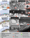
Illustrative model of melanization in C. neoformans. Melanogenesis in C. neoformans is characterized by multiple events that can be grouped into four phases: synthesis, transport, aggregation-deposition, and remodeling. Intracellular biosynthesis of the polymer occurs within melanosomes (nanospheres 10–20 nm in diameter) enclosed in MVBs (60, 81) (A) and laccase-containing vesicles (89) (B, top inset). These organelles could be signaled to start the biopolymer production via incorporation of the mature Qsp1 peptide (64). Active melanization is evidenced by the presence of melanosomes with variable degrees of pigmentation within these lipid bilayer compartments. Melanosomes forming aggregates ∼60 nm in diameter are also noticeable in the cell cytoplasm (B, bottom inset). Melanosomes are transported to the cell wall using an unconventional mechanism that involves invaginations of the plasma membrane (79, 80, 90). Finger-like protrusions of the plasma membrane scoop up melanosomes to transport them from the cytosol to the cell wall (C and D, white arrowheads). Once in the cell wall, aggregation of melanosomes up to 200-nm melanin granules might be promoted by laccase enzyme delivered to the cell wall via MVB fusion with the plasma membrane (46, 81, 89) (E), whereas their arrangement within the fungal cell wall may use as a scaffold concentric membranous sheets reported in basidiomycetes (49, 93, 94) (F, white arrows). Melanosomes anchor to the cell wall via multiple interactions: (i) associative and covalent interactions between them and cell-wall constituents (chitin, chitosan, glucans, and lipids), possibly mediated by Blp1-chitin and CNAG_05312-glucans/chitin/chitosan (16, 18, 19), and (ii) electrostatic interactions modulated by chitosan–melanin charges (14, 31, 32, 34). Regular cell growth requires the cell-wall remodeling to allow budding inducing the melanin scaffold degradation/alteration via secretion of peptidases (96), chitinases (97), and glucanase (62), thus releasing melanin granules and string-like granules (G) into the extracellular environment. Removal of nonpigmented acid-labile cell-wall components exposes the concentric layered arrangement of the melanin granules within the cryptococcal cell wall from melanin ghosts (48, 94) (H). Scale bars, 100 nm (main micrographs), and 50 nm (insets).
Figure 13.
Figure 13.
Summary of C. neoformans' melanin particles visualized with different microscopic approaches. These images are also found in Figs. 2 and 9.

References

    1. Shosuke I., Kazumasa W., Marco d. I, Alessandra N., and Alessandro P. (2011) in Melanins and Melanosomes: Biosynthesis, Biogenesis, Physiological, and Pathological Functions. (Borovansky J., and Patrick A., R., eds) 1st Ed., pp. 167–185, Wiley-VCH Verlag & Co. KGaA, Germany
    1. Hill H. Z. (1992) The function of melanin or six blind people examine an elephant. Bioessays 14, 49–56 10.1002/bies.950140111 - DOI - PubMed
    1. Cordero R. J., and Casadevall A. (2017) Functions of fungal melanin beyond virulence. Fungal Biol. Rev. 31, 99–112 10.1016/j.fbr.2016.12.003 - DOI - PMC - PubMed
    1. Christensen B. M., Li J., Chen C. C., and Nappi A. J. (2005) Melanization immune responses in mosquito vectors. Trends Parasitol. 21, 192–199 10.1016/j.pt.2005.02.007 - DOI - PubMed
    1. White L. P. (1958) Melanin: a naturally occurring cation exchange material. Nature 182, 1427–1428 10.1038/1821427a0 - DOI - PubMed

Publication types