. 2019 Jul 17;93(15):e02307-18.
doi: 10.1128/JVI.02307-18. Print 2019 Aug 1.

Junín Virus Promotes Autophagy To Facilitate the Virus Life Cycle

Affiliations

Junín Virus Promotes Autophagy To Facilitate the Virus Life Cycle

Julieta S Roldán et al. J Virol. .

Abstract

Junín virus (JUNV), a member of the family Arenaviridae, is the etiological agent of Argentine hemorrhagic fever (AHF), a potentially deadly endemic-epidemic disease affecting the population of the most fertile farming land of Argentina. Autophagy is a degradative process with a crucial antiviral role; however, several viruses subvert the pathway to their benefit. We determined the role of autophagy in JUNV-infected cells by analyzing LC3, a cytoplasmic protein (LC3-I) that becomes vesicle membrane associated (LC3-II) upon induction of autophagy. Cells overexpressing enhanced green fluorescent protein (EGFP)-LC3 and infected with JUNV showed an increased number of LC3 punctate structures, similar to those obtained after starvation or bafilomycin A1 treatment, which leads to autophagosome induction or accumulation, respectively. We also monitored the conversion of LC3-I to LC3-II, observing LC3-II levels in JUNV-infected cells similar to those observed in starved cells. Additionally, we kinetically studied the number of LC3 dots after JUNV infection and found that the virus activated the pathway as early as 2 h postinfection (p.i.), whereas the UV-inactivated virus did not induce the pathway. Cells subjected to starvation or pretreated with rapamycin, a pharmacological autophagy inductor, enhanced virus yield. Also, we assayed the replication capacity of JUNV in Atg5 knockout or Beclin 1 knockdown cells (both critical components of the autophagic pathway) and found a significant decrease in JUNV replication. Taken together, our results constitute the first study indicating that JUNV infection induces an autophagic response, which is functionally required by the virus for efficient propagation.IMPORTANCE Mammalian arenaviruses are zoonotic viruses that cause asymptomatic and persistent infections in their rodent hosts but may produce severe and lethal hemorrhagic fevers in humans. Currently, there are neither effective therapeutic options nor effective vaccines for viral hemorrhagic fevers caused by human-pathogenic arenaviruses, except the vaccine Candid no. 1 against Argentine hemorrhagic fever (AHF), licensed for human use in areas of endemicity in Argentina. Since arenaviruses remain a severe threat to global public health, more in-depth knowledge of their replication mechanisms would improve our ability to fight these viruses. Autophagy is a lysosomal degradative pathway involved in maintaining cellular homeostasis, representing powerful anti-infective machinery. We show, for the first time for a member of the family Arenaviridae, a proviral role of autophagy in JUNV infection, providing new knowledge in the field of host-virus interaction. Therefore, modulation of virus-induced autophagy could be used as a strategy to block arenavirus infections.

Keywords: Arenaviridae; Beclin 1; Junín virus; autophagy; innate immunity; proviral role; replication.


Figures

FIG 1
FIG 1
JUNV infection induces the autophagy pathway. (A) A549 cells were treated with MEM (MOCK), MEM plus 100 nM BafA1, 100 nM RAP, EBSS, EBSS plus 100 nM BafA1, or 100 nM WORT or infected with JUNV at an MOI of 1. After 2 h of treatment, the cells were processed for Western blot analysis. *, P < 0.05. (B) A549 cells were mock treated or infected with JUNV at an MOI of 1. At 5 and 11 h p.i., BafA1 was added for 1 h before processing. A rabbit polyclonal anti-LC3 antibody, monoclonal antibodies against the JUNV NP, and a monoclonal antibody against β-tubulin were used, followed by the corresponding peroxidase-conjugated secondary antibodies. The quantifications of the Western blot were conducted as described in Materials and Methods. *, P < 0.05. (C and D) A549 cells were treated with EBSS, RAPA, WORT, or MEM for 2 h and then infected with JUNV at an MOI of 1; 24 h p.i., the cells were processed for Western blot or immunofluorescence analysis. (C) After 24 h, cells were processed for Western blot analysis using monoclonal antibodies against the JUNV NP and a monoclonal antibody against β-tubulin, followed by the corresponding peroxidase-conjugated secondary antibodies. The quantifications of the Western blot analysis were conducted as described in Materials and Methods. *, P < 0.05. (D) At 24 h p.i., supernatants were harvested, and titers were determined by a PFU assay. Also, in order to determine the percentage of infected cells, samples were fixed and processed for immunofluorescence assay to detect viral NP, as described in Materials and Methods, and the nuclei were stained with Hoechst stain (magnification, ×100; *, P < 0.05). The data represent means and SE. In all cases, a representative experiment from three independent experiments is shown.
FIG 2
FIG 2
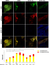
JUNV infection promotes autophagic induction. A549 cells were transfected with a plasmid encoding the mCherry-GFP-LC3 tandem construct or with the empty construct. Twenty-four hours posttransfection, the cells were treated with MEM (MOCK) or MEM plus 100 nM BafA1, and the numbers of red and yellow punctate structures were determined. Two-tailed ANOVA with Tukey’s test contrast was used to determine statistical significance, considering the total number of dots (red plus yellow) under each condition (*, P < 0.05). The ratio of red dots to yellow dots (Ratio R/Y) was also determined as a measure of the autophagy flux. Representative images were taken using a confocal microscope (Olympus FV 1000). The data represent means and SE. In all cases, a representative experiment from three independent experiments is shown.
FIG 3
FIG 3
Efficient JUNV replication requires the autophagy protein Atg5. (A) WT and atg5−/− MEFs were treated with MEM or EBSS for 2 h and then fixed and processed for immunofluorescence assay using an LC3 antibody (magnification, ×400). (B to D) Both types of MEF and the Atg5-complemented atg5−/− MEFs were infected with JUNV at an MOI of 1. At 24 and 48 h p.i., the supernatants were collected, and the cells were fixed and processed for immunofluorescence using LC3 (red) and NP (green) antibodies. The numbers of infected and total cells were determined and then related to the control treatment (panel C) (*, P < 0.05). (C) Titers of the supernatants harvested 24 and 48 h p.i. were determined by PFU assay (*, P < 0.05). (D) Cells were processed for Western blotting 24 and 48 h p.i. LC3 and β-tubulin antibodies were used. The protein band quantification was conducted as described in Materials and Methods. The data represent means and SE. In all cases, a representative experiment from three independent experiments is shown.
FIG 4
FIG 4
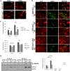
JUNV requires the autophagic protein Beclin 1. A549 cells were transfected with a plasmid encoding a pSuper Beclin 1 (siRNA) construct or the empty construct (control). Twenty-four hours posttransfection, the cells were infected with JUNV at an MOI of 1 or mock treated. Twenty-four hours p.i., the cells were fixed and processed for Western blotting (A) or immunofluorescence analysis (B). (A) Monoclonal antibodies against Beclin 1, JUNV NP, and β-tubulin were used, followed by the corresponding peroxidase-conjugated secondary antibodies. The quantifications of the Western blot analysis were conducted as described in Materials and Methods (*, P < 0.05). (B) A monoclonal antibody against the JUNV NP was used as described in Materials and Methods to detect infected cells. Nuclei were stained with Hoechst stain in order to determine the percentage of infected cells (*, P < 0.05). (C) Supernatants were harvested, and titers were determined by a PFU assay (*, P < 0.05). The data represent means and SE. In all cases, a representative experiment from three independent experiments is shown.
FIG 5
FIG 5
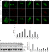
UV-inactivated JUNV infection does not induce LC3-II lipidation. A549 cells were transfected with a plasmid construct encoding EGFP-LC3. (A) Twenty-four hours posttransfection, cells were mock treated or treated with EBSS, 100 nM BafA1, 100 nM RAP, or 100 nM WORT or infected with JUNV or UV-inactivated JUNV (JUNV*) at an MOI of 1. After 2 h of treatment and at 2 or 6 h p.i., the cells were fixed and processed for immunofluorescence assay. An antibody against the JUNV GP was used, and the number of punctate structures was determined. Representative images were taken using a confocal microscope (Olympus FV 1000) (*, P < 0.05). (B) Twenty-four hours posttransfection, cells were mock treated or infected with JUNV or JUNV* at an MOI of 1. After 2 h of treatment and at different times p.i., the cells were processed for Western blotting using antibodies to EGFP, NP, and β-tubulin, followed by the corresponding peroxidase-conjugated secondary antibodies. The quantifications of the Western blot analysis were conducted as described in Materials and Methods (*, P < 0.05). The data represent means and SE. In all cases, a representative experiment from three independent experiments is shown.

References

    1. Buchmeier MJ. 2007. Arenaviridae: the virus and their replication, p 1792–1827. In Knipe DM, Howley PM, Griffin DE, Lamb RA, Martin MA, Roizman B, Straus SE (ed), Fields Virology, 5th ed, vol 2 Lippincott Williams & Wilkins, Philadelphia, PA.
    1. Charrel RN, de Lamballerie X, Emonet S. 2008. Phylogeny of the genus Arenavirus. Curr Opin Microbiol 11:362–368. doi:10.1016/j.mib.2008.06.001. - DOI - PubMed
    1. Briese T, Paweska JT, McMullan LK, Hutchison SK, Street C, Palacios G, Khristova ML, Weyer J, Swanepoel R, Egholm M, Nichol ST, Lipkin WI. 2009. Genetic detection and characterization of Lujo virus, a new hemorrhagic fever–associated arenavirus from Southern Africa. PLoS Pathog 5:e1000455. doi:10.1371/journal.ppat.1000455. - DOI - PMC - PubMed
    1. Frame JD, Gocke DJ, Baldwin JM, Troup JM. 1970. Lassa fever, a new virus disease of man from West Africa. Am J Trop Med Hyg 19:670–676. doi:10.4269/ajtmh.1970.19.670. - DOI - PubMed
    1. Parodi AS, Greenway DJ, Rugiero HR, Frigerio M, De La Barrera JM, Mettler N, Garzon F, Boxaca M, Guerrero L, Nota N. 1958. Concerning the epidemic outbreak in Junin. Dia Med 30:2300–2301. - PubMed

Publication types

Substances

LinkOut - more resources