. 2019 Jun 11;116(24):12007-12012.
doi: 10.1073/pnas.1812056116. Epub 2019 May 22.

Kinase pathway inhibition restores PSD95 induction in neurons lacking fragile X mental retardation protein

Affiliations

Kinase pathway inhibition restores PSD95 induction in neurons lacking fragile X mental retardation protein

Ying Yang et al. Proc Natl Acad Sci U S A. .

Abstract

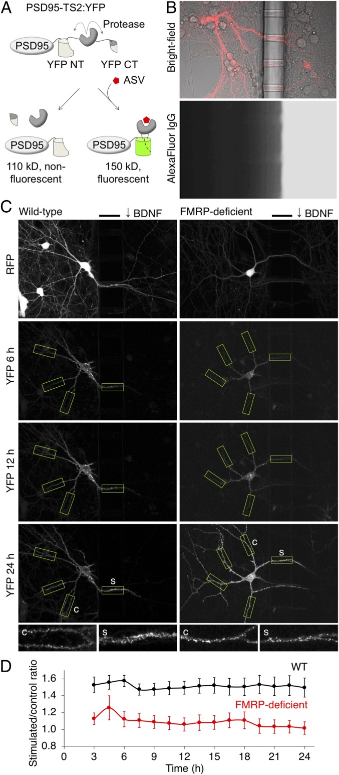
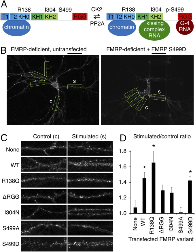
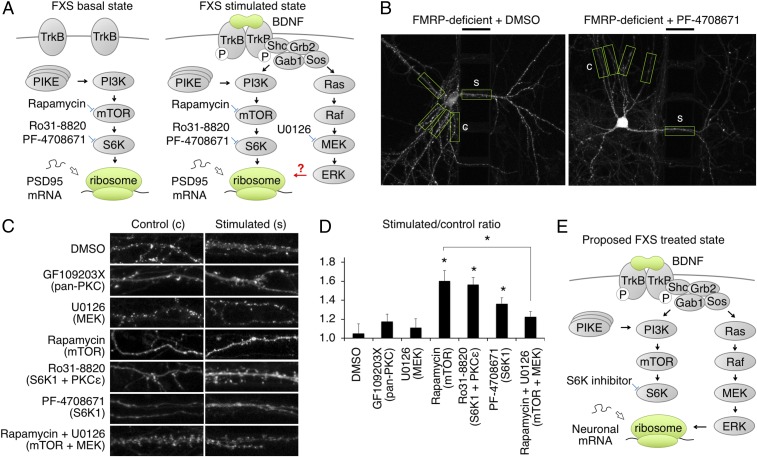
Fragile X syndrome (FXS) is the leading monogenic cause of autism and intellectual disability. FXS is caused by loss of expression of fragile X mental retardation protein (FMRP), an RNA-binding protein that regulates translation of numerous mRNA targets, some of which are present at synapses. While protein synthesis deficits have long been postulated as an etiology of FXS, how FMRP loss affects distributions of newly synthesized proteins is unknown. Here we investigated the role of FMRP in regulating expression of new copies of the synaptic protein PSD95 in an in vitro model of synaptic plasticity. We find that local BDNF application promotes persistent accumulation of new PSD95 at stimulated synapses and dendrites of cultured neurons, and that this accumulation is absent in FMRP-deficient mouse neurons. New PSD95 accumulation at sites of BDNF stimulation does not require known mechanisms regulating FMRP-mRNA interactions but instead requires the PI3K-mTORC1-S6K1 pathway. Surprisingly, in FMRP-deficient neurons, BDNF induction of new PSD95 accumulation can be restored by mTORC1-S6K1 blockade, suggesting that constitutively high mTORC1-S6K1 activity occludes PSD95 regulation by BDNF and that alternative pathways exist to mediate induction when mTORC1-S6K1 is inhibited. This study provides direct evidence for deficits in local protein synthesis and accumulation of newly synthesized protein in response to local stimulation in FXS, and supports mTORC1-S6K1 pathway inhibition as a potential therapeutic approach for FXS.

Keywords: BDNF; FMRP; PSD95; fragile X syndrome; mTORC1.


Conflict of interest statement

The authors declare no conflict of interest.

Figures

Fig. 1.
Fig. 1.
Local expression of new PSD95 in BDNF-stimulated dendritic regions is absent in FMRP-deficient neurons. (A) Diagram of the PSD95-TS2:YFP reporter. After asunaprevir is added, YFP fluorescence is preserved on new PSD95 copies. CT, C terminus; NT, N terminus. (B) Compartmentalized culture system. (B, Top) Superimposed fluorescence and bright-field images showing a transfected neuron extending dendrites through tunnels in the barrier. (B, Bottom) Dye added to one side for 24 h demonstrates separation between compartments and dye diffusion down the tunnels. Barrier width is 50 μm. (C) New PSD95 in WT or FMRP-deficient neurons after local BDNF stimulation. The bars mark the location of the 50-μm barrier. BDNF was added to the right chamber. Boxes indicate regions identified and analyzed by the automated algorithm. Stimulated segments were defined as the 50 μm in the tunnel, and then control segments were defined in the unstimulated compartment at an equidistant path length from the cell body. (C, Insets) Stimulated (s) and control (c) segments. (D) Stimulated/control intensity ratios in FMRP-deficient neurons were significantly lower than in WT neurons at all times (P = 0.03 by mixed-effect repeated-measures ANOVA; n = 31 WT and 24 FMRP-deficient neurons). Error bars represent SEM.
Fig. 2.
Fig. 2.
Translational repression by FMRP is required cell-autonomously for BDNF-induced local expression of new PSD95. (A) Diagram of FMRP protein interactions disrupted by mutations. KH, heterogeneous nuclear ribonucleoprotein K homology domain; T, Tudor domain. (B) Representative images of new PSD95 in FMRP-deficient neurons expressing RFP only (Left) or FMRP S499D (Right) 21–24 h after local BDNF stimulation. The black bars above the images indicate the location of the barrier and provide a 50-μm scale bar. BDNF was applied to the right of the barrier. Boxes indicate stimulated and equidistant control regions identified and analyzed by the automated algorithm. (C) Representative stimulated and control regions for each transfected FMRP variant. Whole-neuron images are in SI Appendix, Fig. S3. (D) Relative intensities of stimulated versus unstimulated control dendritic segments. Error bars represent SEM. Untransfected, n = 10 neurons; WT, n = 11 neurons, *P = 0.008; R138Q, n = 14 neurons, *P = 0.01; ΔRGG, n = 21 neurons, P = 0.11; I304N, n = 17 neurons, P = 0.18; S499A, n = 13 neurons, P = 0.58; S499D, n = 15 neurons, *P = 0.005 [each condition vs. untransfected by Student’s t test with Tukey’s correction, after ANOVA test with F (6, 95) = 5.77, P = 3.7 × 10⁻⁵].
Fig. 3.
Fig. 3.
Inhibition of mTOR or S6K1 rescues BDNF-induced new PSD95 accumulation in FMRP-deficient neurons. (A) Proposed model of mTORC1-S6K1 pathway hyperactivity in FMRP-deficient neurons, occluding induction of local protein synthesis by stimuli. (B) Representative images of new PSD95 in FMRP-deficient neurons 21–24 h after local BDNF stimulation with DMSO (Left) or 10 μM S6K1 inhibitor PF-4708671 (Right). The black bars above the images indicate the location of the barrier and provide a 50-μm scale bar. BDNF was applied to the right of the barrier. Boxes indicate stimulated and equidistant control regions identified and analyzed by the automated algorithm. (C) Representative stimulated and control regions of neurons with DMSO, 2 μM pan-PKC inhibitor GF109203X, 20 μM MEK inhibitor U0126, 10 μM mTORC1 inhibitor rapamycin, 2 μM S6K1/PKC inhibitor Ro31-8820, 10 μM S6K1 inhibitor PF-4708671, or 10 μM rapamycin + 20 μM U0126 added to the stimulated dendrites together with BDNF. (D) Relative intensities of stimulated versus unstimulated control dendritic segments. Error bars represent SEM. DMSO, n = 12 neurons; GF109203X, n = 10 neurons, P = 0.35; U0126, n = 13 neurons, P = 0.67; rapamycin, n = 16 neurons, *P = 0.001; Ro31-8820, n = 12 neurons, *P = 0.0005; PF-4708671, n = 11 neurons, *P = 0.02; rapamycin + U0126, n = 13 neurons, P = 0.13 [each condition vs. DMSO by Student’s t test with Tukey’s correction, after ANOVA test with F (6, 81) = 5.67, P = 5.9 × 10⁻⁵]. The difference between rapamycin and rapamycin + U0126 was also statistically significant (*P = 0.01). (E) Proposed treatment model for FXS.

References

    1. Dahlhaus R., Of men and mice: Modeling the fragile X syndrome. Front. Mol. Neurosci. 11, 41 (2018). - PMC - PubMed
    1. Kazdoba T. M., Leach P. T., Silverman J. L., Crawley J. N., Modeling fragile X syndrome in the Fmr1 knockout mouse. Intractable Rare Dis. Res. 3, 118–133 (2014). - PMC - PubMed
    1. Kooy R. F., Of mice and the fragile X syndrome. Trends Genet. 19, 148–154 (2003). - PubMed
    1. Bassell G. J., Warren S. T., Fragile X syndrome: Loss of local mRNA regulation alters synaptic development and function. Neuron 60, 201–214 (2008). - PMC - PubMed
    1. Richter J. D., Bassell G. J., Klann E., Dysregulation and restoration of translational homeostasis in fragile X syndrome. Nat. Rev. Neurosci. 16, 595–605 (2015). - PMC - PubMed

Publication types

MeSH terms

Substances

LinkOut - more resources