. 2019 May 22;11(493):eaav7325.
doi: 10.1126/scitranslmed.aav7325.

Phagocytosis-shielded lentiviral vectors improve liver gene therapy in nonhuman primates

Affiliations

Phagocytosis-shielded lentiviral vectors improve liver gene therapy in nonhuman primates

Michela Milani et al. Sci Transl Med. .

Abstract

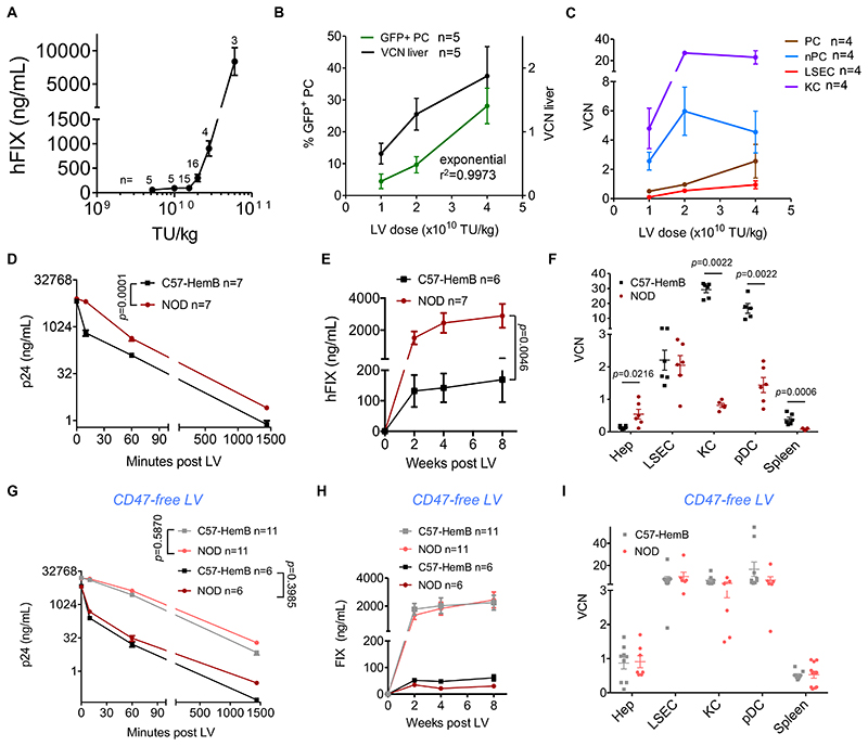
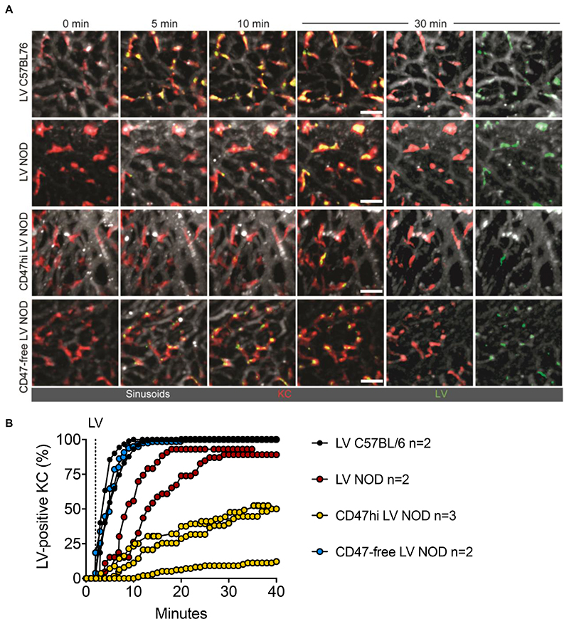
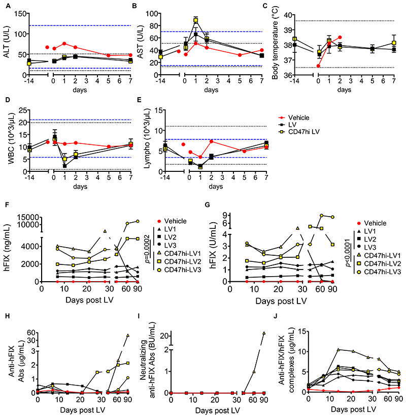
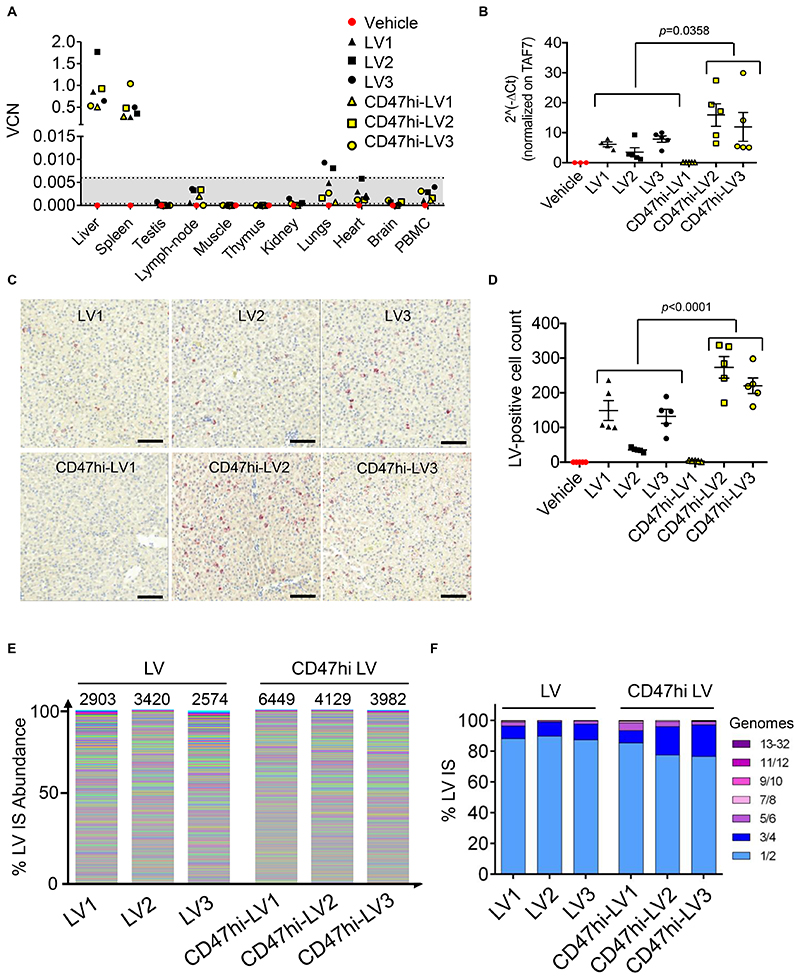
Liver-directed gene therapy for the coagulation disorder hemophilia showed safe and effective results in clinical trials using adeno-associated viral vectors to replace a functional coagulation factor, although some unmet needs remain. Lentiviral vectors (LVs) may address some of these hurdles because of their potential for stable expression and the low prevalence of preexisting viral immunity in humans. However, systemic LV administration to hemophilic dogs was associated to mild acute toxicity and low efficacy at the administered doses. Here, exploiting intravital microscopy and LV surface engineering, we report a major role of the human phagocytosis inhibitor CD47, incorporated into LV cell membrane, in protecting LVs from uptake by professional phagocytes and innate immune sensing, thus favoring biodistribution to hepatocytes after systemic administration. By enforcing high CD47 surface content, we generated phagocytosis-shielded LVs which, upon intravenous administration to nonhuman primates, showed selective liver and spleen targeting and enhanced hepatocyte gene transfer compared to parental LV, reaching supraphysiological activity of human coagulation factor IX, the protein encoded by the transgene, without signs of toxicity or clonal expansion of transduced cells.


Conflict of interest statement

Competing interests

L.N., A.C, A.A., M.M., R.P., T.L., S.P.-W. are inventors on patent applications (Vector Production, P105283GB, P114659GB) submitted by Foundation Telethon (F.T.) and San Raffaele Scientific Institute (S.R.S.I.) or Bioverativ on LV technology related to the work presented in this manuscript. F.T. and S.R.S.I., through SR-Tiget, have established a research collaboration on liver-directed lentiviral gene therapy of hemophilia with Bioverativ.

Figures

Fig. 1
Fig. 1. Role of CD47 in LV biodistribution within the liver of i.v. injected mice.
(A) Mean with standard error of the mean (SEM) of human FIX (hFIX) expression measured in the plasma of C57BL/6 mice treated at the indicated LV doses (n=48, from 8 independent experiments, performed with 3 different LV batches; the n of mice per dose is reported on the top of each point). (B) Mean with SEM of percentage of GFP-positive PC (green line) in liver sections (5-10 optical fields scored from 6-8 non-consecutive sections/mouse), and VCN measured in genomic DNA extracted from whole liver (black line), of mice treated with the indicated dose of LV with hepatocyte-specific expression, 2 months after administration (n=5 per dose cohort). (C) Mean with SEM of VCN measured in fractionated liver PC (brown line) or nPC (light blue line), FACS-sorted LSEC (red line) or KC (purple line) of mice 2 months after LV administration (n=4 per dose cohort). (D,E) Mean with SEM of (D) LV particles (ng HIV Gag p24/mL) measured in serum, and (E) human FIX expression (ng/mL) measured in plasma of C57BL/6 hemophilia B mice (n=6, black line) or NOD mice (n=6, dark red line), treated with LV-FIX at the indicated time after administration. Two-way ANOVA for repeated measures. (F) Single values and mean with SEM of VCN measured in FACS-sorted hepatocytes (Hep), LSEC, KC or pDC, and whole spleen, as indicated, of C57BL/6 hemophilia B mice (n=5-6, black circles) or NOD mice (n=5-6, dark red circles), 2 months after administration (1.2x10¹⁰ TU/kg). Mann-Whitney test. (G,H) Mean with SEM of (G) LV particles or (H) human FIX expression as in (D,E) measured in C57BL/6 hemophilia B mice or NOD mice, treated with LV-FIX produced by CD47-negative 293T cells at 1.2x10¹⁰ TU/kg (C57-HemB n=11 black line; NOD n=11 dark red line) or 2x10¹⁰ TU/kg (C57-HemB n=6 grey line; NOD n=6 pink line). Two-way ANOVA for repeated measures. (I) Single values and mean with SEM of VCN measured as in (F), of C57BL/6 hemophilia B mice (n=9) or NOD mice (n=7-11), 2 months after CD47free LV administration (2x10¹⁰ TU/kg). Mann-Whitney test.
Fig. 2
Fig. 2. Generation and evaluation of CD47hi LV.
(A) Single values and mean with SEM of percentage of GFP-positive differentiated THP-1 cells transduced with LV (n=3, black circles) or CD47hi LV (n=6, yellow circles), at the indicated multiplicity of infection (MOI) analyzed by flow cytometry, 3 days after transduction (2 independent experiments performed with 2 different CD47hi LV batches). (B) Single values and mean with SEM of VCN in 293T cells and primary human macrophages transduced with LV (n=4 for 293T, n=6 for macrophages) or CD47hi LV (n=4 for 293T, n=5 for macrophages) at MOI 10 and analyzed 3 days after transduction (2 independent experiments performed with 2 different CD47hi LV batches produced by transfection into CD47-overexpressing 293T cells or by CD47-overexpressing LV-GFP stable producer cell line and 2 different healthy blood donors). Mann-Whitney test. (C-E) Representative photomicrographs (C) and quantitative analysis (D,E) of LV batches produced by control (LV, black circles), CD47-overexpressing (CD47hi LV, yellow circles), or CD47-negative 293T cells (CD47-free LV, light blue circles), immunostained with anti-CD47 (D) or anti-VSV.G (E) antibodies, as indicated, or as staining control without the primary antibody (ctrl, black triangles) and analyzed by electron microscopy (n=41-70 virions per sample). Kruskal-Wallis test with Dunn’s multiple comparison test. Bar, 100 nm. (F) Single values and mean with SEM of VCN in 293T cells and primary human macrophages (n=6 for 293T, n=15 for macrophages) transduced with LV (black circles) or CD47-free LV (light blue circles) at MOI 10 and analyzed 3 days after transduction (2 independent experiments with 5 different healthy blood donors). Mann-Whitney test. Note that VCN denotes integrated or non-integrated reverse-transcribed LV genome. (G) Single values and mean with SEM of percentage of HIV Gag p24 recovered at 24 hours compared to 10 minutes after LV (n=25, black circles) or CD47hi LV (n=23, yellow circles) administration to NOD mice. Mann-Whitney test. (H,I) Single values and mean with SEM of VCN in FACS-sorted hepatocytes (Hep), LSEC, KC or pDC, and whole spleen, as indicated, of NOD mice (H) injected with LV (n=13-19, n=4 for pDC, black circles) or CD47hi LV (n=11-16, n=4 for pDC, yellow circles) at 1.2-2x10¹⁰ TU/kg (3 independent experiments) or (I) injected with LV (n=6-12) or CD47hi LV (n=7-14) at 4-8x10⁹ TU/kg (3 independent experiments). VCN measured 2 months after LV administration. Mann-Whitney test. (J) Single values and mean with SEM of human FIX expression (ng/mL) measured in plasma of NOD mice injected with LV (n=12) or CD47hi LV (n=8-11) at the indicated vector dose. Mann-Whitney test. (K-O) Mean with SEM of the concentration of (K) MIP-1α, (L) MIP-1β, (M) MCP1, (N) CXCL1 and (O) G-CSF in the serum of NOD mice at the indicated time after administration of LV (n=29 for K-M; n=14 for N,O), CD47hi LV (n=12 for K-M; n=7 for N,O), CD47-free LV (n=11 for K-M; n=7 for N,O) or left untreated (n=17 for K-M; n=12 for N,O). The dashed lines show the mean concentration in untreated cohorts. Kruskal-Wallis test with Dunn’s multiple comparison test. Reported statistics refer to comparison between LV- (red line) or CD47free-LV (blue line) treated and untreated (dashed line) mice 3 hours post LV administration. p<0.05 = *; p<0.01 = **; p<0.001 = ***; p<0.0001 = ****. Complete statistical analysis of data in (K-O) is in Fig. S5.
Fig. 3
Fig. 3. Intravital imaging of LV, CD47hi or CD47-free LV uptake by liver KC in mice.
(A) IV2PM images from 8–12 z-stacks spacing 4 μm of liver of C57BL/6 or NOD mice treated with GFP-labeled LV, CD47hi or CD47-free LV as indicated, at the indicated time (minutes; note that LV i.v. injection starts at min 2). Sinusoids are labeled in white, KC in red. Scale bar, 30 μm. Separate channels (white and red or white and green) are also shown for the 30-min time point. (B) Single values of the percentage of LV-positive KC (yellow stained) over time in C57BL/6 or NOD mice treated with LV, CD47hi or CD47-free LV, as indicated (analyzed KC per mouse n = 43-130).
Fig. 4
Fig. 4. Tolerability and efficacy of i.v. LV gene therapy in NHP.
(A-E) Mean with SEM of the concentration of (A) ALT, (B) AST, (C) body temperature, (D) counts of WBC and (E) lymphocytes of vehicle (n=1, red circles), LV-treated (n=3, black squares) or CD47hi-LV treated NHP (n=3, yellow squares) at the indicated time after administration. The black dashed lines show the mean±3SD calculated on 14 pre-LV samples taken from the same animals; the blue dashed lines show the normal reference values for Macaca fascicularis. (F-J) Concentration of (F) human FIX antigen or (G) human FIX activity measured in the plasma, or (H) total anti-human FIX Abs, or (I) neutralizing anti-human FIX Abs, or (J) anti-human FIX/human FIX immune complexes measured in the serum of vehicle (n=1, red circles), LV-treated (n=3, black symbols) or CD47hi-LV treated NHP (n=3, yellow symbols) at the indicated time after administration; U: units; BU: Bethesda Units. Non-parametric two-way ANOVA on the first 30 days post LV.
Fig. 5
Fig. 5. Selectivity of i.v. LV gene therapy in NHP and IS analysis.
(A) Single values of VCN in the indicated organs of vehicle-, LV- or CD47hi-LV treated NHP at necropsy (90 days post LV). The dashed lines defining the grey area represent the lower limit of detection (0.0004) and the lower limit of quantification (0.006), thus values in the grey area can be detected (different from the negative control), but not reliably quantified (see methods section). (B) Expression analysis by qRT-PCR of WPRE normalized on the endogenous TAF7 gene (2^-ΔCt) on RNA extracted from different liver lobes of LV- or CD47hi-LV treated NHP, as indicated. Mann-Whitney test. (C,D) Counts of LV-RNA positive cells (D, LV-expressing cells) by ISH on liver tissue slices of the indicated NHP (n=5 random fields taken from 5 non-consecutive slides/NHP); representative images are shown in (C). Scale bar 100 μm. Mann-Whitney test. (E,F) Stacked bar plots representing the abundance of each LV IS retrieved from the liver of LV- or CD47hi-LV treated NHP. In (E) each LV IS is represented by a different color with the height in relative proportion with the number of retrieved genomes (frequency) over the total. In (F) the frequency by which individual LV integrations are found in 1 or more genomes are plotted in groups of increasing number of genomes.

References

    1. Dunbar CE, High KA, Joung JK, Kohn DB, Ozawa K, Sadelain M. Gene therapy comes of age. Science. 2018;359 - PubMed
    1. George LA, Sullivan SK, Giermasz A, Rasko JEJ, Samelson-Jones BJ, Ducore J, Cuker A, Sullivan LM, Majumdar S, Teitel J, McGuinn CE, et al. Hemophilia B Gene Therapy with a High-Specific-Activity Factor IX Variant. The New England journal of medicine. 2017;377:2215–2227. - PMC - PubMed
    1. Nathwani AC, Reiss UM, Tuddenham EG, Rosales C, Chowdary P, McIntosh J, Della Peruta M, Lheriteau E, Patel N, Raj D, Riddell A, et al. Long-term safety and efficacy of factor IX gene therapy in hemophilia B. N Engl J Med. 2014;371:1994–2004. - PMC - PubMed
    1. Rangarajan S, Walsh L, Lester W, Perry D, Madan B, Laffan M, Yu H, Vettermann C, Pierce GF, Wong WY, Pasi KJ. AAV5-Factor VIII Gene Transfer in Severe Hemophilia A. The New England journal of medicine. 2017;377:2519–2530. - PubMed
    1. High KA, Anguela XM. Adeno-associated viral vectors for the treatment of hemophilia. Hum Mol Genet. 2016;25:R36-41. - PMC - PubMed

Publication types