. 2019 May 22;9(1):7715.
doi: 10.1038/s41598-019-44156-9.

Exploring Collagen Parameters in Pure Special Types of Invasive Breast Cancer

Affiliations

Exploring Collagen Parameters in Pure Special Types of Invasive Breast Cancer

Rodrigo de Andrade Natal et al. Sci Rep. .

Abstract

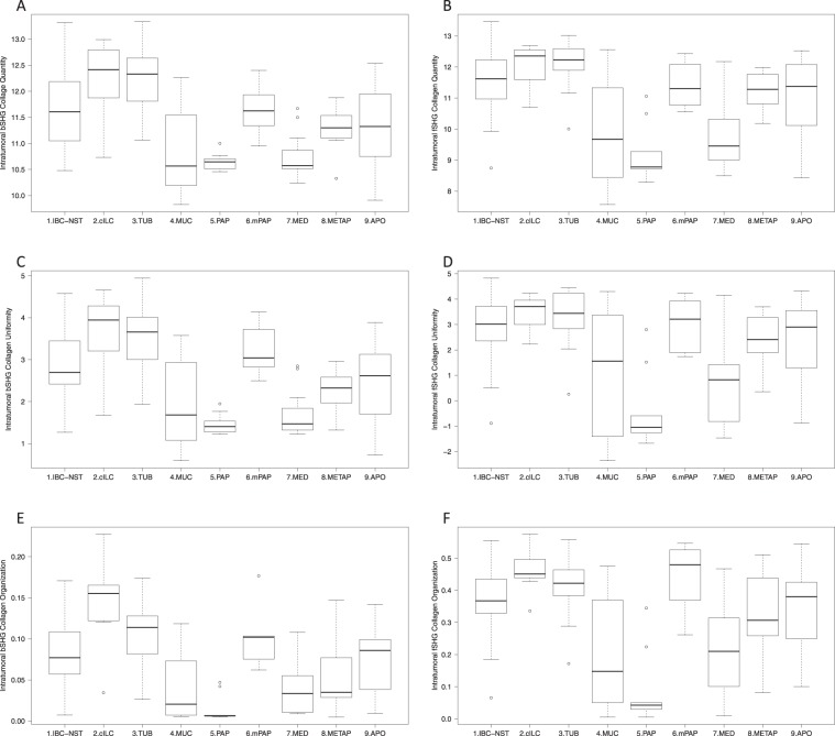
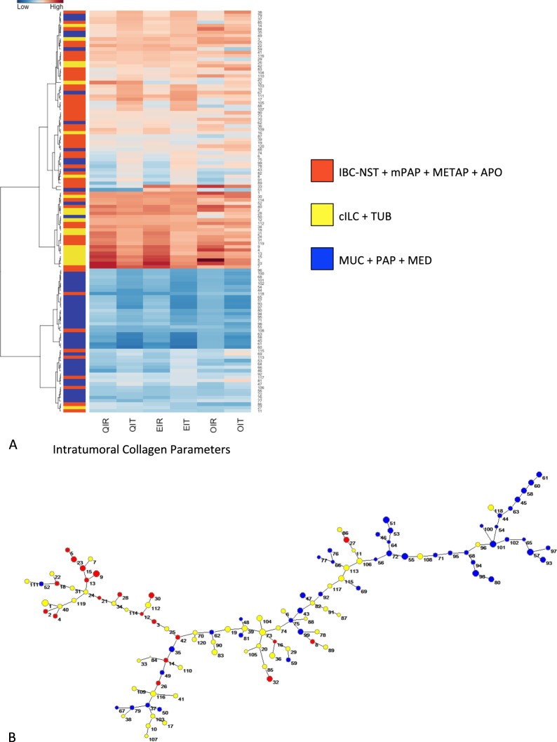
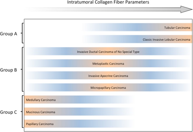
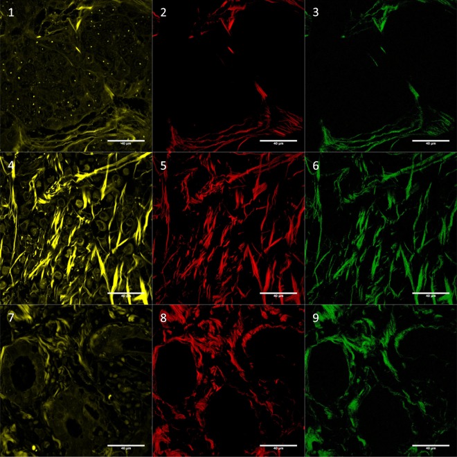
One of the promising tools to evaluate collagen in the extracellular matrix is the second-harmonic generation microscopy (SHG). This approach may shed light on the biological behavior of cancers and their taxonomy, but has not yet been applied to characterize collagen fibers in cases diagnosed as invasive breast carcinoma (BC) of histological special types (IBC-ST). Tissue sections from 99 patients with IBC-ST and 21 of invasive breast carcinoma of no special type (IBC-NST) were submitted to evaluation of collagen parameters by SHG. Tissue microarray was performed to evaluate immunohistochemical-based molecular subtype. In intratumoral areas, fSHG and bSHG (forward-SHG and backward-SHG) collagen parameters achieved their lowest values in mucinous, papillary and medullary carcinomas, whereas the highest values were found in classic invasive lobular and tubular carcinomas. Unsupervised hierarchical cluster analysis and minimal spanning tree using intratumoral collagen parameters allowed the identification of three main groups of breast cancer: group A (classic invasive lobular and tubular carcinomas); group B (IBC-NST, metaplastic, invasive apocrine and micropapillary carcinomas); and group C (medullary, mucinous and papillary carcinomas). Our findings provide further characterization of the tumor microenvironment of IBC-ST. This understanding may add information to build more consistent tumor categorization and to refine prognostication.


Conflict of interest statement

The authors declare no competing interests.

Figures

Figure 1
Figure 1
Boxplots demonstrating the distribution of intratumoral collagen parameters in histological subtypes: (A) bSHG collagen quantity; (B) fSHG collagen quantity; (C) bSHG collagen uniformity; (D) fSHG collagen uniformity; (E) bSHG collagen organization; (F) fSHG collagen organization. Classic invasive lobular (2. cILC) and tubular (3. TUB) carcinomas presented the highest values for intratumoral collagen parameters. Mucinous (4. MUC), papillary (5. PAP) and medullary (7. MED) presented the lowest values. Invasive ductal carcinoma of no special type (1. IBC-NST), micropapillary (6. mPAP), metaplastic (8. METAP) and invasive apocrine (9. APO) presented intermediary values of intratumoral collagen parameters. The statistically significant relations are shown in Supplementary Table 3. [bSHG, fSHG: backward and forward propagation second harmonic generation, respectively].
Figure 2
Figure 2
(A) Unsupervised hierarchical clustering of intratumoral collagen parameters using Ward algorithms and Euclidian distance; the lowest values for intratumoral collagen parameters refer to mucinous (MUC), papillary (PAP) and medullary (MED) breast carcinomas (light blue); the highest values refer to classical invasive lobular (cILC) and tubular carcinomas (TUB) (light yellow); the intermediary values refer to invasive ductal carcinoma of no special type (IBC-NST), micropapillary (mPAP), metaplastic (METAP) and invasive apocrine (APO) (light orange), being the latter group very heterogeneous. QIR: bSHG collagen quantity; QIT: fSHG collagen quantity; EIR: bSHG collagen uniformity; EIT: fSHG collagen uniformity; OIR: bSHG collagen organization; and OIT: fSHG collagen organization. (B) Minimal Spanning Tree (MST) of the Euclidian matrix with three defined intratumoral collagen parameters. The MSP gives a spatial representation of what is shown in the heatplot. Cases presenting similar collagen parameters are linked by a line. As result, cases belonging to the same group (blue, yellow and orange) are concentrated in three branches of the tree: blue in the superior part, yellow in the middle and right region and orange in the left branch.
Figure 3
Figure 3
Distribution of breast cancer histological subtypes per intratumoral collagen parameters. Tubular and invasive lobular breast carcinoma showed the highest intratumoral collagen parameters (quantity, uniformity and organization), while medullary, papillary and mucinous breast carcinoma presented the lowest intratumoral collagen parameters. Invasive ductal, metaplastic, invasive apocrine and micropapillary breast carcinomas presented intermediary intratumoral collagen parameters.
Figure 4
Figure 4
Autofluorescence (01, 04 and 07), fSHG collagen (02, 05 and 08) and bSHG collagen (03, 06 and 09). Images correspond to: invasive ductal carcinoma no special type (01, 02 and 03), classic invasive lobular (04, 05 and 06) and tubular (07, 08 and 09). Autofluorescence was included in this Figure to show tissue architecture. The images were log-transformed. For more examples see Supplementary Fig. 1. [bSHG, fSHG: backward and forward propagation second harmonic generation, respectively].

References

    1. Weigelt B, Geyer FC, Reis-Filho JS. Histological types of breast cancer: how special are they? Mol. Oncol. 2010;4:192–208. doi: 10.1016/j.molonc.2010.04.004. - DOI - PMC - PubMed
    1. Weigelt B, Reis-Filho JS. Histological and molecular types of breast cancer: is there a unifying taxonomy? Nat. Rev. Clin. Oncol. 2009;6:718–30. doi: 10.1038/nrclinonc.2009.166. - DOI - PubMed
    1. Lakhani, S., Ellis, I., Schnitt, S., Tan, P. & van de Vijver, M. WHO Classification of Tumours of the Breast, Fourth Edition - WHO - OMS (2012).
    1. Page DL. Special types of invasive breast cancer, with clinical implications. Am. J. Surg. Pathol. 2003;27:832–5. doi: 10.1097/00000478-200306000-00016. - DOI - PubMed
    1. Azzopardi JG, Ahmed A, Millis RR. Problems in breast pathology. Major Probl. Pathol. 1979;11(i–xvi):1–466. - PubMed

Publication types

MeSH terms