. 2019 May;569(7758):718-722.
doi: 10.1038/s41586-019-1228-x. Epub 2019 May 22.

A conserved PLPLRT/SD motif of STING mediates the recruitment and activation of TBK1

Affiliations

A conserved PLPLRT/SD motif of STING mediates the recruitment and activation of TBK1

Baoyu Zhao et al. Nature. 2019 May.

Abstract

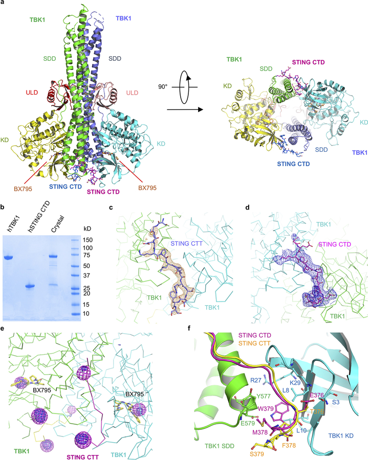
Nucleic acids from bacteria or viruses induce potent immune responses in infected cells1-4. The detection of pathogen-derived nucleic acids is a central strategy by which the host senses infection and initiates protective immune responses5,6. Cyclic GMP-AMP synthase (cGAS) is a double-stranded DNA sensor7,8. It catalyses the synthesis of cyclic GMP-AMP (cGAMP)9-12, which stimulates the induction of type I interferons through the STING-TBK1-IRF-3 signalling axis13-15. STING oligomerizes after binding of cGAMP, leading to the recruitment and activation of the TBK1 kinase8,16. The IRF-3 transcription factor is then recruited to the signalling complex and activated by TBK18,17-20. Phosphorylated IRF-3 translocates to the nucleus and initiates the expression of type I interferons21. However, the precise mechanisms that govern activation of STING by cGAMP and subsequent activation of TBK1 by STING remain unclear. Here we show that a conserved PLPLRT/SD motif within the C-terminal tail of STING mediates the recruitment and activation of TBK1. Crystal structures of TBK1 bound to STING reveal that the PLPLRT/SD motif binds to the dimer interface of TBK1. Cell-based studies confirm that the direct interaction between TBK1 and STING is essential for induction of IFNβ after cGAMP stimulation. Moreover, we show that full-length STING oligomerizes after it binds cGAMP, and highlight this as an essential step in the activation of STING-mediated signalling. These findings provide a structural basis for the development of STING agonists and antagonists for the treatment of cancer and autoimmune disorders.


Conflict of interest statement

Author Information Reprints and permissions information is available at www.nature.com/reprints. The authors declare no competing interests. Readers are welcome to comment on the online version of the paper. Correspondence and requests for materials should be addressed to P.L. (pingwei@tamu.edu).

Figures

Extended Data Fig. 1
Extended Data Fig. 1. Potential phosphorylation sites within the STING C-terminal tail.
a, List of phosphorylated STING peptides (residues 358–379, S358W) identified by LC-MS/MS. Phosphorylated residues are underlined and shown in orange. b, Representative MS/MS spectra of phosphorylated STING peptides (residues 358–379, S358W). b and c fragment ions are shown in red, while y and z fragment ions are shown in blue. Residues phosphorylated are shown in orange. The data are representative of two independent experiments. c, STING and cGAMP dependent activation of IFN-β luciferase reporter in HEK293T cells. The cells were transfected with indicated amounts of pcDNA3.1-wild-type human STING (STING WT) plasmid and stimulated with cGAMP. Luciferase signals from stimulated cells are indicated by orange bars and unstimulated controls by blue bars. The data (mean ± S.E.M) are representative of three independent experiments. Each dot represents a technical replicate (n = 3). The p values were calculated by two-tailed Student’s t test: * p < 0.05, ** p < 0.01, *** p < 0.001, NS-not significant. d, Western blot of HEK293T cells transfected with the plasmids of STING WT or deletion of the nine C-terminal residues. The data are representative of three independent experiments.
Extended Data Fig. 2
Extended Data Fig. 2. Surface plasmon resonance (SPR) binding studies of human STING with TBK1.
a, Domain organization of human STING and truncated forms of STING used in this study. b, SDS-PAGE analyses of human STING (left panel) and TBK1 (right panel) used in the SPR studies. c to l, SPR binding studies of human STING with TBK1 (upper panels). Experiments without cGAMP in the running buffer are indicated. All others were conducted with 1 μM cGAMP in the running buffer. The binding affinity (Kd) was determined by fitting the binding data to a one-site binding model (lower panels). The data of b-l are representative of at least two independent experiments.
Extended Data Fig. 3
Extended Data Fig. 3. Binding studies of human STING mutants with TBK1.
a, SDS-PAGE analysis of human STING mutants and human IRF-3 used in the SPR binding studies. b to l, SPR binding studies of human STING mutants (residue 155 to 379) with human TBK1 (upper panels) in presence of 1 μM cGAMP. The binding affinity (Kd) was determined by fitting the binding data to a one-site binding model (lower panels). m, SPR binding study of STING L374A mutant with IRF-3. n, STING C-terminal tail contains a highly conserved PLPLRT/SD motif. The sequence Logo of STING is generated by WebLogo based on the sequence alignment of STING from mammals. The frequency of occurrence of an amino acid is indicated underneath the sequence. The PLPLRT/SD motif is downstream of the pLxIS motif that is involved in IRF-3 binding. o, SPR binding study of the high affinity phosphomimetic EMW mutant of STING with human TBK1. p, IFN-β luciferase reporter assays of STING S366A and L374A mutants. For each assay, HEK293T cells were transfected with pcDNA3.1-hSTING variants and stimulated with cGAMP. q, Time course IFN-β luciferase reporter assays of HEK293T cells transfected with WT and T376A mutant of STING. The cells were stimulated with cGAMP. r, Western blot showing the phosphorylation of STING, TBK1, and IRF-3 in HEK293T cells transfected with WT STING, the S366A and L374A mutants of STING. The data of b-m, o, and r are representative of at least two independent experiments. The data of p and q (mean ± S.E.M) are representative of three independent experiments. Each dot represents a technical replicate (n = 3 in p and n = 6 in q). The p values were calculated by two-tailed Student’s t test: * p < 0.05, ** p < 0.01, *** p < 0.001, NS-not significant.
Extended Data Fig. 4
Extended Data Fig. 4. Crystal structures of STING in complex with TBK1.
a, Ribbon representation of the structure of human TBK1 bound to human STING CTD EMW mutant (residue 155 to 379, T376E, F378M, S379W). The kinase domains (KD) are in yellow and cyan, the ubiquitin-like domains (ULD) are in pink and red, the scaffold and dimerization domains (SDD) are in green and slate. The STING CTDs are shown by the blue and magenta ball-and-stick models. The TBK1 inhibitor BX795 is shown by the orange stick models. b, SDS-PAGE analysis of crystals of human TBK1 in complex with human STING CTD EMW mutant. The data are representative of two independent experiments. c, Difference map of human STING CTT bound to mouse TBK1 contoured at 2.5 σ. The σA-weighted Fo - Fc map was calculated with STING CTT omitted from the model. STING CTT is shown by the slate ball-and-stick model. TBK1 dimer is shown by the ribbon representation colored in green and cyan. d, Difference map of human STING CTD bound to human TBK1 contoured at 2.5 σ. The σA-weighted Fo - Fc map was calculated with STING CTD omitted from the model. STING CTD is shown by the purple stick model. The TBK1 dimer is shown by the ribbons colored green and cyan. e, Anomalous difference maps of Se-Met derivative of human STING CTT EMW mutant bound to human TBK1. The blue map was calculated with model phases (ϕc) and the magenta map was calculated with experimental phases after density modification (ϕdm). The STING peptide is shown by the magenta ribbon and TBK1 shown by the green and cyan ribbons. f, Superposition of the structures of human STING CTD EMW mutant (magenta) and human STING CTT (yellow) bound to human and mouse TBK1. Mouse TBK1 is shown by the green and cyan cartoon representation. Residues Glu376, Met378, and Trp379 from STING CTD EMW mutant are shown by the magenta ball-and-stick models. Residues Thr376, Phe378, and Ser379 from STING CTT are shown as the yellow ball-and-stick models.
Extended Data Fig. 5
Extended Data Fig. 5. Binding studies of human STING with human TBK1 mutants and characterization of TBK1 knockout HEK293T cells.
a to g, Surface plasmon resonance (SPR) binding studies of phosphorylated human STING C-terminal domain (CTD, residue 155 to 379) with human TBK1 mutants in presence of 1 μM cGAMP. The binding affinity (Kd) was determined by fitting the binding data to a one-site binding model. SDS-PAGE analysis of proteins used in these studies are shown in the inset of panel a. h, Western blot characterization of TBK1 knockout HEK293T cell lines. i, IFN-β luciferase reporter assays using TBK1 knockout cells. For each assay, 0.2 ng pcDNA3.1-human STING plasmids or/and 1.0 ng pcDNA3.1-human TBK1 plasmids were transfected into TBK1 knockout cells. Luciferase signals from stimulated cells are indicated by orange bars and unstimulated controls by blue bars. The data (mean ± S.E.M) are representative of three independent experiments. Each dot represents a technical replicate (n = 3). The p values were calculated by two-tailed Student’s t test: * p < 0.05, ** p < 0.01, *** p < 0.001, NS-not significant. j, Western blot showing the phosphorylation of TBK1, STING, and IRF-3 in TBK1 knockout cells transfected with STING and TBK1 plasmids. TBK1 knockout cells were transfected with 0.2 ng pcDNA3.1-human STING plasmids or/and 1.0 ng pcDNA3.1-human TBK1 plasmids and stimulated with cGAMP. k, Immunoprecipitation and immunoblot of Flag-STING and TBK1 in TBK1 KO cells. The cells were transfected with Flag-STING and TBK1 mutants and stimulated with cGAMP. Flag-STING and TBK1 in the pull-downs and whole cell lysates (WCL) were analyzed by immunoblotting. STING was visualized with FLAG antibody. TBK1 in the pull-downs were detected with an antibody against TBK1. The data of a-h are representative of at least two independent experiments. The western blot data in j and k are representative of three independent experiments.
Extended Data Fig. 6
Extended Data Fig. 6. cGAMP binding induces the oligomerization of full-length STING.
a, Gel-filtration chromatography analyses of full length STING in presence and in absence of 1 μM cGAMP using a Superose 6 column. b, SDS-PAGE analyses of fractions containing full-length STING from gel filtration chromatography. c, Gel filtration chromatography analyses of full length STING in 0.1% DDM or Amphipol A8–35 using a Superose 6 column in presence of 1 μM cGAMP. d, SDS-PAGE analysis of cross-linked full length STING. e, SEC-MALS analysis of full length STING in 0.1% DDM and 1 μM cGAMP. f, Representative cryo-EM micrograph of full-length STING stabilized with Amphipol A8–35. g, Representative 2D averages of full-length STING particles in Amphipol A8–35. h, i, and j, Three views of 12 Å resolution map of STING oligomers. Human STING CTD dimers bound to cGAMP were docked into the map. k, A list of human STING mutants in the transmembrane domain. l, Gel-filtration chromatography analyses of wild type and mutants of full length STING in presence of 1 μM cGAMP using a Superose 6 column. m, SDS-PAGE analyses of fractions of WT STING and STING mutants purified by gel filtration chromatography using a superose 6 column. n, IFN-β luciferase reporter assays showing that the mutations in N-terminal transmembrane domain affect STING mediated signaling. Indicated amounts of pcDNA3.1-human STING plasmids were transfected into HEK293T cells. Luciferase signals from stimulated cells are indicated by orange bars and unstimulated controls by blue bars. The data (mean ± S.E.M) are representative of three independent experiments. Each dot represents a technical replicate (n = 3). The p values were calculated by two-tailed Student’s t test: * p < 0.05, ** p < 0.01, *** p < 0.001, NS-not significant. o, Western blot showing that mutations in the transmembrane domain of STING affect the phosphorylation of STING, TBK1, and IRF-3. HEK293T cells were transfected with indicated amounts of pcDNA3.1-human STING plasmids and stimulated with cGAMP. p, Confocal microscopy images of HEK293T cells transfected with WT STING and STING mutants with or without cGAMP stimulation. Scale bars denote 20 μm. The data of a-e, l, and m are representative of at least two independent experiments. The data of f, g, o, and p are representative of at least three independent experiments.
Extended Data Fig. 7
Extended Data Fig. 7. Proposed model for the recruitment and activation of TBK1 and IRF-3 through the cGAS-STING pathway.
1) cGAS is activated by dsDNA in the cytosol and catalyzes the synthesis of cGAMP from ATP and GTP. 2) cGAMP binding induces the oligomerization of STING at the ER or Golgi membranes. 3) TBK1 is recruited to the STING oligomers via its C-terminal PLPLRT/SD motif and activated by induced proximity in trans. Phosphorylation of STING by TBK1 increases the binding affinity between TBK1 and STING and facilitates further recruitment and activation of TBK1. 4) Activated TBK1 phosphorylates STING at the pLxIS motif, allowing it to recruit IRF-3 to the signaling complex. 5) The proximity of TBK1 and IRF-3 bound to adjacent STING molecules within the cGAMP-STING oligomer causes the phosphorylation of the pLxIS motif of IRF-3. 6) Phosphorylated IRF-3 dissociates from STING, oligomerizes, translocates to the nucleus, and initiates the transcription of IFN-β gene.
Fig. 1
Fig. 1. The nine C-terminal residues of STING are critical for STING-mediated signaling and TBK1 binding.
a, IFN-β luciferase reporter assays in HEK293T cells. The cells were transfected with the indicated pcDNA3.1-hSTING plasmid and stimulated with cGAMP. Luciferase signals from stimulated cells are indicated by orange bars and unstimulated controls by blue bars. The data (mean ± S.E.M) are representative of three independent experiments. Each dot represents a technical replicate (n = 3). The p values were calculated by two-tailed Student’s t test: * p < 0.05, ** p < 0.01, *** p < 0.001, NS - not significant. b, Western blot analyses of cells transfected with STING plasmids containing mutations at the phosphorylation sites and deletion of the nine C-terminal residues. c, Confocal microscopy images of HEK293T cells transfected with wild type or truncated STING with and without cGAMP stimulation. Localization of STING and TBK1 is shown. Scale bars denote 5 μm. d, Immunoprecipitation showing interactions between STING mutants and TBK1. HEK293T cells were transfected with mutants of Flag-STING and stimulated with cGAMP. Total and phosphorylated IRF-3 and TBK1 were detected with IRF-3 and TBK1 antibodies, respectively. STING was visualized with FLAG antibody. e, f, SPR binding studies of phosphorylated human STING C-terminal domain (CTD, residue 155 to 379), ΔC9 (residue 155 to 370), cGAMP-binding domain (residue 155 to 341) and C-terminal tail (residue 342 to 379) with human TBK1 (upper panel) in presence of 1 μM cGAMP. The binding affinity (Kd) was determined by fitting the binding data to a one-site binding model (lower panel).The data of b-f are representative of three independent experiments.
Fig. 2 A
Fig. 2 A. conserved PLPLRT/SD motif within the C-terminal tail of STING mediates TBK1 recruitment and activation.
a, Binding affinities (Kd) of hSTING mutants to TBK1 determined by SPR. b, IFN-β luciferase reporter assays using HEK293T cells transfected with STING mutants. The cells were transfected with pcDNA3.1-hSTING (WT or mutants) and stimulated with cGAMP. ΔC9 represents truncation of the nine C-terminal residues. EMW indicates STING with mutations T376E, F378M, and S379W. Luciferase signals from stimulated cells are represented by orange bars and unstimulated controls by blue bars. c, Western blot analyses of TBK1, STING and IRF-3 phosphorylation in HEK293T cells transfected with WT hSTING or STING mutants and stimulated with cGAMP d, Immunoprecipitation showing the interactions between STING mutants and TBK1. HEK293T cells were transfected with mutants of Flag-STING and stimulated with cGAMP. STING was detected with an antibody against the FLAG tag. e, IFN-β luciferase reporter assay of STING containing an insertion of six residues (GSGSGS) between the pLxIS motif and the PLPLRT/SD motif. f, IFN-β luciferase reporter assays in HEK293T cells expressing hSTING S366A or L374A mutant and co-expressing the S366A and L374A mutants. g, Western blot showing the phosphorylation of STING, TBK1, and IRF-3 in HEK293T cells expressing hSTING S366A or L374A mutant and co-expressing the S366A and L374A mutants. The data of b, e, and f (mean ± S.E.M) are representative of three independent experiments. Each dot represents a technical replicate (n = 3). The p values were calculated by two-tailed Student’s t test: * p < 0.05, ** p < 0.01, *** p < 0.001, NS - not significant. All western blot and immunoprecipitation data are representative of three independent experiments.
Fig. 3
Fig. 3. Crystal structures of STING in complex with TBK1.
a, Ribbon representations of mouse TBK1 in complex with hSTING C-terminal tail (CTT). A side view looking into the active site of TBK1 is shown on the left and a bottom view is shown on the right. The kinase domains (KD) are in yellow and cyan. The ubiquitin-like domains (ULD) are in pink and red. The scaffold and dimerization domains (SDD) are in green and slate. STING CTT are shown by the blue and magenta ball-and-stick models. The TBK1 inhibitor BX795 are shown by the orange stick model. b, The C-terminal tail of STING binds to the interface between the kinase domain (KD) and the scaffold and dimerization domain (SDD) of a TBK1 dimer. The KD and SDD are shown by the cyan and green surfaces respectively. The C-terminal tail of STING is shown by the magenta ball-and-stick model. c, Interactions between STING CTT and TBK1. STING CTT is shown by the magenta ball-and-stick model and TBK1 is shown as ribbons. Residues of TBK1 involved in STING binding are shown by the green and cyan ball-and-stick model. The black dashed lines indicate distances less than 3.5 Å. d, Superposition of the structures of hSTING CTD (yellow ball-and-stick) bound to human TBK1 and hSTING CTT (pink ball-and-stick) bound to mouse TBK1. Mouse TBK1 is shown by the surface representation colored according to surface electrostatic potential with positively charged surface in blue and negatively charged surface in red.
Fig. 4
Fig. 4. Mutations at the TBK1/STING interface affect STING binding and signaling.
a, Binding affinities (Kd) of TBK1 mutants to phosphorylated hSTING CTD determined by SPR. n.d., no data. b, IFN-β luciferase reporter assays using TBK1 knockout HEK293T cells transfected with TBK1 mutants. The cells were transfected with pcDNA3.1-hSTING plasmids and/or pcDNA3.1-hTBK1 plasmids and stimulated with cGAMP. The kinase inactive S172A mutant of TBK1 was used as a negative control. Luciferase signals from stimulated cells are indicated by orange bars and unstimulated controls by blue bars. The data (mean ± S.E.M) are representative of three independent experiments. Each dot represents a technical replicate (n = 3). The p values were calculated by two-tailed Student’s t test: * p < 0.05, ** p < 0.01, *** p < 0.001, NS - not significant. c, Western blot showing the phosphorylation of TBK1, STING, and IRF-3 in TBK1 knockout cells transfected with TBK1 mutants. The cells were transfected with pcDNA3.1-hSTING plasmid plus pcDNA3.1-hTBK1 plasmids and stimulated with cGAMP. The data are representative of three independent experiments.

References

    1. Chen Q, Sun L & Chen ZJ Regulation and function of the cGAS-STING pathway of cytosolic DNA sensing. Nat Immunol 17, 1142–1149, doi:10.1038/ni.3558 (2016). - DOI - PubMed
    1. Roers A, Hiller B & Hornung V Recognition of Endogenous Nucleic Acids by the Innate Immune System. Immunity 44, 739–754, doi:10.1016/j.immuni.2016.04.002 (2016). - DOI - PubMed
    1. Barber GN Innate immune DNA sensing pathways: STING, AIMII and the regulation of interferon production and inflammatory responses. Curr Opin Immunol 23, 10–20, doi:10.1016/j.coi.2010.12.015 (2011). - DOI - PMC - PubMed
    1. Kato H, Takahasi K & Fujita T RIG-I-like receptors: cytoplasmic sensors for non-self RNA. Immunol Rev 243, 91–98, doi:10.1111/j.1600-065X.2011.01052.x (2011). - DOI - PubMed
    1. Paludan SR & Bowie AG Immune sensing of DNA. Immunity 38, 870–880, doi:10.1016/j.immuni.2013.05.004 (2013). - DOI - PMC - PubMed

Publication types

Substances